Soft tissue grafts for dural reconstruction after meningioma surgery

Authors

  • Tomaz Velnar Department of Neurosurgery, University Medical Centre Ljubljana, Ljubljana, Slovenia; Alma Mater Europaea – European Center Maribor (AMEU-ECM), Maribor, Slovenia
  • Lidija Gradisnik Alma Mater Europaea – European Center Maribor (AMEU-ECM), Maribor, Slovenia; Institute of Biomedical Sciences, Faculty of Medicine, University of Maribor, Maribor, Slovenia

DOI:

https://doi.org/10.17305/bjbms.2019.3949

Keywords:

Soft tissue grafts, dura mater, dural reconstruction, brain surgery, cerebrospinal fluid leak, CSF leak, convexity meningioma

Abstract

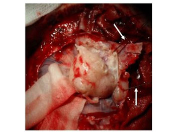
The meninges are involved in various pathologies and are often directly or indirectly severed during surgical procedures, especially the dura mater. This can pose a real challenge for the surgeon, as a proper reconstruction of the meninges is important to prevent complications such as cerebrospinal fluid leak (CSF). A variety of techniques for dural reconstruction have been described, employing natural and artificial materials. A novel technique for dural reconstruction involves soft tissue grafts in the form of fibrous or fibromuscular flaps, which are placed on the dural defects to seal the gaps. These soft tissue grafts represent an appropriate scaffold for cell ingrowth and fibrosis, thus preventing CSF. In this pilot study, we described the application of soft tissue grafts for dural reconstruction in 10 patients who underwent convexity meningioma surgery.

Downloads

Download data is not yet available.
Soft tissue grafts for dural reconstruction after meningioma surgery

Downloads

Additional Files

Published

2019-08-20

How to Cite

1.
Velnar T, Gradisnik L. Soft tissue grafts for dural reconstruction after meningioma surgery. Biomol Biomed [Internet]. 2019Aug.20 [cited 2023Apr.1];19(3):297-303. Available from: https://www.bjbms.org/ojs/index.php/bjbms/article/view/3949

Issue

Section

New and Emerging Methods

INTRODUCTION

The regeneration of lost or damaged tissues is one of the main goals of modern medicine and is particularly important in wound management [1]. Wound healing is influenced by various factors and poor healing outcomes have numerous negative consequences for the organism [1,2]. Wounds that are very slow to heal or which do not heal at all are a significant cause of patient morbidity and mortality and represent a challenge for researchers and clinicians [1]. A new approach for the treatment of complex wounds that are difficult to heal is tissue augmentation, which employs tissue grafts or fillers [3,4]. Although tissue grafts have the potential to treat defects after trauma or operation [5-7], this technique is still in the experimental stage. Soft tissue grafts may be used for tissue reinforcement in the treatment of acute and chronic wounds that cannot be adequately treated using standard-of-care treatments. They can be immediately placed into the wound to facilitate the closure. In the following stages of healing, a tissue graft in the wound acts as a matrix that fills the gap of the wound (or tissue defect) and enables the growth of different types of cells and connective tissue [2,8,9]. Soft tissue augmentation techniques are very effective in stimulating the regeneration of the bone. However, due to complications associated with the survival of tissue in the graft, soft tissue augmentation can be a challenging task. Consequently, these techniques are not widely used despite good results in some studies [10-12].

In neurosurgical operations, dural defects represent a significant problem for both the patient and surgeon, since incomplete closure may lead to cerebrospinal fluid (CSF) leak and associated complications. Impaired healing of dural defects and incomplete closure, associated with extensive and repeated neurosurgical procedures of trauma and tumors [13,14], are difficult to treat and have numerous risks. These dural defects may be reconstructed using a range of artificial or natural materials. However, no uniform strategy has been developed so far regarding the techniques used for the closure of dural defects and resulting CSF fistulas.

In the present study, we evaluated a new soft-tissue grafting technique for the reconstruction of dural defects after meningioma resection, to prevent CSF leak and related complications.

MATERIALS AND METHODS

Patients

This prospective pilot study was initiated at neurosurgery centers in Ljubljana and Maribor, in Slovenia. The National Medical Ethics Committee approved the study protocol (ethical approval number: KME 104/02/16). All patients were informed about the surgical procedure and study protocol, and they signed informed consent to participate in the study.

A total of 10 patients who underwent convexity meningioma surgery between 2016 and 2017 were included in the study. Meningiomas were classified according to the World Health Organization (WHO) classification system as grade I or II. In all 10 patients, the tumor covered large areas of the dura mater, and in two patients, it extended to the cranial bone and subcutaneous tissue. The reconstruction of the dura after tumor removal was challenging in all patients.

Surgical procedure and dural reconstruction

The neurosurgery included a standard procedure for the removal of convexity meningioma and was uneventful in all 10 patients. In two patients in whom the tumor was growing into the bone and infiltrating the subcutaneous tissue of the scalp, the bone was removed and replaced by an artificial implant. The tumor tissue involving the skin was also excised and no plastic reconstruction was needed.

Dural reconstruction was performed in later phases of the operation. Fibrous or fibromuscular tissue was used for soft tissue grafts, measuring in length from 2 cm2 to 4 cm2 (Figure 1). Fibrous tissue grafts were harvested from the inner layer of the skin flap that was formed when the scalp was retracted at the incision site. Alternatively and depending on the location of the primary tumor, fibromuscular tissue grafts were taken from the surface of the temporal muscle.

FIGURE 1: Fibrous (left) and fibromuscular tissue graft (right) thinned after harvesting and ready for implantation (A). The fibromuscular tissue graft before the attachment (B). Bar = 1 cm.

Prior to fibrous graft harvesting, the inner layer of the skin flap was lifted and the inner surface, facing the bone, was exposed as usual during the initial part of the neurosurgical operation, when the skin flap is lifted off the skin. The size of fibrous tissue graft was estimated according to dural involvement. Tissue undermining was performed with blunt scissors and only the fibrous layer was removed. Special care was taken not to damage the subcutis extensively or to penetrate the skin. Minute bleeding after harvesting was stopped by bipolar coagulation or with a wet warm patty. The donor site as well as fibrous tissue grafts after removal were kept moist. The grafts were deposited on a moist patty (Figure 2) and immediately transferred to saline solution, to prevent shrinkage.

FIGURE 2: After removal, the tissue grafts were placed on a moist patty (black arrow) and transferred to saline solution to prevent shrinkage.

The donor site for fibromuscular tissue grafts was the temporal muscle. The surface of the muscle was exposed and the fibromuscular layer was dissected and excised from the remaining muscle, including the fascia and superficial muscle layer of approximately 3-mm thickness. The majority of the muscle thickness was preserved. Bleeding from the muscle donor site that was encountered during this part of the operation was more extensive compared to fibrous graft harvesting, but was easily stopped by bipolar coagulation. The muscle was then covered with a layer of absorbable oxidized cellulose polymer (Surgicel) and protected with wet patties. The donor sites remained covered during the operation. The fibromuscular tissue grafts were immediately transferred to saline solution.

The fibrous tissue grafts were thin and ready to use immediately. On the other hand, the fibromuscular tissue grafts were thick; to enlarge their surface and make them softer they were thinned under pressure. Both types of grafts were stored in saline solution at room temperature until needed. This was done to prevent their contraction, which was especially pronounced in the fibrous grafts due to higher collagen content. Similarly, bipolar coagulation of grafts during harvesting and the exposure of grafts to a strong operating light during storage were avoided, as changes in temperature may cause shrinkage of grafts containing collagen fibers.

In all patients, the tumor-infiltrated dura was excised together with the tumor (Figure 3). The dural leaves were then brought as close as possible with nonresorbable stitches and a fibrous or fibromuscular tissue graft of appropriate size was placed over the defect so that the dural gap was covered completely (Figure 4). The graft was fixed with four zero stitches to the dural leaves in order to prevent sliding on the smooth surface. A Valsalva maneuver was performed to evaluate the integrity of the construct (graft) after the reconstruction. In addition, in some patients, fibrin glue was sprayed over the immobilized tissue grafts, sealing the opening completely. The bone or artificial implant followed after a proper dural reconstruction with the tissue grafts and the skin was closed in layers.

FIGURE 3: Magnetic resonance imaging of a typical convexity meningioma with large areas of dural involvement. The invasion of meningioma into the bone and subcutaneous tissue is evident (thick arrow), as it is meningiomatous dural tail (thin arrows) that needs to be removed together with the tumor, posing additional problem for the dural reconstruction (A). Intraoperative image of meningioma removal. Holding sutures are placed to aid with careful elevation of the tumor (arrows). The dura was excised and later reconstructed with soft tissue grafts (B).
FIGURE 4: The reconstruction of a dural gap with a fibromuscular graft (A). A fibromuscular tissue graft is in place, covering the dura mater defect. Fibrin glue was additionally sprayed over the construct to achieve a complete seal. The reconstruction of a dural gap with a fibrous graft (B). Black arrows indicate the rim of the graft attached to the dural surface, covering the defect. Black silk sutures of the approximated (sutured) dural leaves are visible. Bar = 1 cm.

RESULTS

Clinical and surgical data of 10 patients included in this pilot study are presented in Table 1. There were six men and four women, and the mean age of patients was 48 years (range: 31–75 years). Based on histopathological analysis, six patients (five men and one woman) were diagnosed with WHO grade I meningioma, while four patients (one man and three women) were diagnosed with WHO grade II meningioma. No patient had WHO grade III tumor. The tumor size ranged from 2.5 cm to 6 cm (mean 4 cm).

TABLE 1: Clinical and surgical data of patients with meningioma

In four patients, the reoperation was necessary. Three patients with grade II meningioma (one man and two women) were operated more than once due to tumor recurrence. Soft tissue grafts were used in each operation. During the reoperation, we found that the soft tissue grafts were well integrated with the dura and that fibrosis was adequate in the areas of dural reconstruction, in all three patients. One patient with grade I meningioma had a postoperative CSF leak. In this patient, a standard dural closure with fibrin patch and glue was used during the first operation. Due to the postoperative CSF leak, another reconstructive operation with soft tissue graft was required to cover the dural gaps successfully.

The soft tissue grafts obtained from the galea and temporal muscle included fibrous and fibromuscular tissue layers, respectively. The temporal muscle was the donor site for fibromuscular grafts in four patients in which the surgical approach included pterional and frontal craniotomies. In six patients, the tumors were located in the parietal and occipital regions and therefore fibrous grafts were harvested from subgaleal tissue. The skin flap that was formed when the scalp was retracted and the temporal muscle that was separated from the bone at the incision site provided donor sites that was large enough for soft tissue grafts. No surgical complications were encountered during the harvesting of soft tissue grafts. The tissue layers separated from each other well during the operation and no excess bleeding was present. With a meticulous surgical technique, it was possible to collect the grafts in one piece, without penetrating or otherwise damaging the grafts. Due to careful storage and manipulation, the size of the grafts remained unchanged and their shrinkage was negligible. Tissue folding that prevents the covering of dural defects was not observed. Since the size of the remaining dural leaves was approximated after the excision of the tumor and involved dura, no size limitations were encountered during reconstruction and the grafts were large enough to cover the defects.

During the course of treatment, we did not observe any early or late complications. There were no signs of CSF leakage or infection and the wounds were healing by the first intention. No complications related to the donor site were reported.

DISCUSSION

This clinical pilot study was designed to evaluate the usefulness of soft tissue grafts in the reconstruction of complicated dural defects after convexity meningioma surgery. We based our research on the previous experience of using soft tissue grafts in vitro [15]. Due to promising results obtained in that basic study, here we investigated the efficacy of soft tissue grafts in surgical practice. Our results confirmed that the fibrous and fibromuscular tissue grafts are useful for covering smaller dural gaps, not exceeding the size of 4 cm2. In this study, however, we were not able to harvest soft tissue grafts for larger dural defects, as the size of grafts was limited by the donor site.

The idea of using tissue grafts in the reconstruction of defects originates from wound healing research [10,16]. Most wounds will eventually heal by secondary intention, even without special treatment. However, wounds with extensive damage, soft tissue loss, and large chronic wounds will heal very slowly or not at all. To promote or accelerate healing, cover and close tissue defects, and improve the outcomes of patients after the injury, tissue engineering and reconstruction techniques may be employed [9]. Currently, free tissue transfer and pedicle flaps are used [13]. A new reconstructive technique is tissue augmentation with tissue grafts, which is suitable not only in neurosurgery but in other surgical specialties as well [13]. The main areas of application are maxillofacial and trauma surgery, especially to facilitate the healing of hard (bony) tissues in bone reconstruction and fracture repair [17-19]. Nevertheless, soft tissue augmentation techniques are still in the experimental phase and not widely used in surgery. Soft tissue grafts are sometimes utilized in plastic and reconstructive surgery and, experimentally, in wound healing [15,16]. In this study, we tested the efficacy of soft tissue grafts in the surgical reconstruction of the dura using a similar approach as in wound healing research.

Primary and secondary brain tumors that require surgical intervention represent frequent pathologies of the central nervous system (CNS) [3,4,8,9]. Most brain tumors are in the form of infiltrative and expansive lesions that destruct and compress brain tissue and, therefore, present a challenge for a surgeon, especially in the later stages of surgery when the reconstruction is needed. Some tumors may erode the adjoining structures, particularly the dura and cranial bone and, in some cases, adjacent soft tissue [1,13,14]. Meningiomas, and to a lesser extent other tumors, are well-known for infiltrating the dura mater, which requires excision and appropriate reconstruction of the dura. Both the tumor location and invasion into the neighboring tissue may be so problematic that radical resection and reconstruction cannot be easily accomplished [1,13,14]. In these conditions, the reconstruction is challenging because of tissue deficiency [15,20]. While CSF leak may complicate any brain tumor surgery when opening the subarachnoid space, the risk of CSF leak is higher at some locations, such as the posterior fossa, skull base and spinal cord. CSF leak may also occur after large convexity meningiomas with the dura and cranial bone involvement. In such cases, the reconstruction of missing tissue may be difficult [21-23]. Numerous alternatives for the reconstruction of dural defects exists, including autografts, allografts, xenografts and artificial replacement materials. Each of these techniques has some advantages and disadvantages and is optimal for specific conditions [24-26].

The choice of surgical reconstruction technique is primarily determined by the degree of CSF leak, in addition to the location, size and shape of the dural defect, as well as by the surgeon’s preferences and experience [27]. Autografts and allograft transplants represent an attractive option [24,28,29]. Autografts enable the reconstruction of large dural defects [26,20]. The survival and healing of this viable tissue is easier and less prone to infection. Autografts do not trigger the immune response and, therefore, do not cause excessive inflammation that can interfere with the healing process and result in impaired wound healing and associated complications [30]. Moreover, autografts are relatively resistant to postoperative radiation, which may be required after the resection of WHO grade III and, occasionally, WHO grade II meningiomas [28,26,30,31]. Various types of tissues can be used for autografts, including fascia lata, muscle, galea and adipose tissue. These tissue grafts may be an efficient alternative to fibrous and fibromuscular grafts [28,32]. All these autografts can be either sutured to the dural defects or attached with fibrin glue, facilitating the sealing of any potential opening between the dura and the graft. The grafts may be harvested from various locations of the patient body and are thus easily obtainable, in most cases with no trauma to other body regions where the tissue was harvested. These sites mean additional, albeit limited resections for the donor tissue. [33,34]. Viable tissue near the incision site represents another source of autografts, as demonstrated in our study.

For allografts, fascia lata and cadaveric dura mater are most often used. Compared to artificial materials, fascia lata is very flexible and can be adjusted to the surface. However, the risk of infection is higher with this type of allograft and immunological reactions against foreign tissue are possible, complicating the recovery and slowing down the healing process [30]. Cadaveric dura mater allografts and bovine pericardium xenografts may also be employed for dural reconstruction. However, the major shortcoming of these grafts is the increased risk of immune reactions and infection. In addition, the cost of allografts may be high and their availability may be limited, as they are not always available from biobanks [35,36]. Commercially available artificial replacement materials include expanded polytetrafluoroethylene dural substitutes, two-component fibrin glue, and collagen sponges [37-39]. These products are easily obtainable, although they may be available at a higher price. Their use is time-saving, straightforward, and enables good brain protection and dural covering, resulting in an optimal healing outcome. However, as with all artificial materials, the risk of infections and host versus graft immune reactions is always present [36-38].

Although grafts from viable tissues are very efficient in surgical reconstructions, their use is not without limitations and risks. This is especially true for large tissue grafts, as their harvesting may contribute to longer operative time and associated higher infection rate, postoperative complications at the donor site, longer recovery, or may lead to plastic reconstruction [33,36,40]. In our study, the grafts were up to 4 cm2 in size and were obtained directly from the flaps formed during the operation, so we did not experience difficulties during the operation or in the recovery period. Problems, however, may arise with small skin or muscle flaps and when large areas of the brain need to be covered. Therefore, a preoperative planning is essential in these procedures [24,28,36].

Because large areas of the dura sometimes need to be removed after neurosurgical operations, a proper reconstruction is mandatory. These dural defects will not heal or close on their own. As a result, CSF leak may occur and accumulate in the subcutaneous tissue, further delaying the healing of the wound [28,21]. In addition, other complications may occur, such as an external CSF leak and infection. Fistula formation is one of many unwanted complications after the resection, especially in the later phases of treatment when oncological therapy with radiation is necessary. Associated with CSF leak and impaired wound healing is a higher incidence of meningitis, which develops due to the exposure of the CNS to microbes. According to some studies, the risk of meningitis is significantly increased in patients with postoperative CSF leak, i.e., up to 14-fold [41,42], and the majority of meningitis cases after skull base operations are associated with an obscure leak [42]. A proper intraoperative reconstruction of the dura is, therefore, required to prevent CSF leak.

A number of artificial and industrial materials are available for the reconstruction of dural defects [39,43-45]. These include various fibrin adhesives and covering materials, which can be placed on dural gaps. They are especially useful in endoscopic surgery of the cranial base and to repair the dura of the convexity of the brain. Disadvantages of artificial materials are a higher risk of infection and biodegradability of some materials [39,40,44].

Tissue augmentation with soft tissue grafts used in our study for dural covering proved to be an efficient and straightforward procedure with good results. We performed this procedure immediately after the tumor resection. This soft tissue augmentation technique is time-efficient and does not require additional donor sites to be used. The fibrous and fibromuscular tissue grafts were ideal for dural covering in our case, as they were thin, adjustable and had a good strength. After it is obtained from the subcutaneous tissue or muscle, the graft is soft and can be additionally adjusted in shape to properly fit the dural defect. The adhesion of soft tissue grafts to the dural surface was adequate in all 10 cases, enabling the closure of dural defects. It created a watertight seal between the extradural space and the brain. During the healing phases the scarring was abundant, which led to the complete sealing of the openings. The minute openings in the dura may be potentially dangerous, if CSF leakage into the subcutaneous tissue or CSF liquorrhea occur [41,43,45]. In this study, we did not observe wound healing complications, nor the signs of CSF leak or CNS infection after the surgery. The reoperations, required in four patients, were also successful.

Both types of grafts utilized in the study were intended for the covering of dural defects. The surgical technique for harvesting the grafts, storage, manipulation and mode of application of grafts were similar for fibrous and fibromuscular grafts used in this study. The fibrous grafts could immediately be used as they were already thin, while the fibromuscular grafts were additionally thinned under pressure before their use, to achieve a good adhesion and complete dural closure. Thick tissue does not adhere well and is more difficult to suture. The surgical approach and location of the defect were the only factors determining the type of graft to be used in our study. In cases when the skin incision before the trepanation involved the temporal muscle, we collected fibromuscular tissue grafts due to the abundance and good accessibility of the muscular layer. In other areas of the cranial vault, fibrous tissue grafts were harvested form the lifted skin flap, as an additional incision of the skin over the temporal muscle to collect a graft would not be practical.

This study was experimental in design and, therefore, performed on a very small number of patients with convexity meningiomas, with the aim to determine whether fibrous and fibromuscular tissue grafts can successfully cover dural defects and promote the healing of the dura. Overall, our results confirm the findings of previous in vitro studies [15,19]. Although the small sample size is a significant limitation of our study, it is worth mentioning that we did not encounter any particular technical problem with the soft-tissue grafting technique. The technique was quick, efficient, and resulted in a good outcome in all 10 cases. For further evaluation of soft tissue grafts in dural reconstruction, studies involving a larger number of patents, other types of brain tumors and various tumor locations such as the skull base, posterior fossa and spine are necessary. Even though we obtained good results with fibrous and fibromuscular tissue grafts in this study, this technique is not without limitations and risks. The placement of a tissue graft on the dura is the most important step, as the graft needs to seal all dural gaps and to be fixed on the surface. However, the size of tissue grafts is limited by the donor site [24,36]. In addition, care must be taken not to harvest excess tissue from the temporal muscle or galea or damage the skin surface.

CONCLUSION

The results of our pilot study support the potential of tissue augmentation in the reconstruction of dural defects. The fibrous and fibromuscular tissue grafts integrated well with the dura, promoted scarring, and prevented postoperative CSF leakage. These soft tissue grafts seem to be a suitable alternative to artificial materials or fascia lata and may also be used in a combination with available reconstruction options. The results of our study suggest that the soft-tissue grafting technique may be safely integrated into the clinical practice and tested on a larger number of patients. Our preliminary results should encourage more studies on soft tissue augmentation in dural reconstruction.

DECLARATION OF INTERESTS

The authors declare no conflict of interests.

REFERENCES

  1. , (). General principles of wound healing. Surg Clin North Am. https://doi.org/10.1016/S0039-6109(05)70566-1
  2. (). Wound healing and aging. Dermatol Clin. https://doi.org/10.1016/S0733-↻(18)30811-8
  3. , (). Which hard tissue augmentation techniques are the most successful in furnishing bony support for implant placement?. Int J Oral Maxillofac Implants.
  4. , (). Facial defect augmentation with a dermal-fat graft. Oral Surg Oral Med Oral Pathol. https://doi.org/10.1016/0030-4220(85)90054-4
  5. , (). Bone augmentation techniques. J Periodontol. https://doi.org/10.1902/jop.2007.060048
  6. , (). Multitier technique for bone augmentation using intraoral autogenous bone blocks. Implant Dent. https://doi.org/10.1097/ID.0b013e3180327595
  7. (). Tissue engineering solutions for traumatic bone loss. J Am Acad Orthop Surg.
  8. (). Physiology of the acute wound. Clin Plast Surg.
  9. , , , , , (). Surgical mortality and selected complications in 273 consecutive craniotomiaes for intracranial tumors in pediatric patients. Neurosurgery. https://doi.org/10.1227/NEU.0b013e31823bcc61
  10. , , , (). Autologous fat grafting and facial reconstruction. J Craniofac Surg. https://doi.org/10.1097/SCS.0b013e318241e1de
  11. , , (). Soft-tissue reconstruction of the face:A comparison of dermal-fat grafting and vascularized tissue transfer. Ann Plast Surg. https://doi.org/10.1097/00000637-199211000-00002
  12. , , (). Augmentation of facial soft-tissue defects with Alloderm dermal analysis. Ann Plast Surg. https://doi.org/10.1097/00000637-199811000-00009
  13. , , , , (). Endoscopic endonasal versus open repair of anterior skull base CSF leak, meningocele, and encephalocele:A systematic review of outcomes. J Neurol Surg A Cent Eur Neurosurg. https://doi.org/10.1055/s-0032-1325636
  14. , , , , (). Reevaluation of surgery for the treatment of brain metastases:Review of 208 patients with single or multiple brain metastases treated at one institution with modern neurosurgical techniques. Neurosurgery.
  15. , , , (). Is tissue augmentation a reality in biosurgery?An experimental study of endothelial cell invasion into tissue filler. Int Wound J. https://doi.org/10.1111/j.1742-481X.2012.00980.x
  16. (). Experimental assessment of the revascularization of acellular human dermis for soft-tissue augmentation. Plast Reconstr Surg. https://doi.org/10.1097/00006534-200103000-00016
  17. (). Acute wounds:An overview of the physiological healing process. Nurs Times.
  18. , , , , , (). Clinical approach to wounds:Debridement and wound bed preparation including the use of dressings and wound-healing adjuvants. Plast Reconstr Surg. https://doi.org/10.1097/01.prs.0000225470.42514.8f
  19. , , (). Wound healing:An overview. Plast Reconstr Surg. https://doi.org/10.1097/01.prs.0000222562.60260.f9
  20. , , (). Soft tissue wounds and principles of healing. Emerg Med Clin North Am. https://doi.org/10.1016/j.emc.2006.12.002
  21. , , , , , (). Risk factors for the development of serious medical complications after resection of meningiomas. J Neurosurg. https://doi.org/10.3171/2010.6.JNS091974
  22. , , (). Brain meningioma invading and destructing the skull bone:Replacement of the missing bone in vivo. Radiol Oncol. https://doi.org/10.2478/v10019-011-0036-1
  23. , , , , , (). Surgical strategies for petroclival meningioma in 57 patients. Chin Med J (Engl).
  24. , (). In situ cranioplasty for hyperostosing meningiomas of the cranial vault. Can J Neurol Sci. https://doi.org/10.1017/S0317167100011082
  25. , , , (). Sphenoorbital meningiomas:Surgical limitations and lessons learned in their long-term management. J Neurosurg. https://doi.org/10.3171/jns.2005.103.3.0491
  26. , , , , (). Use of free composite graft for a large defect in the anterior skull base. J Craniofac Surg. https://doi.org/10.1097/00001665-199801000-00016
  27. , , , , , (). Challenging reconstructive techniques for skull base defect following endoscopic endonasal approaches. Acta Neurochir (Wien). https://doi.org/10.1007/s00701-011-0941-5
  28. , , , , , (). Dural suturing for repair of cerebrospinal fluid leak in transnasal transsphenoidal surgery. Acta Neurochir (Wien). https://doi.org/10.1007/s00701-009-0406-2
  29. , , (). Contemporary skull base reconstruction. Curr Opin Otolaryngol Head Neck Surg. https://doi.org/10.1097/01.moo.0000131445.27574.be
  30. , , , , (). Craniofacial neurosurgical approach for extensive hyperostotic meningioma. J Craniofac Surg. https://doi.org/10.1097/00001665-199307000-00004
  31. , (). Bone graft materials. An overview of the basic science. Clin Orthop Relat Res. https://doi.org/10.1097/00003086-200002000-00003
  32. , (). Options for the management of forehead and scalp defects. Facial Plast Surg Clin North Am. https://doi.org/10.1016/j.fsc.2009.05.001
  33. , , (). Mass effect due to hypertrophic pericranial flap in the reconstruction of dural defect. World Neurosurg. https://doi.org/10.1016/j.wneu.2015.07.070
  34. , , (). Scalp and forehead reconstruction. Semin Plast Surg. https://doi.org/10.1055/s-0038-1642638
  35. , , , , (). Duraplasty using autologous fascia lata reinforced by on-site pedicled muscle flap:Technical note. J Craniofac Surg. https://doi.org/10.1097/SCS.0b013e31819b968f
  36. , (). Mechanical properties of solvent-dehydrated bovine pericardium xeno graft for dura mater repair. J Appl Biomater Biomech.
  37. , , , , , (). Materials for osteo-dural reconstruction in the cranium. An update. [Article in French]. Neurochirurgie.
  38. , (). Subdural patch graft technique for watertight closure of large dural defects in extended transsphenoidal surgery. Neurosurgery. https://doi.org/10.1227/01.NEU.0000108780.72365.DC
  39. , , (). Collagen sponge repair of small cerebrospinal fluid leaks obviates tissue grafts and cerebrospinal fluid diversion after pituitary surgery. Neurosurgery.
  40. , , , (). BioGlue for prevention of postoperative cerebrospinal fluid leaks in transsphenoidal surgery:A case series. Surg Neurol. https://doi.org/10.1016/j.surneu.2006.06.043
  41. , , (). Three-layered osteodural plasty for severe anterior skull base and facial injuries. Report of eleven cases. Neurol Neurochir Pol. https://doi.org/10.1016/j.pjnns.2013.03.003
  42. , , , , , (). Neurosurgical outcomes in a modern series of 400 craniotomies for treatment of parenchymal tumors. Neurosurgery. https://doi.org/10.1097/00006123-199805000-00054
  43. , , , , (). Association between cerebrospinal fluid leak and meningitis after skull base surgery. Otolaryngol Head Neck Surg. https://doi.org/10.1177/0194599811411534
  44. , (). Acute wound healing:An overview. Clin Plast Surg. https://doi.org/10.1016/S0094-1298(02)00070-6
  45. , , , , , (). Extensive traumatic anterior skull base fractures with cerebrospinal fluid leak:Classification and repair techniques using combined vascularized tissue flaps. J Neurosurg. https://doi.org/10.3171/2015.4.JNS1528