Recombinant deoxyribonucleoside kinase from Drosophila melanogaster can improve gemcitabine based combined gene/chemotherapy for targeting cancer cells

Authors

  • Mahak Fatima Institute of Biochemistry and Biotechnology, University of the Punjab, Lahore, Pakistan
  • Muhammad Mubashar Iqbal Ahmed Institute of Biochemistry and Biotechnology, University of the Punjab, Lahore, Pakistan
  • Faiza Batool Institute of Biochemistry and Biotechnology, University of the Punjab, Lahore, Pakistan https://orcid.org/0000-0002-9045-1774
  • Anjum Riaz Institute of Biochemistry and Biotechnology, University of the Punjab, Lahore, Pakistan
  • Moazzam Ali Institute of Biochemistry and Biotechnology, University of the Punjab, Lahore, Pakistan
  • Birgitte Munch-Petersen Department of Science and Environment, Roskilde University, Roskilde, Denmark https://orcid.org/0000-0003-0057-3688
  • Zeeshan Mutahir Institute of Biochemistry and Biotechnology, University of the Punjab, Lahore, Pakistan

DOI:

https://doi.org/10.17305/bjbms.2019.4136

Keywords:

Drosophila melanogaster deoxyribonucleoside kinase, suicide gene therapy, breast cancer, drug resistance, gemcitabine

Abstract

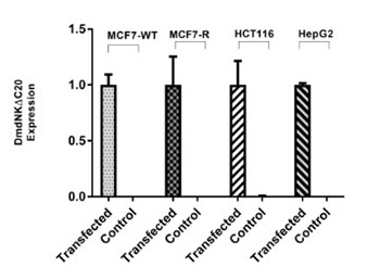
A recombinant deoxyribonucleoside kinase from Drosophila melanogaster with a deletion of the last 20 amino acid residues (named DmdNKΔC20) was hypothesized as a potential therapeutic tool for gene therapy due to its broad substrate specificity and better catalytic efficiency towards nucleosides and nucleoside analogs. This study was designed to evaluate the effect of DmdNKΔC20 for sensitizing human cancer cell lines to gemcitabine and to further investigate its role in reversal of acquired drug resistance in gemcitabine-resistant cancer cell line. The DmdNKΔC20 gene was delivered to three different cancer cell lines, including breast, colon and liver cancer cells, using lipid-mediated transfection reagent. After transfection, gene expression of DmdNKΔC20 was confirmed by quantitative reverse transcription PCR (qRT-PCR) and the combined effect of DmdNKΔC20 and gemcitabine based cytotoxicity was observed by cell viability assay. We further evolved a gemcitabine-resistant breast cancer cell line (named MCF7-R) through directed evolution in the laboratory, which showed 375-fold more resistance compared with parental MCF7 cells. Upon transfection with DmdNKΔC20 gene, MCF7-R cells showed 83-fold higher sensitivity to gemcitabine compared with the control group of MCF7-R cells. Moreover, we observed 79% higher expression of p21 protein in transfected MCF7-R cells, which may indicate induction of apoptosis. Our findings highlight the importance and therapeutic potential of DmdNKΔC20 in combined gene/chemotherapy approach to target a wide range of cancers, particularly gemcitabine-resistant cancers.

Downloads

Download data is not yet available.
Recombinant deoxyribonucleoside kinase from Drosophila melanogaster can improve gemcitabine based combined gene/chemotherapy for targeting cancer cells

Downloads

Additional Files

Published

2019-11-08

How to Cite

1.
Fatima M, Iqbal Ahmed MM, Batool F, Riaz A, Ali M, Munch-Petersen B, Mutahir Z. Recombinant deoxyribonucleoside kinase from Drosophila melanogaster can improve gemcitabine based combined gene/chemotherapy for targeting cancer cells. Biomol Biomed [Internet]. 2019Nov.8 [cited 2023Mar.28];19(4):342-9. Available from: https://www.bjbms.org/ojs/index.php/bjbms/article/view/4136

Issue

Section

Molecular Biology

INTRODUCTION

Nucleoside analogs are being effectively used in clinics for the management of cancer, either alone or in combination with other drugs [1,2]. Gemcitabine, also known as 2’, 2’-difluoro-deoxycytidine (dFdC), is a potent nucleoside analog used against a wide range of solid tumors [1,3-5]. Like other nucleoside analogs, gemcitabine is transported to the cells by human equilibrative and concentrative membrane transporters, where it is phosphorylated to the monophosphate form (dFdMP) by deoxycytidine kinase (dCK) [6]. dFdMP is further phosphorylated to cytotoxic diphosphate (dFdCDP) and triphosphate (dFdCTP) forms by intrinsic nucleotide kinases [7]. The diphosphorylated form of gemcitabine (dFdCDP) inhibits ribonucleotide reductase (RNR) causing a decrease in intracellular dCTP concentration. Consequently, increased phosphorylation of gemcitabine to its di and triphosphate forms occurs, leading to accelerated incorporation of dFdCTP into replicating DNA [8]. The triphosphorylated form of gemcitabine incorporates into DNA, thereby causing chain termination and apoptosis [5,9].

Akin to other nucleoside analog-based chemotherapies, the clinical significance of dFdC is limited by the development of drug resistance, either through intrinsic factors such as hypoxia, extracellular matrix transitions (EMT), and reactive oxygen species (ROS)-induced changes, or by acquiring alterations in drug activation and metabolic pathways [10-16]. dCK plays an important role in the activation of gemcitabine through phosphorylation, and an altered expression of dCK has been correlated with the development of gemcitabine resistance both in vitro and in vivo [17-21]. Besides dCK, several other targets of gemcitabine metabolism have been associated with acquired resistance to gemcitabine [22,23]. One of those targets is cytidine deaminase (CDA), for which increased expression was associated with decreased sensitivity of cancer cells to gemcitabine both in vitro and in vivo. CDA converts dFdC to difluorodeoxyuridine (dFdU), thus contributing to gemcitabine resistance in cancer cells [24].

Different therapeutic approaches have been proposed to overcome resistance due to nucleoside kinase deficiency, such as the introduction of monophosphate derivatives of the nucleoside analogs into the cells or nucleoside kinase enzymes by gene therapy [20,25-30]. Gene-directed enzyme prodrug therapy (GDEPT), also called suicide gene therapy, has been shown to be more effective for specific targeting of cancer cells as compared to standard chemotherapy [31]. Suicide gene therapy has been employed successfully in conjunction with human dCK [22-25,27,28,31,32]; however, a non-human dNK, known as Herpes simplex 1 thymidine kinase (HSV1-TK), has also served as an archetype for this approach [33,34].

The kinetic properties of the kinases used in GDEPT approach are one of the limiting factors, therefore, a better catalytic enzyme that could render more sensitivity to nucleoside analogs in cancer cells is desired. The landmark discovery of a multisubstrate nucleoside kinase from Drosophila melanogaster (called DmdNK), in 1998, ushered the kinases research into a new era. Based on its unique capacity to catalyze more efficiently the phosphorylation of all natural nucleosides and its substantial activity against a wide range of nucleoside analogs, DmdNK was distinguished among all nucleoside kinases for its potential use in suicide gene therapy [35,36]. The amino acid alignment data and structural knowledge of DmdNK show its structural similarity with non-TK1 family of nucleoside kinases, which include mammalian TK2, dCK, and dGK [36,37]. Munch-Petersen et al. further studied recombinant DmdNK and generated its C-terminus truncated versions called DmdNKΔC10, DmdNKΔC20, and DmdNKΔC30 by deleting the last 10, 20, and 30 amino acid residues, respectively [37]. The deletion of the last 20 amino acid residues from the C-terminus increased the specific activity by 2-fold, whereas the deletion of the last 30 C-terminal residues resulted in an almost inactive enzyme. Furthermore, DmdNKΔC20 was shown to be more stable than the wild-type or full-length DmdNK [37].

DmdNK and its improved variants, produced by site-directed mutagenesis, have been tested for their optimum application in suicide gene therapy [38-40]. Both viral (adeno- and lentivirus-based vectors) and non-viral delivery systems have been used for transfecting different cancer cells with DmdNK and its mutants, to study the combined cytotoxic effect of gene/prodrug and to reverse drug resistance in various cancer cell lines [9,41-43]. To the best of our knowledge, DmdNKΔC20 has not been reported neither for sensitizing human cancer cell lines to gemcitabine nor for the reversal of gemcitabine resistance in drug-resistant cancer cells so far. Therefore, we studied the effect of DmdNKΔC20 to sensitize different human cancer cell lines to gemcitabine, and we further elaborated its role in the reversal of acquired gemcitabine resistance due to dCK deficiency in the breast cancer cell line MCF7-R.

MATERIALS AND METHODS

Chemicals

Gemcitabine, MTT (methylthiazolyldiphenyl-tetrazolium bromide), and all other standard chemicals were purchased from Sigma-Aldrich, USA unless mentioned otherwise. Cytarabine (araC) used in this study was obtained from Pfizer. GeneJet RNA purification kit (cat. # K0731), RevertAid first strand cDNA synthesis kit (cat. # K1622), and SYBR Green (cat. # K0221) were purchased from Thermo Fisher Scientific, USA. DMEM cell culture medium supplemented with Glutamax and sodium pyruvate (cat. # 1880272), fetal bovine serum (FBS), penicillin/streptomycin, Opti-MEM™ (cat. # 1758537), trypsin, and cell culture grade PBS were obtained from Gibco® (Thermo Fisher Scientific, USA). Lipofectamine 3000® was purchased from Invitrogen (Thermo Fisher Scientific, USA). Primary antibodies were obtained from Santa Cruz Biotechnology, USA as follows: dCK (sc-81245), CDA (sc-365292), p21 (sc-756), and β-actin (sc-130656). The following secondary antibodies were used from Invitrogen: goat anti-mouse IgG, horseradish peroxidase conjugate (Catalog No G21040) and goat anti-rabbit IgG, horseradish peroxidase conjugate (Catalog No G21234).

Cell culture

Breast cancer cell line MCF7, colon cancer cell line HCT116, and liver cancer cell line HePG2 were kindly provided by Professor Staffan Johansson (Department of Medical Biochemistry and Microbiology, Uppsala University, Sweden). MCF7 cells resistant to 2 µM gemcitabine (named MCF7-R) were evolved by exposing wild-type MCF7 (MCF7-wt) to gradually increasing concentrations of gemcitabine over a period of 7 months. MCF7-R cells were viable in a medium containing 2 µM gemcitabine. The cancer cell lines used in this study were maintained in DMEM supplemented with 10% (v/v) FBS and penicillin/streptomycin (1%). Cells were grown in culture flasks with vented caps at 37°C in an incubator with 5% CO2.

Cell viability assay

Cell viability was analyzed by standard MTT assay. Cells from respective cell lines were seeded in triplicate with a seeding density of 5000–7500 cells per well in 100 µl of fresh culture medium in 96-well plates. After 24 hours of incubation at 37°C, the culture medium was replaced with 100 µl of fresh medium containing different concentrations of either araC or gemcitabine depending on the experiment. In the presence of the candidate drug, cells were further incubated for 72 hours at 37°C under 5% CO2. After the incubation, 10 µl of MTT (5 mg/ml) was added and the cells were further incubated at 37ºC for 4 hours. This resulted in the formation of formazan crystals due to mitochondrial dehydrogenase activity of viable cells; afterwards, the medium was replaced with 100 µl of acidified isopropanol (0.04N HCl in isopropanol) to dissolve the formazan crystals. The absorbance was recorded at 492 nm using Labtech LT-4500 plate reader. IC50 was defined as the drug concentration that reduces cell proliferation to 50% and calculated using equation 1. Survival curves were generated and analyzed by the non-linear regression function of GraphPad Prism 7 for Windows (GraphPad Software, La Jolla California USA). Resistance ratio was calculated as the ratio between IC50 of resistant cells to the IC50 of wild-type MCF7 cells.

Transfection of cancer cells with DmdNKΔC20

DmdNKΔC20 cloned in pGEX-2T vector was kindly provided by the late Professor Jure Piskur (Department of Biology, Lund University, Sweden). DmdNKΔC20 was restricted with BamH1 and EcoR1 and cloned into pcDNA3 vector. For transfection studies, 70–80% confluent cancer cells were used. The cancer cells were transfected with pcDNA3 vector containing DmdNKΔC20 using Lipofectamine® 3000 reagent following the protocol described in Lipofectamine® 3000 reagent kit. For control groups, empty pcDNA3 vector was used for transfecting the cancer cells using the same protocol. The transfected cells were incubated overnight before seeding into 96-well plates for cell viability assay (described earlier). Moreover, the total mRNA and protein of both groups of transfected cells were isolated 72 hours after the transfection for subsequent quantitative reverse transcription PCR (qRT-PCR) and western blot analyses.

Quantitative reverse transcription PCR (qRT-PCR)

The total mRNA was extracted from cells using GeneJet RNA purification kit according to the manufacturer’s protocol and quantified by NanoDrop™ 2000/2000c spectrophotometer (Thermo Fisher Scientific). After DNase-I treatment of template, 1 µg of cDNA was synthesized using the protocol of RevertAid first strand cDNA synthesis kit and used as a template in qRT-PCR employing SYBR Green chemistry for the detection of quantified product. The reaction was performed in a thermal cycler (BioRad-CFX) with amplification profile as follows: initial denaturation at 95ºC for 10 minutes followed by 40 cycles at 95ºC for 15 seconds, 58ºC for 20 seconds, and 72ºC for 20 seconds. The sequences of primers used for gene expression analysis in this study are provided in Table S1.

Western blot analysis

Total protein was extracted from transfected and control MCF7 groups using Laemmli method [44]. Proteins were separated by SDS-PAGE and transferred to polyvinylidene difluoride (PVDF) membrane for detection. Ponceau S staining was performed to verify the successful blotting of proteins on the PVDF membrane. Membranes were blocked using 5% skimmed milk in TBST. After blocking, membranes were incubated with primary antibody against actin (1:250 dilution), dCK (1:50 dilution), CDA (1:1000 dilution), and p21 (1:500 dilution) and further incubated with HRP-conjugated secondary antibodies (1:10,000 dilution). Protein signals were detected by chemiluminescence and exposed to Kodak films for quantification of the intensity of detected bands.

Statistical analysis

Each experiment was performed in triplicate and the results are expressed as the mean ± standard error (SE). Statistical analyses including Student’s t-test were performed using IBM SPSS Statistics for Windows, Version 23.0. (IBM Corp., Armonk, NY). Transfected and control groups were compared by paired t-test analysis using GraphPad Prism 7. Experiments with p < 0.05 were considered statistically significant and **** represents p < 0.0001.

RESULTS

Development and characterization of 2 µM gemcitabine-resistant cells

Gemcitabine-resistant MCF7 (MCF7-R) cells were evolved by exposing wild-type MCF7 cells to gradually increasing concentrations of gemcitabine over a period of 7 months. The resulting MCF7-R cells showed 375-fold higher resistance to gemcitabine compared with wild-type (MCF7-wt) cells. Moreover, MCF7-R cells showed a significant cross-resistance to araC with a resistance ratio of 39-fold. The gene and protein expressions of enzymes dCK and CDA were estimated by qRT-PCR and western blot analysis, respectively (Figure 1). An altered expression of both dCK and CDA was observed in MCF7-R cells in comparison to the wild-type counterpart. The gene expression of dCK was decreased to 2.7-fold in MCF7-R compared with wild-type MCF7, whereas the expression of CDA was 187-fold higher in MCF7-R cells compared with near basal level expression of this enzyme in MCF7 wild-type. Beta-2 microglobulin (B2M) gene expression was used as an internal control to normalize the expression of dCK and CDA. Similar changes in protein expression of the two enzymes were also observed in western blot analysis.

FIGURE 1: (A) Gene expression of deoxycytidine kinase (dCK) and cytidine deaminase (CDA) in MCF7-wt and MCF7-R cell line was measured by quantitative reverse transcription PCR (qRT-PCR). Error bars represent standard error (SE). Two-tailed p values obtained by t-test, **** is p < 0.0001, *** is p < 0.001. (B) Protein expression of dCK and CDA was quantified by western blotting in MCF7-wt and MCF7-R cells, ß-actin was used as an internal control. An altered expression of both dCK and CDA was observed in MCF7-R cells in comparison to the wild-type counterpart. The gene expression of dCK was decreased to 2.7-fold in MCF7-R compared with wild-type MCF7, whereas the expression of CDA was 187-fold higher in MCF7-R cells compared with near basal level expression of this enzyme in MCF7 wild-type.

Determination of IC50 for gemcitabine and araC before transfection of DmdNKΔC20

IC50 for gemcitabine and araC were determined by MTT assay for breast, colon and hepatic cancer cell lines. The survival curves were generated and analyzed by non-linear regression analysis using GraphPad Prism 7 (Figure 2). IC50 values for the cell lines determined in this study are shown in Table 1.

FIGURE 2: Cell viability of MCF7-wt, MCF7-R, HepG2, and HCT116 in the presence of gemcitabine and cytarabine (araC) was analyzed by MTT assay; each experiment was performed in triplicate. X-axis represents drug concentration and Y-axis represents cell survival in the presence of drug. Error bars represent standard error (SE).
TABLE 1: IC50 values (in mM) for nucleoside analog effect on different cancer cell lines before transfecting DmdNKΔC20

Effect of DmdNKΔC20 on wild-type and gemcitabine-resistant human cancer cell lines

The combined cytotoxic effect of suicide gene (DmdNKΔC20) and gemcitabine was studied using breast cancer (MCF7-wt and MCF7-R), colon cancer (HCT116) and hepatic cancer (HepG2) cell lines. The qRT-PCR results confirmed the expression of DmdNKΔC20 in transfected samples of each cell line. Human B2M expression was used as a reference for normalization (Figure 3).

FIGURE 3: Expression of DmdNKΔC20 in human cancer cell lines after transfection, measured by quantitative reverse transcription PCR (qRT-PCR). Error bars represent standard error (SE).

MTT assay was performed to calculate the IC50 value for gemcitabine in each transfected cell line and compared with respective controls. Survival curves were generated (Figure 4) and data were analyzed by non-linear regression using GraphPad Prism 7.

FIGURE 4: Cell viability of MCF7-wt, MCF7-R, HepG2, and HCT116 cancer cells after transfection with DmdNKΔC20; each experiment was performed in triplicate. Error bars represent standard error (SE). The results were further analyzed by Prism’s paired t-t est analysis. **** represents p < 0.0001. A significant increase in the sensitivity of transfected cell lines to gemcitabine upon transfection of DmdNKΔC20 was observed, as shown by a decrease in IC50 compared with control group.

A significant increase in the sensitivity of transfected cell lines to gemcitabine was observed, as shown by a decrease in IC50 compared with control group. The IC50 and folds of sensitivity of each transfected cell line for gemcitabine are described in Table 2.

TABLE 2: IC50 values (in mM) and increase folds in the sensitivity to gemcitabine of each cell line transfected with either empty vector (control) or with DmdNKΔC20

Furthermore, gemcitabine-resistant MCF7-R cells transfected with DmdNKΔC20 became 83-fold more sensitive to gemcitabine than control, showing significant reversal of gemcitabine resistance upon transfection with DmdNKΔC20. A comparison of IC50 ratios of control and transfected MCF7-R cells provided extremely significant statistical results (paired t-test was performed as mentioned in Table 2).

Detection of p21 protein as apoptotic marker

Previous studies showed the role of p21 protein in growth arrest and induction of apoptosis by either p53 dependent or independent activation pathways and indicated that its upregulation in cancer cell lines is probably a consequence of anticancer treatment [45-48]. The western blot analysis using cell lysates showed 79% higher level of p21 protein expression in transfected MCF7-R cells compared with the control, which may indicate the induction of apoptosis as described earlier (Figure 5). The p21 specific primary antibody expression was normalized against ß-actin.

FIGURE 5: Western blot was performed on DmdNKΔC20 transfected and control MCF7-R cells grown in the presence of 2 µM gemcitabine for 72 hours. The p-21 protein was highly expressed in transfected MCF7-R cells as compared to control, which possibly indicates the induction of apoptosis as a result of transfection.

DISCUSSION

Gemcitabine is a potent nucleoside analog, which has been shown to have substantial anticancer activity against a wide range of solid tumors [5,9]. Due to its low toxicity profile, gemcitabine has also been recommended as an effective drug for use in suicide gene therapy [49]. The truncated variant of Drosophila nucleoside kinase, called DmdNKΔC20, has been reported to be highly efficient for the phosphorylation of gemcitabine, and its crystal structure bound with gemcitabine further supports this character [40]. Knecht et al. have previously shown 44- and 4-fold reversal of gemcitabine resistance in ovarian cancer cell line (AG6000) and glioblastoma cell line (U-87 MG), respectively, upon transduction with a full-length DmdNK [40]. In this study, we explored the efficacy of DmdNKΔC20 to sensitize different cancer cell lines to gemcitabine in vitro and we further studied the reversal of gemcitabine resistance in the breast cancer cell line MCF7-R upon transfection with DmdNKΔC20.

In line with our hypothesis and the results from earlier studies, a significant increase in the sensitivity to gemcitabine was observed in the wild-type MCF7, HCT116 and HepG2 cancer cell lines transfected with DmdNKΔC20, as a result of the combined enzyme/prodrug effect (Table 2). Moreover, due to the expression of DmdNKΔC20, the gemcitabine-resistant cancer cells MCF7-R exhibited 83-fold increase in sensitivity to gemcitabine when compared with the corresponding control (Table 2). MCF7-R cells showed lower dCK and higher CDA expression which favored and conferred higher gemcitabine resistance in the evolved MCF7-R cells. However, after transfection with DmdNKΔC20, MCF7-R cells appeared to become more sensitive to gemcitabine by losing their drug resistance phenotype. The observed effect is considerably higher than previously reported for human uterine sarcoma cell line Messa 10K, where a full-length DmdNK was transfected to sensitize human cancer cell lines to various nucleosides [9,41-43, 50-52]. A possible explanation for the increased sensitivity to gemcitabine is the lower Km value of DmdNKΔC20 for this analog and unique interaction of fluoride atoms of gemcitabine [40]. The strong binding affinity of DmdNKΔC20 towards gemcitabine enhances the phosphorylation of gemcitabine that may result in an increased apoptosis with high p21 protein expression in transfected cells, as observed in Figure 5.

The results shown in this study are comparable to previously reported results in which a full-length DmdNK with different nucleoside analogs has been used [5,9,20,26,27,53-55]. However, our findings emphasize the role of the truncated version of Drosophila melanogaster enzyme, DmdNKΔC20, in sensitizing different cancer cells to gemcitabine and elaborate its role in the reversal of gemcitabine resistance in MCF7 cancer cells in vitro. These findings may be useful for future studies aiming to explore the potential use of DmdNKΔC20 in combined enzyme/prodrug therapeutic approach in treating a wide range of cancers, particularly gemcitabine-resistant cancers.

Acknowledgements

ACKNOWLEDGMENTS

The startup research grant from Higher Education Commission (HEC), Pakistan, and annual research grants from University of the Punjab, Lahore, to the corresponding author are gratefully acknowledged for supporting this study.

DECLARATION OF INTERESTS

The authors declare no conflict of interests.

SUPPLEMENTAL DATA

TABLE S1: Primers used in qRT-PCR experiments

REFERENCES

  1. , (). The systemic treatment of advanced and metastatic bladder cancer. Lancet Oncol. https://doi.org/10.1016/S1470-2045(03)01168-9
  2. (). Gemcitabine:Progress in the treatment of pancreatic cancer. Oncology. https://doi.org/10.1159/000055290
  3. , (). Gemcitabine as a single-agent treatment for ovarian cancer. Gynecol Oncol. https://doi.org/10.1016/S0090-8258(03)00340-8
  4. (). Role of gemcitabine in the treatment of advanced and metastatic breast cancer. Oncology. https://doi.org/10.1159/000069315
  5. , , , , (). Increased expression of the large subunit of ribonucleotide reductase is involved in resistance to gemcitabine in human mammary adenocarcinoma cells. Mol Cancer Ther. https://doi.org/10.1158/1535-7163.MCT-05-0121
  6. , , , , , (). Pretreatment deoxycytidine kinase levels predict in vivo gemcitabine sensitivity. Mol Cancer Ther.
  7. , , , (). Gemcitabine intercellular diffusion mediated by gap junctions:New implications for cancer therapy. Mol Cancer. https://doi.org/10.1186/1476-4598-9-141
  8. , , (). Understanding ribonucleotide reductase inactivation by gemcitabine. Chemistry. https://doi.org/10.1002/chem.200700260
  9. , , (). Gemcitabine resistance due to deoxycytidine kinase deficiency can be reverted by fruitfly deoxynucleoside kinase, dmdNK, in human uterine sarcoma cells. Cancer Chemother Pharmacol. https://doi.org/10.1007/s00280-006-0195-8
  10. , , , , , (). Deregulation of the EGFR/PI3K/PTEN/Akt/mTORC1 pathway in breast cancer:Possibilities for therapeutic intervention. Oncotarget. https://doi.org/10.18632/oncotarget.2209
  11. , , , , , (). Involvement of Akt and mTOR in chemotherapeutic- and hormonal-based drug resistance and response to radiation in breast cancer cells. Cell Cycle. https://doi.org/10.4161/cc.10.17.17119
  12. , , , (). Cancer drug resistance:An evolving paradigm. Nat Rev Cancer. https://doi.org/10.1038/nrc3599
  13. , , , , , (). Drug resistance in cancer:An overview. Cancers (Basel). https://doi.org/10.3390/cancers6031769
  14. , , , , , (). Mechanisms of overcoming intrinsic resistance to gemcitabine in pancreatic ductal adenocarcinoma through the redox modulation. Mol Cancer Ther. https://doi.org/10.1158/1535-7163.MCT-14-0420
  15. , , , , , (). Complex roles of the stroma in the intrinsic resistance to gemcitabine in pancreatic cancer:Where we are and where we are going. Exp Mol Med. https://doi.org/10.1038/emm.2017.255
  16. , , , , , (). The role of six1. signaling in paclitaxel-dependent apoptosis in MCF-7 cell line. Bosn J Basic Med Sci. https://doi.org/10.17305/bjbms.2016.674
  17. , , , (). Cytotoxic activity of 2',2'-difluorodeoxycytidine, 5-aza-2'-deoxycytidine and cytosine arabinoside in cells transduced with deoxycytidine kinase gene. Biochem Biophys Res Commun. https://doi.org/10.1016/S0006-291X(02)00413-8
  18. , , , , , (). Tumor uptake and elimination of 2',2'-difluoro-2'-deoxycytidine (gemcitabine) after deoxycytidine kinase gene transfer:Correlation with in vivo tumor response. Clin Cancer Res.
  19. , , , , (). Mechanisms of resistance to 5-aza-2'-deoxycytidine in human cancer cell lines. Blood. https://doi.org/10.1182/blood-2008-02-140038
  20. , , , , , (). DCK is frequently inactivated in acquired gemcitabine-resistant human cancer cells. Biochem Biophys Res Commun. https://doi.org/10.1016/j.bbrc.2012.03.122
  21. , , , , , (). Acquisition of chemoresistance to gemcitabine is induced by a loss-of-function missense mutation of DCK. Biochem Biophys Res Commun. https://doi.org/10.1016/j.bbrc.2015.07.080
  22. , , (). Determinants of resistance to 2',2'-difluorodeoxycytidine (gemcitabine). Drug Resist Updat. https://doi.org/10.1016/S1368-7646(02)00002-X
  23. , , , , , (). Gene therapy based on gemcitabine chemosensitization suppresses pancreatic tumor growth. Mol Ther. https://doi.org/10.1016/j.ymthe.2006.07.010
  24. , , (). Pharmacogenomics of gemcitabine:Can genetic studies lead to tailor-made therapy?. Br J Cancer. https://doi.org/10.1038/sj.bjc.6603860
  25. , , , (). Gemcitabine-based chemogene therapy for pancreatic cancer using ad-dCK:UMK GDEPT and TS/RR siRNA strategies. Neoplasia. https://doi.org/10.1593/neo.81686
  26. , , , , , (). Sensitization of ara-C-resistant lymphoma cells by a pronucleotide analogue. Int J Cancer. https://doi.org/10.1002/ijc.11339
  27. , , , , , (). Characterization of a gemcitabine-resistant murine leukemic cell line:Reversion of in vitro resistance by a mononucleotide prodrug. Clin Cancer Res. https://doi.org/10.1158/1078-0432.CCR-04-0506
  28. , , (). Retroviral transfer of deoxycytidine kinase into tumor cell lines enhances nucleoside toxicity. Cancer Res.
  29. , , (). Development of gene therapy in association with clinically used cytotoxic deoxynucleoside analogues. Cancer Gene Ther. https://doi.org/10.1038/cgt.2009.25
  30. , , , , , (). Plant thymidine kinase 1:A novel efficient suicide gene for malignant glioma therapy. Neuro Oncol. https://doi.org/10.1093/neuonc/nop067
  31. , , , , , (). The “bystander effect”: Tumor regression when a fraction of the tumor mass is genetically modified. Cancer Res.
  32. , , , , , (). Viral vector transduction of the human deoxycytidine kinase cDNA sensitizes glioma cells to the cytotoxic effects of cytosine arabinoside in vitro and in vivo. Nat Med. https://doi.org/10.1038/nm0596-567
  33. , , , , , (). In vivo gene transfer with retroviral vector-producer cells for treatment of experimental brain tumors. Science. https://doi.org/10.1126/science.1317968
  34. , , , (). In vitro evidence that metabolic cooperation is responsible for the bystander effect observed with HSV tk retroviral gene therapy. Hum Gene Ther. https://doi.org/10.1089/hum.1993.4.6-725
  35. , , (). Four deoxynucleoside kinase activities from Drosophila melanogaster are contained within a single monomeric enzyme, a new multifunctional deoxynucleoside kinase. J Biol Chem. https://doi.org/10.1074/jbc.273.7.3926
  36. , , , , (). Cloning and characterization of the multisubstrate deoxyribonucleoside kinase of Drosophila melanogaster. J Biol Chem. https://doi.org/10.1074/jbc.274.34.23814
  37. , , , , (). Functional expression of a multisubstrate deoxyribonucleoside kinase from Drosophila melanogaster and its C-terminal deletion mutants. J Biol Chem. https://doi.org/10.1074/jbc.275.9.6673
  38. , , , , , (). Adenovirus-mediated Drosophila melanogaster deoxyribonucleoside kinase mutants combined with gemcitabine harbor a safe cancer treatment profile. Int J Oncol. https://doi.org/10.3892/ijo.2010.887
  39. , , , , , (). Drosophila deoxyribonucleoside kinase mutants with enhanced ability to phosphorylate purine analogs. Gene Ther. https://doi.org/10.1038/sj.gt.3302982
  40. , , , , , (). Drosophila melanogaster deoxyribonucleoside kinase activates gemcitabine. Biochem Biophys Res Commun. https://doi.org/10.1016/j.bbrc.2009.03.041
  41. , , , , (). Lentivirus-mediated expression of Drosophila melanogaster deoxyribonucleoside kinase driven by the hTERT promoter combined with gemcitabine:A potential strategy for cancer therapy. Int J Mol Med.
  42. , , , , , (). Cytotoxic effects of adenovirus- and lentivirus-mediated expression of Drosophila melanogaster deoxyribonucleoside kinase on bcap37. abreast cancer cells. Oncol Rep. https://doi.org/10.3892/or.2012.2194
  43. , , , , , (). Synergistic antitumor effect of adenovirus armed with Drosophila melanogaster deoxyribonucleoside kinase and nucleoside analogs for human breast carcinoma in vitro and in vivo. Drug Des Devel Ther. https://doi.org/12.10.2147/DDDT.S81717
  44. (). Cleavage of structural proteins during the assembly of the head of bacteriophage T4. Nature. https://doi.org/10.1038/227680a0
  45. , , , (). Induction of HSP70 gene expression by modulation of Ca(+2) ion and cellular p53 protein by curcumin in colorectal carcinoma cells. Mol Carcinog. https://doi.org/10.1002/(SICI) 1098-2744(199612)17:4<224: AID-MC6>3.0.CO;2-D
  46. , , , , (). Cell cycle-independent regulation of p21Waf1/Cip1 and retinoblastoma protein during okadaic acid-induced apoptosis is coupled with induction of Bax protein in human breast carcinoma cells. Cell Growth Differ.
  47. , , (). MiR-145 inhibits cell proliferation of human lung adenocarcinoma by targeting EGFR and NUDT1. RNA Biol. https://doi.org/10.4161/rna.8.1.14259
  48. , (). The role of the cyclin-dependent kinase inhibitor p21 in apoptosis. Mol Cancer Ther.
  49. , , , , (). Role of gemcitabine in cancer therapy. Future Oncol. https://doi.org/10.1517/14796694.1.1.7
  50. , , , , (). Conditionally replicating adenovirus SG500-expressed mutant dm-dNK gene for breast cancer therapy. Int J Oncol. https://doi.org/10.3892/ijo.2012.1657
  51. , , (). Retroviral transduction of cancer cell lines with the gene encoding drosophila melanogaster multisubstrate deoxyribonucleoside kinase. J Biol Chem. https://doi.org/10.1074/jbc.M006212200
  52. , , , , , (). The multisubstrate deoxyribonucleoside kinase of Drosophila melanogaster as a therapeutic suicide gene of breast cancer cells. J Gene Med. https://doi.org/10.1002/jgm.1573
  53. , , , , , (). Resistance to gemcitabine in a human follicular lymphoma cell line is due to partial deletion of the deoxycytidine kinase gene. BMC Pharmacol. https://doi.org/10.1186/1471-2210-4-8
  54. , , , , , (). Sensitivity to ionizing radiation and chemotherapeutic agents in gemcitabine-resistant human tumor cell lines. Int J Radiat Oncol Biol Phys. https://doi.org/10.1016/S0360-3016(02)02891-2
  55. , , , (). Deoxycytidine kinase is overexpressed in poor outcome breast cancer and determines responsiveness to nucleoside analogs. Breast Cancer Res Treat. https://doi.org/10.1007/s10549-011-1477-3