Predictors of early cardiac changes in patients with type 1 diabetes mellitus: An echocardiography-based study

Authors

DOI:

https://doi.org/10.17305/bjbms.2019.4250

Keywords:

Type 1 diabetes mellitus, T1DM, body mass index, BMI, left ventricular mass, left atrial volume, diastolic dysfunction, tissue Doppler imaging

Abstract

In patients with type 1 diabetes mellitus (T1DM) imaging studies have demonstrated an increased prevalence of left ventricular diastolic dysfunction and increased left ventricular mass (LVM) unrelated to arterial hypertension and ischemic heart disease. The aim of our study was to identify potential predictors of early subclinical changes in cardiac chamber size and function in such patients. Sixty-one middle-aged asymptomatic normotensive patients with T1DM were included in the study. Conventional and tissue Doppler echocardiography was performed and fasting serum levels of glucose, glycated hemoglobin (HbA1c), lipids, and creatinine were measured. We found moderate bivariate correlations of body mass index (BMI) with left atrial volume (r = 0.47, p < 0.01), LVM (r = 0.42, p < 0.01), left ventricular relative wall thickness (r = 0.32, p = 0.01), and all observed parameters of diastolic function of both ventricles. The five-year average value of HbA1c weakly correlated with the Doppler index of left ventricular filling pressure E/e´sept (r = 0.27, p = 0.04). We found no significant association of diabetes duration, five-year trend of HbA1c, serum lipids, and glomerular filtration rate with cardiac structure and function. After adjusting for other parameters, BMI remained significantly associated with left atrial volume, LVM as well as with the transmitral Doppler ratio E/A. In our study, BMI was the only observed parameter significantly associated with subclinical structural and functional cardiac changes in the asymptomatic middle-aged patients with T1DM.

Downloads

Download data is not yet available.
Predictors of early cardiac changes in patients with type 1 diabetes mellitus: an echocardiography-based study

Downloads

Additional Files

Published

2019-06-18

How to Cite

1.
Šuran D, Kanič V, Naji F, Krajnc I, Čokolič M, Zemljič E, Sinkovič A. Predictors of early cardiac changes in patients with type 1 diabetes mellitus: An echocardiography-based study. Biomol Biomed [Internet]. 2019Jun.18 [cited 2023Sep.25];19(4):384-91. Available from: https://www.bjbms.org/ojs/index.php/bjbms/article/view/4250

Issue

Section

Translational and Clinical Research

INTRODUCTION

Diabetes mellitus has been increasingly recognized as an important risk factor for heart failure unrelated to arterial hypertension, ischemic, and valvular heart disease [1,2]. The entity was first described in 1972 as diabetic cardiomyopathy [3]. Many different mechanisms have been proposed and the etiology appears to be multifactorial [4,5]. Most studies have included patients with type 2 diabetes mellitus (T2DM), but the link between type 1 diabetes mellitus (T1DM) and heart failure has also been documented [6-9]. In our previous work, we showed that even asymptomatic middle-aged adults with T1DM have some structural and functional changes in both ventricles compared to the age- and gender-matched healthy controls [10]. However, only limited data exist on the contribution of diabetes duration, long-term glycemic control, body mass index (BMI) as well as the frequently coexisting dyslipidemia and renal dysfunction to early adverse changes in the heart of patients with T1DM [11,12]. The aim of our study was to find potential predictors of early structural and functional changes in asymptomatic middle-aged T1DM patients without ischemic, hypertensive, and valvular heart disease.

MATERIALS AND METHODS

Study design and participants

The study was conducted at the Department of Cardiology and Angiology, University Medical Centre Maribor, Slovenia. It was approved by the National Medical Ethics Committee of the Republic of Slovenia (Medical Ethics Committee approval number 130/10/13). All participants signed informed consent to participate in the study.

We prospectively enrolled 61 asymptomatic patients with T1DM who attended diabetic outpatient clinic of the University Medical Centre Maribor, Slovenia for more than 5 years and met the inclusion/exclusion criteria. The inclusion criteria were age between 30 and 55 years and the duration of T1DM of more than 5 years. The exclusion criteria were: arterial hypertension; ischemic heart disease; clinical signs of heart failure at enrolment and in the past; bundle branch block or non-sinus rhythm in the electrocardiogram (ECG) and advanced nephropathy (glomerular filtration rate [GFR] below 60 mL/min). Based on the echocardiographic results, we further excluded patients with left ventricular ejection fraction below 50%, left ventricular wall motion abnormalities, valvular stenoses of any degree, more than mild valvular regurgitations, and low-quality echocardiographic image.

Before enrolment, all medical records of the included patients were verified and medical history obtained. Anthropometric measurements (weight and height) were taken during the physical examination in the morning after overnight fasting. Rigid stadiometer and digital scales were used to measure height and weight, respectively. BMI was calculated as weight in kilograms divided by the square of height in meters. Patients with BMI ≥ 25 were classified as overweight and BMI ≥ 30 was defined as obesity. Patients were asked to bring home blood pressure measurements to the enrolment visit. Home blood pressure measurements were obtained by patients using an automated blood pressure monitor twice daily 7 consecutive days prior to the enrolment. Ambulatory blood pressure was obtained in the presence of the investigator at enrolment using an automated blood pressure monitor. The average value of the two consecutive ambulatory measurements was saved for statistical analysis. All blood pressure measurements were obtained after 5 minutes of rest in the seated position. The presence of arterial hypertension was defined as the average home blood pressure ≥135/85 mmHg and the office blood pressure ≥140/90 mmHg. All patients with arterial hypertension including those receiving antihypertensive medications were excluded from the study.

The standard 12-lead ECG was recorded in the supine position under resting conditions. Patients with ischemic heart disease were excluded based on medical history, resting ECG changes, and the presence of left ventricular regional wall motion abnormalities.

Echocardiography

Two-dimensional echocardiography was performed in all patients by a single experienced echocardiographer on the same ultrasound machine (iE33, Philips Medical Systems, WA, USA) and included detailed two-dimensional, pulsed wave Doppler and tissue Doppler imaging (TDI) evaluation. All measurements were based on the recommendations of the American Society of Echocardiography and the European Association of Cardiovascular Imaging [13-15].

Biplane area-length formula was used to calculate the maximal end-systolic volume of the left atrium. Linear method (Cube formula) based on left ventricular measurements in the parasternal long axis view was used to calculate left ventricular mass (LVM). Relative wall thickness was calculated as 2 times the posterior wall thickness divided by the left ventricular end-diastolic diameter. Left ventricular ejection fraction was calculated by the biplane modified Simpson’s method [13,14]. Two-dimensional measurements were indexed to the body surface area (BSA).

Peak mitral and tricuspid inflow velocities were measured by pulsed wave Doppler [15]. All Doppler measurements of the right ventricle were added the suffix - t.

Peak myocardial systolic and diastolic velocities were measured by TDI at the septal part of the mitral annulus (s´sept, e´sept, a´sept) and at the lateral part of the tricuspid annulus (s´t, e´t, a´t). Myocardial acceleration during isovolumetric contraction (IVA) was calculated for both ventricles using TDI as the ratio between the peak isovolumetric velocity and the acceleration time. Acceleration time was defined as the time interval from baseline to the peak isovolumetric velocity [15]. Three consecutive Doppler measurements were averaged for statistical analysis. All Doppler measurements were performed at the frame rate ≥150 Hz.

Laboratory measurements

Blood samples were obtained in the fasting state from all patients. Serum levels of glycated hemoglobin (HbA1c), glucose, creatinine, triglycerides, and total cholesterol were measured. The results of HbA1c obtained at 6-month follow-up visits in the preceding 5 years were averaged to assess the long-term glycemic control.

HbA1c was measured by the ion-exchange high-performance liquid chromatography (Bio-Rad Laboratories, PA, USA) [16]. The isotope dilution mass spectrometry traceable enzymatic assay (Siemens Healthcare Diagnostics Inc., Newark, NJ, USA) was performed to measure serum creatinine. GFR was calculated by the Modification of Diet in Renal Disease (MDRD) equation and indexed to the normalized BSA of 1.73 m2 [16]. Triglycerides were measured by the enzymatic method (Siemens HealthCare Diagnostics Inc., Newark, NJ, USA) and total serum cholesterol by the cholesterol esterase enzymatic assay (Siemens HealthCare Diagnostics Inc., Newark, NJ, USA) [16].

Statistical analysis

IBM SPSS Statistics for Windows, Version 22.0. (IBM Corp., Armonk, New York, USA) was used for statistical analysis. Continuous variables were shown as means ± standard deviations. Shapiro–Wilk test confirmed the normal distribution of continuous variables. Categorical variables were presented as percentages. The degree of association between clinical/laboratory data and echocardiography measurements was demonstrated using the Pearson’s correlation coefficient (r). Kendall’s tau was used to evaluate the trend of HbA1c during the 5-year period. Multiple linear regression analysis was performed to assess whether statistically significant bivariate correlations of BMI with cardiac chamber size and function continues after adjustment for other observed confounders. The value of p < 0.05 was considered statistically significant for all calculations.

RESULTS

Sixty-one patients with a mean age of 44.1 ± 5.3 years were enrolled in the study. The study group consisted of 34 men (55.7%) and 27 women (44.3%). Thirty patients (49.2%) were treated with continuous subcutaneous insulin infusion and 31 patients (50.8%) with multiple daily injections of insulin. Mean diabetes duration was 17.9 ± 7.8 years. Thirty patients (49.1%) were overweight and 13 patients (21.3%) were obese with a mean BMI of 26.9 ± 3.9. Mean systolic and diastolic blood pressure values were 125.2 ± 9.6 mmHg and 78.7 ± 6.4 mmHg, respectively. Mean fasting glucose level was 6.7 ± 2.7 mmol/L. Mean HbA1c at enrolment and the mean 5-year average were 7.4 ± 1.1% and 7.2 ± 0.7%, respectively. The mean trend of HbA1c expressed by Kendall’s tau was 0.0 ± 0.4. Mean GFR was 105.0 ± 23.3 mL/min/1.73 m2, mean serum levels of triglycerides and total cholesterol were 1.0 ± 0.6 mmol/L and 4.8 ± 0.8 mmol/L, respectively. Demographic and clinical data are presented in Table 1.

TABLE 1: Demographic, clinical, and laboratory data of patients (N=61)

Echocardiographic measurements are presented in Table 2. The mean LVM indexed to BSA was 74.2 ± 13.2 g/m2, and the mean left atrial volume indexed to BSA was 29.6 ± 3.7 ml/m2. Mean left ventricular relative wall thickness was 0.41 ± 0.05. The mean values of IVA for the left and the right ventricle were 235.2 ± 71.7 cm/s2 and 283.4 ± 79.4 cm/s2, respectively. Mean e´sept and E/e´sept were 8.9 ± 1.9 cm/s and 9.2 ± 1.9, respectively and mean e´t and E/e´t were 13.4 ± 2.9 cm/s and 4.0 ± 1.1 cm/s, respectively.

TABLE 2: Echocardiographic parameters of patients (N=61)

Bivariate correlations of clinical and laboratory variables with cardiac chamber size are presented in Table 3. For all observed parameters, except BMI, indexed values of chamber size were used for correlation analysis. The correlations with Doppler parameters are presented in Table 4. Only BMI showed moderate correlations with all echocardiographic variables of the left atrial and the left ventricular size: left atrial volume (r = 0.47, p < 0.01), left ventricular end-diastolic diameter (r = 0.27, p = 0.04), left ventricular relative wall thickness (r = 0.32, p = 0.01), and LVM (r = 0.42, p < 0.01). BMI also moderately correlated with all observed Doppler parameters of left ventricular diastolic function (E/A, r = –0.39, p < 0.01; e´sept, r = –0.31, p = 0.02; E/e´sept, r = 0.31, p = 0.01) and right ventricular diastolic function (e´t, r = –0.29, p = 0.02; E/e´t, r = 0.31, p = 0.02). The 5-year average value of HbA1c weakly correlated with the Doppler index of left ventricular filling pressure E/e´sept (r = 0.27, p = 0.04). We found no significant correlations of fasting glucose, diabetes duration, HbA1c trend, GFR, serum cholesterol, and triglycerides with the observed echocardiographic parameters (Table 3 and 4).

TABLE 3: Correlation analysis of clinical and laboratory parameters with cardiac chamber size
TABLE 4: Correlation analysis of clinical and laboratory parameters with echocardiographic functional parameters

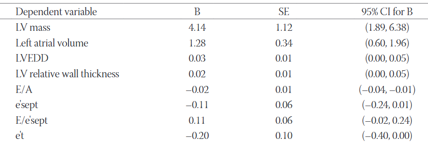
In the multiple linear regression model after adjusting for age, diabetes duration and the 5-year average value of HbA1c, BMI remained significantly associated with LVM, left atrial volume, and the mitral inflow Doppler ratio E/A (Table 5). The association of BMI with e´t and E/e´t met borderline significance (p = 0.050 for e´t and p = 0.047 for E/e´t).

TABLE 5: Association of BMI with cardiac chamber size and function after adjustment for age, diabetes duration, and 5-year average value of glycated hemoglobin

DISCUSSION

In our study, BMI was the only variable consistently associated with early changes in cardiac structure and function in asymptomatic normotensive middle-aged patients with T1DM. Average 5-year glycemic control was weakly associated with the noninvasive Doppler index of left ventricular filling pressure E/e´.

In studies including patients with T1DM left ventricular hypertrophy was an inconsistent finding and was more prevalent in patients with microalbuminuria [8,17]. On the other hand, left ventricular hypertrophy has been much more frequently observed in patients with T2DM, as supported by a considerable amount of evidence [18,19]. Less myocardial hypertrophy in T1DM compared with T2DM might be at least partly explained by the reduced insulin secretion capacity in T1DM, since insulin stimulates growth and hypertrophy of myocardial cells [20]. While chronic hyperglycemia promotes accumulation of advanced glycation end products and subsequent myocardial stiffening, in T2DM insulin resistance and hyperinsulinemia predominantly increase LVM [20,21]. Since left ventricular structural changes evaluated by imaging studies were often mild in patients with T1DM, some authors even questioned the existence of type 1 diabetes-specific cardiomyopathy [22,23]. However, more recent studies confirmed left ventricular diastolic dysfunction in up to 30.7% of middle-aged patients with T1DM, which is comparable with the 15 years older general population [8,10,17,24]. Furthermore, recent observational data confirmed a higher incidence of heart failure and mortality in patients with T1DM compared with the general population, but little is known about early predictors of cardiac abnormalities in these patients [25,26].

Recently published Swedish registry-based data have demonstrated an increased mortality of patients with T1DM compared with matched controls even in patients with HbA1c below 6.9% [26]. It seems that, besides glycemic control, other potentially even more important and poorly recognized contributors to adverse outcome exist in these patients. In the echocardiography-based study of Wai et al. increasing age and BMI were suggested as the two most important predictors of cardiac dysfunction progression in patients with T1DM, while the mean observational time was 3.8 years [27]. Epidemiological data also revealed a link between obesity and hospitalizations for heart failure in patients with diabetes mellitus [28,29]. Due to the volume overload and metabolic derangements, obesity has been linked to left ventricular hypertrophy and dysfunction even in non-diabetic populations [30]. Similarly, in our patients, BMI was associated with LVM and left atrial volume, both of which have been proven to predict adverse cardiac events [29,31].

Few imaging studies evaluated the impact of long-term glycemic control on cardiac function in patients with T1DM. Some of them demonstrated the reduction of LVM as well as the reversal of the left ventricular diastolic dysfunction by improved glycemic control [11,12]. On the other hand, in a subpopulation of the Diabetes Control and Complications Trial/Epidemiology of Diabetes Interventions and Complications study (DCCT/EDIC) only a mild effect of long-term glycemic control on LVM and volume was demonstrated by cardiac magnetic resonance over years [32]. Similarly, we found no significant effect of 5-year glycemic control on cardiac structure, but we confirmed a weak association of glycemic control with the Doppler index of left ventricular filling pressure E/e´. Furthermore, we found no significant correlation of diabetes duration and the 5-year trend of HbA1cwith left ventricular size and function. This might be due to the good average long-term glycemic control achieved by the frequent use of continuous subcutaneous insulin infusion and the absence of an overall trend in HbA1c over years (mean Kendall’s tau = 0.0 ± 0.4). Based on our results and previous findings, we assume that in well-controlled T1DM chronic hyperglycemia per se induces only mild structural and functional cardiac changes, but makes the heart more vulnerable and susceptible to further and more extensive injury due to obesity and other noxious stimuli. This might be the reason for a better association of BMI than diabetes duration and HbA1c with cardiac remodeling in our patients. However, our results may not be applied to patients with poor glycemic control. An increased susceptibility of diabetic heart to more extensive damage has already been demonstrated in case of ischemic and reperfusion injury, which resulted in a significantly larger ischemic area in patients with diabetes compared with the non-diabetic controls [33].

Although autoantibodies in T1DM were assumed to cause dilated phenotype with eccentric left ventricular remodeling and systolic dysfunction, convincing evidence is lacking [8,34]. As left ventricular ejection fraction has low sensitivity to detect early left ventricular systolic impairment, we also used IVA as a relatively novel and less load-dependent TDI-derived parameter of systolic function [35]. Consistent with the results of most previous studies, we found no significant correlation of observed variables with the parameters of left and right ventricular systolic function [24,36]. Recently, few small-sized studies have demonstrated impaired global longitudinal deformation of the left ventricle in patients with T1DM analyzed by the speckle tracking echocardiography [37]. However, left ventricular deformation imaging might be an even more sensitive echocardiographic technique to detect early changes in systolic function than TDI-derived IVA.

Cardiac steatosis is a common finding in diabetes mellitus due to metabolic derangements. The uptake of fatty acids by myocardial cells exceeds the capacity of mitochondrial oxidation. The myocardial lipid overload produces lipotoxic intermediates that increase the production of reactive oxygen species and promote myocardial cell apoptosis [38]. In our patients, serum triglycerides and total cholesterol levels were not significantly associated with cardiac structure and function. This might be explained by the relatively normal lipid profile, especially low serum triglycerides.

In the observational study of Vestberg et al., advanced chronic kidney failure was related to increased hospitalizations for heart failure in patients with T1DM [39]. We found a weak association of glomerular filtration rate with left and right atrial size but no correlation with observed functional parameters. The reason might be the exclusion of patients with advanced renal disease (GFR <60 mL/min/1.73 m2).

There are limitations of our study. The number of eligible patients was limited due to the restrictive exclusion criteria. However, we decided to select patients with T1DM without major comorbidities influencing cardiac chamber size and function. Coronary angiography was not performed to exclude coronary artery disease due to ethical issues. We excluded all symptomatic patients and patients with a history of ischemic heart disease, resting ECG changes or left ventricular wall motion abnormalities. Although the threshold of T1DM duration for the study entry was 5 years, the time of T1DM diagnosis was quite variable. Furthermore, we could not provide reliable data of weight stability and duration of obesity. Only selected laboratory data were collected as the emphasis of the study was to identify potentially reversible factors for the development of diabetic cardiomyopathy. For instance, inflammatory parameters which might accelerate the development of diabetic heart disease were not studied. Another important limitation of the study is the use of non-corrected multiple testing in a relatively small sample. The study was cross-sectional and generated hypotheses, which warrant further research. The Doppler assessment of diastolic parameters has limitations, especially the biphasic distribution of the Doppler index E/A as well as the non-linear correlation of E/e´ with the invasive measurements of left ventricular filling pressure.

CONCLUSION

In our study, only BMI was associated with early structural and functional changes in the heart of middle-aged asymptomatic patients with T1DM. Long-term glycemic control was associated with the noninvasive Doppler index of left ventricular filling pressure, but not with cardiac chamber size. Based on our results, we hypothesize that overweight and obese middle-aged patients with T1DM might be more prone to the development of cardiomyopathy and that screening of this subset of patients with echocardiography should be recommended. However, as our study is cross-sectional and hypothesis-generating, further studies are needed to confirm the causal relationship between obesity and cardiac dysfunction in patients with T1DM.

DECLARATION OF INTERESTS

The authors declare no conflict of interests.

REFERENCES

  1. , , , (). Diabetic cardiomyopathy;summary of 41 years. Korean Circ J. https://doi.org/10.4070/kcj.2015.45.4.266
  2. , , , , , (). Glycaemic control and incidence of heart failure in 20,985 patients with type 1 diabetes:An observational study. Lancet. https://doi.org/10.1016/s0140-6736(11)60471-6
  3. , , , , , (). New type of cardiomyopathy associated with diabetic glomerulosclerosis. Am J Cardiol. https://doi.org/10.1016/0002-9149(72)90595-4
  4. (). Echocardiographic feature of diabetic cardiomyopathy:Where are we now?. Cardiovasc Diagn Ther. https://doi.org/10.21037/cdt.2018.01.03
  5. , , (). Diabetic cardiomyopathy:An update of mechanisms contributing to this clinical entity. Circ Res. https://doi.org/10.1161/circresaha.117.311586
  6. , , , , , (). Impact of diabetes mellitus on hospitalization for heart failure, cardiovascular events, and death:Outcomes at 4 years from the reduction of atherothrombosis for continued health (REACH) registry. Circulation. https://doi.org/10.1161/circulationaha.114.014796
  7. , , , (). Molecular and metabolic mechanisms of cardiac dysfunction in diabetes. Life Sci. https://doi.org/10.1016/j.lfs.2012.10.028
  8. , , , , , (). Prevalence of systolic and diastolic dysfunction in patients with type 1 diabetes without known heart disease:The Thousand and 1 Study. Diabetologia. https://doi.org/10.1007/s00125-014-3164-5
  9. , , (). Diabetic cardiomyopathy:Does the type of diabetes matter?. Int J Mol Sci. https://doi.org/10.3390/ijms17122136
  10. , , (). Tissue Doppler imaging is a sensitive echocardiographic technique to detect subclinical systolic and diastolic dysfunction of both ventricles in type 1 diabetes mellitus. BMC Cardiovasc Disord. https://doi.org/10.1186/s12872-016-0242-2
  11. , , , , , (). Effect of glycemic control on left ventricular diastolic function in type 1 diabetes mellitus. Am J Cardiol. https://doi.org/10.1016/j.amjcard.2005.07.110
  12. , , , , (). Improved glycemic control induces regression of left ventricular mass in patients with type 1 diabetes mellitus. Int J Cardiol. https://doi.org/10.1016/j.ijcard.2003.04.012
  13. , , , , , (). Recommendations for cardiac chamber quantification by echocardiography in adults:An update from the American Society of Echocardiography and the European Association of Cardiovascular Imaging. J Am Soc Echocardiogr. https://doi.org/10.1016/j.echo.2014.10.003
  14. , , , , , (). Guidelines for the echocardiographic assessment of the right heart in adults:A report from the American Society of Echocardiography endorsed by the European Association of Echocardiography, a registered branch of the European Society of Cardiology, and the Canadian Society of Echocardiography. J Am Soc Echocardiogr. https://doi.org/10.1016/j.echo.2010.05.010
  15. , , , , , (). Recommendations for the evaluation of left ventricular diastolic function by echocardiography:An update from the American Society of Echocardiography and the European Association of Cardiovascular Imaging. J Am Soc Echocardiogr. https://doi.org/10.1016/j.echo.2016.01.011
  16. , , , , , (). Management of hyperglycemia, dyslipidemia, and albuminuria in patients with diabetes and CKD:A systematic review for a KDOQI clinical practice guideline. Am J Kidney Dis. https://doi.org/10.1053/j.ajkd.2012.07.017
  17. , , (). Prevalence of left ventricular hypertrophy in type 1 diabetic patients with diabetic nephropathy. Diabetologia. https://doi.org/10.1007/s001250051116
  18. , , , , , (). Association between diabetes mellitus and left ventricular hypertrophy in a multiethnic population. Am J Cardiol. https://doi.org/10.1016/j.amjcard.2008.02.082
  19. , , (). The epidemiology of left ventricular hypertrophy in type 2 diabetes mellitus. Diabetologia. https://doi.org/10.1007/s00125-005-1896-y
  20. , (). Insulin signaling pathways and cardiac growth. J Mol Cell Cardiol. https://doi.org/10.1016/j.yjmcc.2008.03.008
  21. (). Cross-linking of glycated collagen in the pathogenesis of arterial and myocardial stiffening of aging and diabetes. J Hypertens. https://doi.org/10.1097/00004872-200301000-00002
  22. , , , , , (). Diabetes-specific cardiomyopathy in type 1 diabetes mellitus:No evidence for its occurrence in the era of intensive insulin therapy. Eur Heart J. https://doi.org/10.1093/eurheartj/ehm361
  23. , , , , , (). Medalists with extreme duration of type 1 diabetes exhibit only mild diastolic dysfunction and myocardial fibrosis. Diabetes Care. https://doi.org/10.2337/dc14-2100
  24. , , , , , (). Tissue Doppler imaging must be performed to detect early left ventricular dysfunction in patients with type 1 diabetes mellitus. Eur J Echocardiogr. https://doi.org/10.1093/ejechocard/jep086
  25. , , , , , (). Long-term excess risk of heart failure in people with type 1 diabetes:A prospective case-control study. Lancet Diabetes Endocrinol. https://doi.org/10.1016/s2213-8587(15)00292-2
  26. , , (). Glycemic control and excess mortality in type 1 diabetes. N Engl J Med. https://doi.org/10.1056/NEJMc1415677
  27. , , , , , (). Prevalence, predictors and evolution of echocardiographically defined cardiac abnormalities in adults with type 1 diabetes:An observational cohort study. J Diabetes Complications. https://doi.org/10.1016/j.jdiacomp.2013.09.013
  28. , , , , , (). Comparison of left ventricular mass in normotensive type 2 diabetes mellitus patients with that in the nondiabetic population. J Cardiovasc Dis Res. https://doi.org/10.4103/0975-3583.78597
  29. , , , , , (). Relationship between overweight and obesity with hospitalization for heart failure in 20,985 patients with type 1 diabetes:A population-based study from the Swedish National Diabetes Registry. Diabetes Care. https://doi.org/10.2337/dc12-2007
  30. , , , , , (). The impact of obesity on the left ventricle:The Multi-Ethnic Study of Atherosclerosis (MESA). JACC Cardiovasc Imaging. https://doi.org/10.1016/j.jcmg.2009.10.012
  31. , , , , , (). Left atrial size and heart failure hospitalization in patients with diastolic dysfunction and preserved ejection fraction. J Cardiovasc Echogr. https://doi.org/10.4103/2211-4122.199064
  32. , , , , , (). Effects of prior intensive versus conventional therapy and history of glycemia on cardiac function in type 1 diabetes in the DCCT/EDIC. Diabetes. https://doi.org/10.2337/db12-0546
  33. , , , , (). et al Infarct size, ejection fraction, and mortality in diabetic patients with acute myocardial infarction treated with thrombolytic therapy. Am Heart J. https://doi.org/10.1016/j.ahj.2007.06.020
  34. , (). Clinical diabetic cardiomyopathy:A two-faced disease with restrictive and dilated phenotypes. Eur Heart J. 1727a-1727chttps://doi.org/10.1093/eurheartj/ehv134
  35. , , , , , (). Can isovolumic acceleration be used in clinical practice to estimate ventricular contractile function?Reproducibility and regional variation of a new noninvasive index. J Am Soc Echocardiogr. 431, e1-6https://doi.org/10.1016/j.echo.2010.01.008
  36. , , , , , (). Early diastolic impairment of diabetic heart:The significance of right ventricle. Int J Cardiol. https://doi.org/10.1016/j.ijcard.2006.02.003
  37. , , , , , (). Left and right ventricular systolic function impairment in type 1 diabetic young adults assessed by 2D speckle tracking echocardiography. Eur Heart J Cardiovasc Imaging. https://doi.org/10.1093/ehjci/jev164
  38. , , , (). Adiposity of the heart, revisited. Ann Intern Med. https://doi.org/10.7326/0003-4819-144-7-200604040-00011
  39. , , , , , (). Decreased eGFR as a risk factor for heart failure in 13 781 individuals with type 1 diabetes. J Diabetes Sci Technol. https://doi.org/10.1177/1932296815596174