A preliminary study of microRNA expression in different types of primary melanoma

Authors

  • Ioana Gencia Department of Dermatology, "Victor Babeş" University of Medicine and Pharmacy; University Clinic of Dermatology and Venereology, Municipal Clinical Emergency Hospital of Timisoara, Timisoara, Romania https://orcid.org/0000-0002-2917-8051
  • Flavia Baderca Department of Microscopic Morphology, "Victor Babeş" University of Medicine and Pharmacy, Timisoara, Romania https://orcid.org/0000-0002-5755-179X
  • Stefania Avram Department of Dermatology, "Victor Babeş" University of Medicine and Pharmacy; University Clinic of Dermatology and Venereology, Municipal Clinical Emergency Hospital of Timisoara, Timisoara, Romania https://orcid.org/0000-0002-3784-902X
  • Armand Gogulescu Department of Balneology, Medical Rehabilitation and Rheumatology, "Victor Babeş" University of Medicine and Pharmacy, Timisoara, Romania
  • Anca Marcu Department of Biochemistry and Pharmacology, "Victor Babeş" University of Medicine and Pharmacy, Timisoara, Romania https://orcid.org/0000-0003-3875-8143
  • Edward Seclaman Department of Biochemistry and Pharmacology, "Victor Babeş" University of Medicine and Pharmacy, Timisoara, Romania https://orcid.org/0000-0003-1613-1884
  • Catalin Marian Department of Biochemistry and Pharmacology, "Victor Babeş" University of Medicine and Pharmacy, Timisoara, Romania https://orcid.org/0000-0002-7749-1384
  • Caius Solovan Department of Dermatology, "Victor Babeş" University of Medicine and Pharmacy; University Clinic of Dermatology and Venereology, Municipal Clinical Emergency Hospital of Timisoara, Timisoara, Romania https://orcid.org/0000-0002-3784-902X

DOI:

https://doi.org/10.17305/bjbms.2019.4271

Keywords:

microRNA, primary melanoma, miScript miRNA PCR Array

Abstract

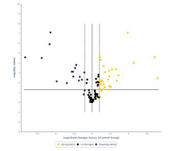
MicroRNAs (miRNAs) have been proven to regulate the development and progression of cancer through various mechanisms. The aim of the present study was to compare miRNA expression between primary melanomas from different sites. We analyzed the expression of 84 miRNAs in 27 primary melanoma and 5 nevus formalin-fixed paraffin-embedded (FFPE) samples using the Human Cancer PathwayFinder miScript miRNA PCR Array. The FFPE samples were obtained from the archives of the Municipal Clinical Emergency Hospital of Timisoara and included 10 cutaneous melanomas, 10 uveal melanomas, 7 mucosal melanomas, and 5 cutaneous nevi. Out of 84 miRNAs, 11 miRNAs showed altered expression in all types of melanoma compared with the nevi. Among these, miR-155-5p, miR-9-5p, miR-142-5p, miR-19a-3p, miR-134-5p, and miR-301a-3p were upregulated, while miR-205-5p, miR-203a-3p, miR-27b-3p, miR-218-5p, and miR-23b-3p were downregulated. The highest similarity in miRNA expression pattern was found between uveal and mucosal melanoma groups, i.e., 15 miRNAs had altered expression in both groups. Overall, we identified several miRNAs with significantly altered expression in primary melanomas, including those reported for the first time in this type of cancer. Among them, mir-9-5p, mir-203a-3p, mir-19a-3p, mir-27b-3p, and mir-218-5p showed altered expression in all three melanoma types vs. nevi. Further research should explore the potential of these miRNAs in melanoma.

Downloads

Download data is not yet available.
A preliminary study of microRNA expression in different types of primary melanoma

Downloads

Additional Files

Published

2020-05-01

How to Cite

1.
Gencia I, Baderca F, Avram S, Gogulescu A, Marcu A, Seclaman E, Marian C, Solovan C. A preliminary study of microRNA expression in different types of primary melanoma. Biomol Biomed [Internet]. 2020May1 [cited 2023Nov.30];20(2):197-208. Available from: https://www.bjbms.org/ojs/index.php/bjbms/article/view/4271

Issue

Section

Molecular Biology

INTRODUCTION

Primary melanoma, a malignant neoplasm of melanocytes, can be highly aggressive and has an increasing incidence worldwide [1]. In 2016, 76,380 new cases of melanoma were estimated in the United States (US) from which approximately 10,130 cases would be fatal [2]. Romania and other countries from Central and Eastern Europe show a higher incidence of 111 primary melanomas at advanced stage compared with Western Europe, which may be due to the lack of proper health education in these countries, among other reasons [3]. Based on this information, we consider melanoma to be one of the most important research areas in our country.

Melanoma can occur in any tissue that contains melanocytes. These cells are predominantly present in the skin, but they can also be found in organs such as the eyes, inner ear, and brain (the substantia nigra and locus coeruleus) as well as in the mucosal lining of the leptomeninges, oral cavity, esophagus, rectum, anal canal, nasal cavity, paranasal sinuses, larynx, vagina, and cervix [4-13]. In each of these tissues melanocytes have different functions and are influenced by different local factors [13].

Primary melanomas originating from different sites show characteristic tumor mutational burden [14-20]. According to the site of involvement, primary melanomas are classified as: cutaneous melanoma (CM), uveal melanoma (UM), mucosal melanoma (MM), and melanoma of the internal organs.

Numerous genomic studies showed different mutational patterns in melanoma, which was followed by the investigation of epigenetic factors involved in melanoma development. MicroRNAs (miRNAs) are small noncoding RNAs (∼22 nt in length) that can regulate the development and progression of cancer through various mechanisms. Different miRNAs have been shown to be upregulated or downregulated in melanoma, which suggests their use as diagnostic and prognostic biomarkers as well as therapeutic targets [21]. Furthermore, circulating miRNAs can be used for non-invasive diagnosis and prognosis of early metastatic disease, representing a more cost-effective method for monitoring patients and deciding about the treatment. These biomarkers have a higher sensitivity for detection in early stages of disease than the current imaging techniques (e.g., computed tomography [CT], positron emission tomography [PET] scan, etc.), and they are less invasive compared with tumor excision and sentinel lymph node biopsy, used for staging of melanoma [22].

Several studies investigated miRNA expression changes in melanomas, most notably in cutaneous and uveal types, however, there is little data on miRNA expression in primary mucosal melanoma. To the best of our knowledge, only two studies have investigated changes in miRNA expression in conjunctival melanomas [23,24] and none in mucosal melanomas involving other sites. Therefore, the current study is the first to analyze miRNA expression in primary mucosal melanomas involving mucosal surfaces other than the conjunctiva. Here, we compared miRNA expression among three primary melanoma types (CM, UM, and MM) and control cutaneous nevi. We identified several miRNAs with significantly altered expression in primary melanomas, including those reported for the first time in this type of cancer.

MATERIALS AND METHODS

Tissue samples

We obtained 27 primary melanoma and 5 nevus formalin-fixed paraffin-embedded (FFPE) samples from the archives of the Pathology Department at the Municipal Clinical Emergency Hospital of Timisoara. The FFPE samples included 10 cutaneous melanomas (stage III-IV), 10 uveal melanomas, 7 mucosal melanomas (2 intestinal mucosa, 2 genital mucosa, 1 nasal mucosa, and 2 oral mucosa) and 5 cutaneous nevi (Table 1). To reduce genomic and transcriptomic changes that occur because of environmental factors, we collected the control nevus samples from younger individuals.

All participants signed informed consent to participate in the study, and the study was approved by the Institutional Ethics Committee (approval number 1-015922/2019). Patients did not receive any treatment prior to tumor excision.

TABLE 1: Characteristics of patients with primary melanoma (n = 27) and controls (n = 5)

Real-time polymerase chain reaction (real-time PCR)

MiRNAs were purified from FFPE samples using a miRNeasy FFPE Kit (Qiagen, MD, US), according to the manufacturer’s instructions. We analyzed the expression of 84 miRNAs using the Human Cancer PathwayFinder miScript miRNA PCR Array (Qiagen, MD, US), on an ABI 7900HT real-time PCR instrument (Thermo Fisher Scientific, MA, US).

Real-time PCR data were analyzed using the online QIAGEN GeneGlobe Data Analysis Center, with a threshold of 2 for fold changes of miRNA expression in melanoma samples. Afterwards, we compared these results with miRNA expression in control nevus samples. We corrected the results using the Benjamini-Hochberg procedure to account for any possible false discovery rate. Moreover, we used the Qiagen Ingenuity pathway analysis (IPA) tool to identify pathways and diseases associated with miRNAs with altered expression.

RESULTS

The distribution of upregulated and downregulated miRNAs in the three primary melanoma types compared with control nevi is presented in Figure 1-3. MiRNAs with most significant changes in their expression in the primary melanoma samples compared with nevus group are presented in Table 2. We used these results to generate a cluster dendrogram that highlights the segregation of miRNA expression according to the 4 studied groups (Figure 4). The samples clustered together for each melanoma type and for nevi, showing similar RNA expression patterns in all three repetitions of miRNA expression analysis.

FIGURE 1: Volcano plot representing the microRNAs (miRNAs) in cutaneous melanomas compared with cutaneous nevi. Upregulated (yellow) and downregulated (blue) miRNAs with a fold change >2 vs. nevi, and miRNAs that were not significantly differently expressed (black, fold change <2 vs. nevi).
FIGURE 2: Volcano plot representing the microRNAs (miRNAs) in uveal melanomas compared with cutaneous nevi. Upregulated (yellow) and downregulated (blue) miRNAs with a fold change >2 vs. nevi, and miRNAs that were not significantly differently expressed (black, fold change <2 vs. nevi).
FIGURE 3: Volcano plot representing the microRNAs (miRNAs) in mucosal melanomas compared with cutaneous nevi. Upregulated (yellow) and downregulated (blue) miRNAs with a fold change >2 vs. nevi, and miRNAs that were not significantly differently expressed (black, fold change <2 vs. nevi).
TABLE 2: MicroRNAs (miRNAs) with most significant changes in their expression in primary melanoma samples (n = 27) compared with cutaneous nevi (n = 5)
FIGURE 4: Cluster analysis of the changes in microRNA (miRNA) expression in 4 analyzed groups (cutaneous nevi, cutaneous melanoma, uveal melanoma, and mucosal melanoma). The samples (the top of the figure) clustered together for each melanoma type and for nevi, showing similar RNA expression patterns (the right of the figure) in all three repetitions of miRNA expression analysis.

Out of 84 miRNAs, 11 miRNAs showed altered expression in all types of melanoma compared with the nevi. Among these, miR-155-5p, miR-9-5p, miR-142-5p, miR-19a-3p, miR-134-5p, and miR-301a-3p were upregulated, while miR-205-5p, miR-203a-3p, miR-27b-3p, miR-218-5p, and miR-23b-3p were downregulated. The highest similarity in miRNA expression pattern was found between uveal and mucosal melanoma groups, i.e., 15 miRNAs had altered expression in both groups.

Using the IPA, we determined the diseases, pathways, and biological functions associated with the miRNAs with altered expression. The IPA revealed that the dysregulated miRNAs are involved in many physiological and pathological processes. Table S1 shows tumor stages, tumor sites, and diseases associated with the dysregulated miRNAs for each melanoma type. It is worth noting that both primary and metastatic disease are common to all three melanoma types. We used IPA for each data set, identifying the targets of the miRNAs that were significantly overexpressed or underexpressed in our study (p < 0.05). Matching our results against the IPA data sets we generated pathway networks for each data set and, by identifying shared molecules, we created merged networks for each type of melanoma (Figure S1-S3). The targets common to all three types of melanoma were Smad2/3, insulin, sirtuin 1 (SIRT1), and tumor protein p53. The other identified targets were either unique to each type of melanoma (35 targets in MM, 29 targets in CM, and 38 targets in UM) or common to at least 2 of the analyzed melanoma types (MM and CM: Dickkopf WNT signaling pathway inhibitor 1 [DKK1], G protein signaling modulator 2 [GPSM2], one cut homeobox 2 [ONECUT2], protein regulator of cytokinesis 1 [PRC1], reversion-inducing-cysteine-rich protein with kazal motifs [RECK], and secretory carrier membrane protein 1 [SCAMP1]; CM and UM: protein kinase B [PKB or Akt], and insulin receptor [INSR]; UM and MM: interferon alpha [IFN-α], pro-inflammatory cytokines, parathyroid hormone like hormone [PTHLH], retinoblastoma [Rb] protein, and telomerase reverse transcriptase [TERT]).

TABLE S1: Tumor stages, tumor sites, and diseases associated with dysregulated microRNAs (miRNAs) for each melanoma type, according to the Qiagen Ingenuity pathway analysis (IPA)
FIGURE S1: The Qiagen Ingenuity pathway analysis (IPA) in cutaneous melanoma (n = 10)
FIGURE S2: The Qiagen Ingenuity pathway analysis (IPA) in mucosal melanoma (n = 7)
FIGURE S3: The Qiagen Ingenuity pathway analysis (IPA) in uveal melanoma (n = 10)

DISCUSSION

In this study, we analyzed the expression of 84 miRNAs in cutaneous, uveal, and mucosal melanomas compared with miRNA expression in control cutaneous nevi. Out of 84 miRNAs, 11 miRNAs showed altered expression in all types of melanoma compared with the nevi. Among these miRNAs, only miR-155-5p, miR-142-5p, miR-205-5p, miR-23b-3p, miR-134-5p, and miR-301a-3p were previously studied in melanoma. Thus, for the first time, here we reported altered expression of miR-9-5p, miR-203a-3p, miR-19a-3p, miR-27b-3p, and miR-218-5p in the three melanoma types vs. nevi.

We found that miR-155-5p was upregulated in all three types of melanoma. Similarly, a previous study reported miR-150 and miR-155 to be upregulated in primary and metastatic melanoma compared with nevi [25]. In addition, miR-155-5p was suggested to play a role in the development of other solid and hematopoietic cancers [26-29]. Nevertheless, in melanoma cell lines, ectopic expression of miR-155-5p had an anti-proliferative and pro-apoptotic effect [30], and higher miR-155-5p expression in metastatic melanoma could predict longer post-recurrence survival [25,31]. The expression of miR-155-5p increases during inflammatory response, especially during lymphocyte proliferation, and some authors suggest this to support the role of miR-155 in melanoma progression [32,33].

In this study, miR-205-5p was downregulated in melanoma vs. nevus samples, which is consistent with previous studies [34,35]. MiR-205 was shown to act as a tumor suppressor, inhibiting melanoma cell proliferation and inducing apoptosis by targeting vascular endothelial growth factor (VEGF) and transcription factor E2F1 in vitro as well as in vivo [35-37].

MiR-142-5p was upregulated in our melanoma samples compared with control nevi, and previous research indicated that miR-142 is one of five miRNAs with important clinical implications and a high prognostic value in metastatic melanoma [38].

MiR-23b-3p was downregulated in our melanoma samples vs. control nevi. This miRNA was shown to have altered expression in different malignant tumors, including melanoma, with clinical, therapeutic, and prognostic significance [34,39].

We showed for the first time that miR-9-5p is upregulated in melanoma compared with nevi. MiR-9-5p promotes cell proliferation and metastasis in non-small cell lung cancer (NSCLC) and colorectal cancer [40,41]. It is involved in the differentiation of B lymphocytes into plasma cells by the negative regulation of the transcription factor PR domain zinc finger protein 1 (PRDM1/BLIMP-1), showing lower levels as the lymphocytes differentiate. This explains high levels of miR-9-5p in primary large-B cell lymphomas [42]. On the other hand, miR-9 is downregulated in human ovarian cancer compared with normal ovary, and the overexpression of miR-9 inhibits cell growth in ovarian cancer in vitro through the negative regulation of nuclear factor NF-kappa-B p105 (NFκB1) [42,43]. MiR-9-5p may represent a new prognostic marker in melanoma and possibly a new therapeutic target.

We found that miR-19a-3p is upregulated in melanoma vs. nevi, which is another novel finding in this cancer type. In gastric cancer, miR-19a-3p had a negative prognostic impact and promoted cell malignancy [44].

MiR-134-5p was upregulated in our melanoma samples compared with nevi. Previous studies showed that miR-134-5p is downregulated in NSCLC cells [45] and nasopharyngeal carcinoma cells [46] and that it has a role in inhibiting tumor progression. Interestingly, another study on melanoma showed that miR-134-5p is downregulated in melanoma patients compared with healthy controls [47], and this discrepancy with our results should be further investigated.

We reported in this study upregulation of miR-301a-3p in melanoma vs. nevi. MiR-301a was previously reported to be upregulated in melanoma samples compared to benign melanocytic lesions [48]. In addition, in hepatocellular carcinoma cell lines, miR-301a-3p overexpression was shown to stimulate cell proliferation, invasion, and chemoresistance [49].

For the first time, we showed that miR-203a-3p was downregulated in melanoma compared with nevi. The overexpression of miR-203a-3p in colorectal cancer cell lines inhibited cell proliferation and reduced chemoresistance [50] and similarly, in nasopharyngeal carcinoma, overexpressed miR-203a-3p inhibited cell proliferation, migration, and invasion in vitro as well as xenograft tumor growth and lung metastasis in vivo [51]. On the other hand, in hepatocellular carcinoma cells, miR-203a-3p.1 overexpression was reported to be oncogenic [52].

MiR-27b-3p and miR-218-5p were another two downregulated miRNAs in our case study that are reported for the first time in melanoma. MiR-27b-3p was also downregulated in lung cancer tissues [53] and in breast cancer tissues and cell lines, where it was associated with chemoresistance [54]. However, miR-27b-3p expression was increased in Dox-resistant anaplastic thyroid cancer (ATC) cells [55]. MiR-218-5p expression was reduced in gastric cancer [56], NSCLC [57], and gallbladder cancer (GBC) tissue [58]; moreover, miR-218-5p overexpression reversed the resistance of GBC cells to gemcitabine [58].

Among miRNAs with altered expression in at least one type of melanoma (Table 3), we focused on those previously studied in melanoma. Although some studies reported that miR-127-5p and miR-15a-5p were not significantly differently expressed in melanoma compared to melanocytic nevi [59,60], our results showed these miRNAs to be overexpressed in both cutaneous and mucosal melanomas vs. nevi. In addition, miR-335-5p was reported to be significantly differentially expressed in the melanoma cell line MML-1 compared to benign nevi [33], but our results showed a significant overexpression of miR-335-5p only in uveal melanoma group compared to control nevi.

TABLE 3: MicroRNAa (miRNAs) with altered expression in at least one group of the studied melanoma types (n = 27) that were or were not investigated before

The main limitation to our study is the small sample size. Thus, although we consider some of our findings to be groundbreaking in melanoma research, they still need to be confirmed in large-scale studies. To this end, we propose analyzing miRNAs in each melanoma sample separately and in combination with follow-up data, so to determine changes in miRNA expression specific to each cell type and site of origin, the impact of local factors, and the prognostic potential of miRNAs in melanoma. To achieve this, we plan to conduct a prospective study in the future.

CONCLUSION

Overall, we identified several miRNAs with significantly altered expression in primary melanomas, including those reported for the first time in this type of cancer. Mir-9-5p, miR-203a-3p, miR-19a-3p, miR-134-5p, miR-301a-3p, miR-155-5p, miR-142-5p, miR-205-5p, miR-23b-3p, miR-27b-3p, and miR-218-5p had altered expression in all three melanoma types. Further research should explore the potential of these miRNAs in melanoma. The primary contribution of this study is to demonstrate that despite originating from the same cell type, the three melanoma types are still separate entities characterized by different miRNA expression patterns.

Acknowledgements

ACKNOWLEDGMENTS

This project was financed by the Victor Babes University of Medicine and Pharmacy Timisoara, through the project for young researchers PII-C5-TC-2017-02 C.I.

REFERENCES

  1. , , (). Melanoma:clinical, evolutive and histopathological characteristics of a series of 136 cases. An Bras Dermatol. https://doi.org/10.1590/abd1806-4841.201⇝0
  2. , , (). Cancer statistics 2016. CA Cancer J Clin. https://doi.org/10.3322/caac.21332
  3. , , , , , (). Risk factors for melanoma and skin health behaviour:an analysis on Romanian melanoma patients. Oncol Lett. https://doi.org/10.3892/ol.2018.9737
  4. , (). The immunohistochemical identification of human oral mucosal melanocytes. Arch Oral Biol. https://doi.org/10.1016/S0003-9969(96)00113-6
  5. (). Human melanocyte biology, toxicology, and pathology. J Environ Sci Health C Environ Carcinog Ecotoxicol Rev. https://doi.org/10.1080/10590500500234970
  6. , , , (). Melanosis of the esophagus. Cancer. https:// doi.org/ 10.1002/1097-0142 (196301)16:1<48:AID- CN CR 282 01 60 107>3.0. CO ;2- M
  7. , , (). Primary malignant melanoma of the rectum. Evidence for origination from rectal mucosal melanocytes. Cancer. https:// doi.org/10.10 02/109 7-0142 (1988 0401)61:7 <13 64:AID - CNCR2 820 6 10715 >3.0 .CO ;2-B
  8. , (). The presence of melanocytes in the nasal cavity. Ann Otol Rhinol Laryngol. https://doi.org/10.1177/000348947408300414
  9. , , (). Melanocytes in the nasal cavity and paranasal sinus. Incidence and distribution in Japan. Acta Pathol Jpn. https://doi.org/10.1111/j.1440-1827.1987.tb00427.x
  10. , , , (). The presence of melanocytes in the human larynx. Laryngoscope. https://doi.org/10.1288/00005537-197205000-00009
  11. , , (). Melanoblasts in vaginal mucosa. Origin for primary malignant melanoma. Cancer. https://doi.org/ 10.1002/ 1097 - 0142 (196407) 17:7 <912: AID-CNCR2820170711 >3.0.CO;2-F
  12. , , , (). Foci of stromal melanocytes (so-called blue naevus) of the uterine cervix in Japanese women. Virchows Arch A Pathol Anat Histopathol. https://doi.org/10.1007/BF01600162
  13. , (). What are melanocytes really doing all day long…?:from the ViewPoint of a keratinocyte:Melanocytes - cells with a secret identity and incomparable abilities. Exp Dermatol. https://doi.org/10.1111/j.1600-0625.2009.00912.x
  14. , , , , , (). KIT gene mutations and copy number in melanoma subtypes. Clin Cancer Res. https://doi.org/10.1158/1078-0432.CCR-08-0575
  15. , , , , , (). Conjunctival melanomas harbor BRAF and NRAS mutations and copy number changes similar to cutaneous and mucosal melanomas. Clin Cancer Res. https://doi.org/10.1158/1078-0432.CCR-13-0163
  16. , , , (). Classifying melanocytic tumors based on DNA copy number changes. Am J Pathol. https://doi.org/10.1016/S0002-9440(10)63536-5
  17. , , (). KIT mutations in Russian patients with mucosal melanoma. Melanoma Res. https://doi.org/10.1097/CMR.0b013e32834bf398
  18. , , , , , (). KIT mutation in anal melanomas correlates with KIT protein expression and is sensitive to specific kinase inhibition. Int J Cancer. https://doi.org/10.1002/ijc.22681
  19. , , , , , (). Comparison of molecular abnormalities in vulvar and vaginal melanomas. Mod Pathol. https://doi.org/10.1038/modpathol.2013.211
  20. , , , , , (). Genome sequencing of mucosal melanomas reveals that they are driven by distinct mechanisms from cutaneous melanoma. J Pathol. https://doi.org/10.1002/path.4204
  21. , , , , , (). MicroRNAs as potential diagnostic and prognostic biomarkers in melanoma. Eur J Cancer. https://doi.org/10.1016/j.ejca.2015.10.009
  22. , , , (). MicroRNAs in cutaneous melanoma:role as diagnostic and prognostic biomarkers. J Cell Physiol. https://doi.org/10.1002/jcp.26395
  23. , , , , , (). Characterization of the melanoma miRNAome by deep sequencing. PLoS One. https://doi.org/10.1371/journal.pone.0009685
  24. , , , , , (). MicroRNA expression profile in conjunctival melanoma. Invest Ophthalmol Vis Sci. https://doi.org/10.1167/iovs.16-19862
  25. , , , , , (). Melanoma MicroRNA signature predicts post-recurrence survival. Clin Cancer Res. https://doi.org/10.1158/1078-0432.CCR-09-2721
  26. , , , , , (). MicroRNA gene expression deregulation in human breast cancer. Cancer Res. https://doi.org/10.1158/0008-5472.CAN-05-1783
  27. , , , , , (). MicroRNA profiling of clear cell renal cell cancer identifies a robust signature to define renal malignancy. J Cell Mol Med. https://doi.org/10.1111/j.1582-4934.2009.00705.x
  28. , , , , , (). Aberrant expression of oncogenic and tumor-suppressive microRNAs in cervical cancer is required for cancer cell growth. PLoS One. https://doi.org/10.1371/journal.pone.0002557
  29. , , , (). Inositol phosphatase SHIP1 is a primary target of miR-155. Proc Natl Acad Sci USA. https://doi.org/10.1073/pnas.0902636106
  30. , , , , , (). Altered expression of selected microRNAs in melanoma:antiproliferative and proapoptotic activity of miRNA-155. Int J Oncol.
  31. , , , , (). MicroRNA and cutaneous melanoma:from discovery to prognosis and therapy. Carcinogenesis. https://doi.org/10.1093/carcin/bgs205
  32. , , (). MiR-155:on the crosstalk between inflammation and cancer. Int Rev Immunol. https://doi.org/10.1080/08830180903093796
  33. , , , , , (). Small RNA deep sequencing discriminates subsets of extracellular vesicles released by melanoma cells - Evidence of unique microRNA cargos. RNA Biology. https://doi.org/10.1080/15476286.2015.1056975
  34. , , , , , (). In-depth characterization of microRNA transcriptome in melanoma. PLoS One. https://doi.org/10.1371/journal.pone.0072699
  35. , , , , (). Differential expression of microRNAs during melanoma progression:miR-200c, miR-205 and miR-211 are downregulated in melanoma and act as tumour suppressors. Br J Cancer. https://doi.org/10.1038/bjc.2011.568
  36. , , , , , (). miRNA-205 suppresses melanoma cell proliferation and induces senescence via regulation of E2F1 protein. J Biol Chem. https://doi.org/10.1074/jbc.M111.227611
  37. , , , , , (). Chemically modified synthetic microRNA-205 inhibits the growth of melanoma cells in vitro and in vivo. Mol Ther. https://doi.org/10.1038/mt.2013.70
  38. , , , , , (). Identification, review, and systematic cross-validation of microRNA prognostic signatures in metastatic melanoma. J Invest Dermatol. https://doi.org/10.1038/JID.2015.355
  39. , , , , (). Functional role of microRNA-23b-3p in cancer biology. Microrna. https://doi.org/10.2174/2211536607666180629155025
  40. , , , , (). MiR-9-5p promotes cell growth and metastasis in non-small cell lung cancer through the repression of TGFBR2. Biomed Pharmacother. https://doi.org/10.1016/j.biopha.2017.11.105
  41. , , , , , (). Overexpression of microRNAs miR-9, -98, and -199 correlates with the downregulation of HK2 expression in colorectal cancer. Mol Biol (Mosk). https://doi.org/10.1134/S0026893318020140
  42. , , , , , (). MicroRNA profiling of primary cutaneous large B-cell lymphomas. PLoS One. https://doi.org/10.1371/journal.pone.0082471
  43. , , , , , (). MicroRNA-9 inhibits ovarian cancer cell growth through regulation of NF-kappaB1. FEBS J. https://doi.org/10.1111/j.1742-4658.2009.07237.x
  44. , , , , , (). Downregulated PITX1 modulated by miR-19a-3p promotes cell malignancy and predicts a poor prognosis of gastric cancer by affecting transcriptionally activated PDCD5. Cell Physiol Biochem. https://doi.org/10.1159/000489590
  45. , , , (). Long non-coding RNA prostate cancer-associated transcript 7 (PCAT7) induces poor prognosis and promotes tumorigenesis by inhibiting mir-134-5p in non-small-cell lung (NSCLC). Med Sci Monit. https://doi.org/10.12659/MSM.907904
  46. , , , , (). Long non-coding RNA PCAT7 regulates ELF2 signaling through inhibition of miR-134-5p in nasopharyngeal carcinoma. Biochem Biophys Res Commun. https://doi.org/10.1016/j.bbrc.2017.07.093
  47. , , , , , (). The circulating transcriptome as a source of biomarkers for melanoma. Cancers (Basel). https://doi.org/10.3390/cancers11010070
  48. , , , , , (). Comparative microarray analysis of microRNA expression profiles in primary cutaneous malignant melanoma, cutaneous malignant melanoma metastases, and benign melanocytic nevi. Cell Tissue Res. https://doi.org/10.1007/s00441-012-1514-5
  49. , , , , (). MicroRNA-301a-3p suppressed the progression of hepatocellular carcinoma via targeting VGLL4. Pathol Res Pract. https://doi.org/10.1016/j.prp.2018.09.008
  50. , , , , , (). LncRNA HOTAIR is a prognostic biomarker for the proliferation and chemoresistance of colorectal cancer via miR-203a-3p-mediated Wnt/ß-catenin signaling pathway. Cell Physiol Biochem. https://doi.org/10.1159/000489110
  51. , , , , , (). MiR-203a-3p suppresses cell proliferation and metastasis through inhibiting LASP1 in nasopharyngeal carcinoma. J Exp Clin Cancer Res . https://doi.org/10.1186/s13046-017-0604-3
  52. , , , , , (). miR-203a-3p.1 targets IL-24 to modulate hepatocellular carcinoma cell growth and metastasis. FEBS Open Bio. https://doi.org/10.1002/2211-5463.12248
  53. , , , (). Antitumor effect of miR-27b-3p on lung cancer cells via targeting Fzd7. Eur Rev Med Pharmacol Sci.
  54. , , , , , (). miR-27b-3p inhibits proliferation and potentially reverses multi-chemoresistance by targeting CBLB/GRB2 in breast cancer cells. Cell Death Dis. https://doi.org/10.1038/s41419-017-0211-4
  55. , , , , , (). miR-27b-3p is involved in doxorubicin resistance of human anaplastic thyroid cancer cells via targeting peroxisome proliferator-activated receptor gamma. Basic Clin Pharmacol Toxicol. https://doi.org/10.1111/bcpt.13076
  56. , , , , , (). miR-218 suppresses gastric cancer cell cycle progression through the CDK6/Cyclin D1/E2F1 axis in a feedback loop. Cancer Lett. https://doi.org/10.1016/j.canlet.2017.06.006
  57. , , , , , (). Tumor-suppressive miR-218-5p inhibits cancer cell proliferation and migration via EGFR in non-small cell lung cancer. Oncotarget. https://doi.org/10.18632/oncotarget.ↀ
  58. , , , , , (). miR-218-5p restores sensitivity to gemcitabine through PRKCE/MDR1 axis in gallbladder cancer. Cell Death Dis. https://doi.org/10.1038/cddis.2017.178
  59. , , , , , (). Silencing of a large microRNA cluster on human chromosome 14q32 in melanoma:biological effects of mir-376a and mir-376c on insulin growth factor 1 receptor. Mol Cancer. https://doi.org/10.1186/1476-4598-11-44
  60. , , , , , (). MiR-497-5p, miR-195-5p and miR-455-3p function as tumor suppressors by targeting hTERT in melanoma A375 cells. Cancer Manag Res. https://doi.org/10.2147/CMAR.S163335

Conflict of interest statement: The authors declare no conflict of interests