In vitro toxicity model: Upgrades to bridge the gap between preclinical and clinical research

Authors

  • Eneko Madorran Institute of Anatomy, Histology and Embryology, Faculty of Medicine, University of Maribor, Maribor, Slovenia; Department of Pharmacology, Faculty of Medicine, University of Maribor, Maribor, Slovenia https://orcid.org/0000-0001-6524-4119
  • Andraž Stožer Institute of Physiology, Faculty of Medicine, University of Maribor, Maribor, Slovenia https://orcid.org/0000-0003-2097-5502
  • Sebastjan Bevc Department of Pharmacology, Faculty of Medicine, University of Maribor, Maribor, Slovenia; Department of Nephrology, Clinic for Internal Medicine, University Medical Center Maribor, Maribor, Slovenia https://orcid.org/0000-0003-2313-1559
  • Uroš Maver Department of Pharmacology, Faculty of Medicine, University of Maribor, Maribor, Slovenia; Institute of Biomedical Sciences, Faculty of Medicine, University of Maribor, Maribor, Slovenia https://orcid.org/0000-0002-2237-3786

DOI:

https://doi.org/10.17305/bjbms.2019.4378

Keywords:

Toxicity models, cell-based models, in vitro, physiological models, bioanalysis, drug approval, toxicity testing, toxicity prediction, drug toxicity, preclinical research

Abstract

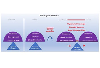
The Centers for Disease Control and Prevention (CDC) provides extensive data that indicate our need for drugs to maintain human population health. Despite the substantial availability of drugs on the market, many patients lack specific drugs. New drugs are required to tackle this issue. Moreover, we need more reliable models for testing drug toxicity, as too many drug approval failures occur with the current models. This article briefly describes various approaches of the currently used models for toxicity screening, to justify the selection of in vitro cell-based models. Cell-based toxicity models have the best potential to reliably predict drug toxicity in humans, as they are developed using the cells of the target organism. However, currently, a large gap exists between in vitro cell-based approach to toxicity testing and the clinical approach, which may be contributing to drug approval failures. We propose improvements to in vitro cell-based toxicity models, which is often an insight approach, to better match this approach with the clinical homeostatic approach. This should enable a more accurate comparison of data between the preclinical as well as clinical models and provide a more comprehensive understanding of human physiology and biological effects of drugs.

Downloads

Download data is not yet available.
In vitro toxicity model: Upgrades to bridge the gap between preclinical and clinical research

Downloads

Additional Files

Published

2020-05-01

How to Cite

1.
Madorran E, Stožer A, Bevc S, Maver U. In vitro toxicity model: Upgrades to bridge the gap between preclinical and clinical research. Biomol Biomed [Internet]. 2020May1 [cited 2023Dec.4];20(2):157-68. Available from: https://www.bjbms.org/ojs/index.php/bjbms/article/view/4378

Issue

Section

Reviews

INTRODUCTION

In 2016, the Centers for Disease Control and Prevention (CDC) recorded 883.7 million physician office visits in the United States (US). Among those visits, 653.5 million visits (73.9%) involved drug therapy [1]. Moreover, 80.4% of hospital emergency department visits involved drug therapy [2]. These two figures illustrate our need for drugs to maintain human population health. In addition, every patient needs a specific drug. Despite the substantial availability of drugs on the market, many patients still lack specific drugs. New drugs are required to tackle this issue. We also need more reliable models for testing drug toxicity, as too many drug approval failures occur with the current models. On the one hand, safe drugs are categorized as unsafe [3] while, on the other hand, unsafe drugs are placed on the market (until 2017, 417 commercial drugs were withdrawn because they caused severe adverse reactions in patients, including death) [4-6]. Furthermore, the current drug approval process is time and money consuming, which is reflected in the time required to place a new drug on the market (approximately 10 years) and overall development costs that often exceed a billion dollars [7].

The above figures indicate that the process of drug development and approval is not efficient and one of the main reasons may be our lack of knowledge. For instance, looking back at the history of cell biology, in 1838, Theodor Schwann made three conclusions about cells6, one of which turned out to be wrong a hundred years later [8,9]. Similarly to what happened with the first cell theory, our assumptions about the current drug approval process may be incorrect8. With this regard, developing more reliable models for testing drug toxicity could improve the reliability of drug approval process.

Although several different features of drug approval process should be addressed concurrently to improve its reliability, the authors of this review will focus on the gap between preclinical and clinical research in drug toxicity testing. Despite our lack of a complete understanding of cell physiology, the majority of preclinical research is based on specific cellular pathways, while clinical research focuses on homeostasis monitoring [10,11]. Therefore, the preclinical and clinical research endpoints are essentially too far apart to create a continuous monitoring throughout the drug approval process.

In the present article, we will briefly describe various approaches of the currently used models for toxicity screening and discuss the differences between them, to justify the selection of in vitro cell-based models as the toxicity models with the best potential to reliably predict drug toxicity in humans. We propose several improvements to in vitro cell-based toxicity models (also cell-based toxicity models) to better match this approach with clinical homeostatic approach. The improvements, described in detail in the following sections, are primarily focused on narrowing the differences between preclinical and clinical research, but may also have a wider impact on the field.

BOX1: Visual Abstract

TOXICITY MODELS

Toxicity may be defined as “the degree to which a substance (a toxin or poison) can harm humans or animals” [12]. There are several interpretations of this definition, mainly related to its extended meaning, which are the basis for many different approaches used in measuring drug toxicity [13].

Toxicity models have been developed to measure harmful effects of substances on the human organism. Currently, there are three main categories of toxicity models in preclinical research, i.e., in silico, in vivo, and in vitro models [14], and one category in clinical research, divided into three phases [15]. The current drug approval process manages these categories as needed [15].

In this section, we will briefly describe the premises of various toxicity models and the differences between them. We will further discuss the most significant failures of drug approval process when predicting drug toxicity, where the differences between the models may contribute to these failures. This overview is the basis on which we have developed improvements to in vitro cell-based toxicity models, presented in the subsequent sections.

In silico toxicity model

In silico means: “performed on a computer or via computer simulation” [14]. This toxicity model is mainly used to predict how drugs interact with the body. The short duration of tests and reduced costs are the main advantages of in silico toxicity models [14]. Several in silico toxicity models exist with different approaches and, accordingly, there are many ways of their classification [16].

Since the simulation in in silico toxicity models is performed by computer, an algorithm is built to transform a given input into an output [17]. However, as mentioned above, we still do not have a complete knowledge of biochemical and physiological processes in the human body [11], thus we cannot develop effective algorithms to predict drug toxicity. In most cases, there is not enough data to perform a computer simulation and get a representative output with in silico toxicity models.

In vivo toxicity model

In vivo tests refer to experiments that use living organisms as models. The major advantage of in vivo models is the ability to investigate the effects of drugs on physiological and biochemical reactions in living organisms as well as the availability of large and coordinated databases [14]. It is assumed that, if properly assessed, the effect of any compound observed in laboratory animals is also relevant to humans [13]. Among many endpoints of in vivo toxicity testing, the most commonly used is the “No Observed Adverse Effect Level (NOAEL)”, which must be determined prior to the launch of the clinical phase [13].

The in vivo toxicity model is the only model that uses an organism as a whole, including all physiological reactions and biochemical interactions, and thus is the only model that provides information on drug distribution in the organism and possible interactions of the drug with non-target organs. As such, in vivo models arguably provide the most representative models for testing drug toxicity. Nevertheless, there are some limitations to the current in vivo toxicity models, especially considering the significant biological differences that exist between the human and other animal organisms [18]. These differences are observed from the molecular to cellular and organ level (Figure 1) [19-21]. An example of an important functional difference between humans and rodents is blood pH, where the normal blood pH in mouse (7.3–7.4) partially overlaps with the pathophysiological blood pH in humans (<7.35 and >7.45) [22,23]. Moreover, small differences at any scale may cause significant changes in organisms, such as single point mutations (SPM) or aseptic conditions [24,25]. Any failure in predicting drug toxicity with in vivo models is most likely due to these differences between animals and humans. Moreover, fewer improvements are possible to in vivo models as they are close to their toxicity assessment potential (i.e., the model has limitations that currently cannot be overcome).

FIGURE 1: Anatomy of the human and the mouse pancreas. (A) The parts of the human pancreas and neighboring organs. (B) The parts of the mouse pancreas and neighboring organs. A microscopic view of the human (C) and mouse (D) pancreas, showing differences in the lobule size between humans and mice. On the other hand, the islets of Langerhans have a similar size (E and F). Cell type composition and distribution significantly differ between the human and mouse pancreas. Moreover, the number of Langerhans islets in the human pancreas significantly differ from that observed in the mouse pancreas. Reprinted with permission by Taylor and Francis from [26].

In vitro toxicity model

In vitro tests use a specific part (e.g., organ, tissue, or cell) of a given organism to study it under a controlled environment, by which the noise of the surrounding internal or external environment is considerably reduced. The advantages of in vitro toxicity models are reduced time and costs and representativeness (as they are developed using the cells or tissues of the target organism) [14]. Among the many in vitro toxicity models available we will focus on cell-based toxicity models in the following sections. Cell-based toxicity models are most likely to take into account the known metabolic networks at the cellular level, compared to other in vitro models [11].

The complexity of recently designed in vitro cell-based toxicity models reaches those of multiple organs, and in some cases, these models are more accurate than in vivo toxicity models [27]. However, the majority of current in vitro cell-based toxicity models do not represent the target organism accurately because they are simplified to such an extent that they lose one or more vital characteristics of the organism. Most in vitro cell-based toxicity models of organs do not include diverse cell lines characteristic for the target organ. Thus, even when a substance does not induce toxicity in a particular cell line, it may be toxic to other cell lines within the target organ. A good example that illustrates this situation are in vitro toxicity models based on human liver cells, where most models are based on hepatocytes, despite a significant role of fibroblasts in liver pathological processes [28]. Moreover, even if a substance induces toxicity in a single cell line it may not be toxic to the whole organ, because other cells in the organ may compensate the imbalance.

While all the processes in the human organism are interconnected, in vitro toxicity models mainly focus on biomolecules involved in particular cellular pathways (Figure 2). In vitro toxicity research that focuses only on a specific cell impairment may not be accurate, because it neglects the self-repair ability of cells, including the repair mechanisms at the DNA level [29]. Moreover, various cells synchronously (according to the physiological rhythm) contribute to the organ function and homeostasis. The response of an organ to an input dramatically changes if the physiological rhythm is altered [30]. Likewise, the same input may result in different outputs depending on the cell population size (e.g., this happens with organ-on-a-chip models, where the cell population size is too small) (Figure 2).

FIGURE 2: The potential effects (outcomes) of an input depending on different cellular pathways it may undergo: (A) A cell may produce different outcomes in response to the same input. In the figure, four different outputs of the same input are represented; (B) with the same input, the output of target cells may change depending on the number of cells. Therefore, the cell population size should be large enough so we can reliably identify the output with the highest probability.

Most experiments with in vitro toxicity models are conducted within a limited time period and are ended after a pre-defined period, rather than continuously monitored. Thus, a substance may induce toxicity to a cell in a given moment and this may be self-repaired later, overall resulting in non-toxic effects. Moreover, the homeostasis of an organism may be recovered even after an acutely high (possibly toxic) exposure [29]. On the other hand, a substance may not induce toxicity to a cell in a given moment but may induce toxicity after a certain period.

Clinical toxicity model

Clinical testing of drug toxicity is the last step in the drug approval processes, because it is indisputable that the model that best mimics the target organism is the target organism itself. Clinical research monitors the body’s biochemical balance and metabolism, and its main endpoint is homeostatic imbalance. Numerous clinical studies have shown that certain biomolecules are present in higher or lower concentrations in different patients, and these biomolecules are used as benchmarks in monitoring homeostasis [10,31].

Comparison of different categories of toxicity models

The approaches to toxicity testing significantly differ among various categories of toxicity models. Consequently, it is challenging to gather information for a systematic comparison of the models (Figure 3) [32-37]. Moreover, heterogeneous endpoints may also be the reason for observed contradictory effects among the models [32-34,36,37] as well as for many drug approval failures (Table 1).

FIGURE 3: Doses, durations, and endpoints of different toxicity models. In silico models are represented with a grey color, in vivo models are represented with a blue color, in vitro models are represented with a green color, and clinical models are represented with a red color.
TABLE 1: List of recent first‑in‑man trial failures with drugs allegedly considered to be safe that should have been classified as unsafe after animal testing

PROPOSED IMPROVEMENTS TO IN VITRO CELL-BASED TOXICITY MODELS

As discussed in the previous sections, the current drug approval process may not accurately predict drug toxicity and, thus, needs to be improved. Reducing the gap between toxicity models may improve the reliability of the current drug approval process. Among various toxicity models, the in vitro cell-based model has the highest potential to be compatible with the clinical model. Nevertheless, there are important issues and limitations related to the current cell-based toxicity models that we need to address to achieve the desired level of reliability. The following paragraphs provide an overview of the most important improvements of cell-based toxicity models that should be considered in the future models (also summarized in Table 2).

TABLE 2: Summary of the suggested improvements to in vitro cell‑based toxicity models

The in vitro cell-based toxicity model should monitor homeostatic processes, similarly to the clinical models. While monitoring homeostatic processes, any observed homeostatic imbalance should be considered as a consequence of the substance’s toxicity.

Cell culture medium should be reduced to support the cell homeostatic mechanisms and facilitate the monitoring of cells with clinical instrumentation, because cells are often too diluted in culture medium (low cell density). Modifying these two elements should enable monitoring of homeostatic imbalance in cell-based toxicity models. In addition, the degree of how well the cell-based toxicity model represents the clinical model should be improved, to enable a reliable comparison between preclinical and clinical data. To increase the representativeness of in vitro cell-based toxicity models we should consider the following:

  • I. The nutrient content of culture medium should correspond to the in vivo conditions to avoid changes in cells due to variation in nutrient availability.

  • II. Culture medium material may induce changes in cell shape, subsequently modifying other cell features. Hence, cell-based toxicity models should use materials that support cells to keep their original shape.

  • III. The toxicokinetic characteristics of cell-based toxicity models should be compatible with those of the human organism.

  • IV. Cell death assays should focus on determining the number of dead cells rather than the number of viable cells, to improve the reliability of cell viability assays.

  • V. The endpoints of cell-based toxicity models should include homeostatic imbalance and cell viability.

  • VI. The cell-based and clinical toxicity model should have the same drug dosage parameters to facilitate the comparison between preclinical and clinical data.

  • VII. The improved cell-based toxicity model should be tested with different drugs to determine the model’s reliability.

The following subsections describe the above-mentioned points in more detail and discuss the limitations of the improved in vitro cell-based toxicity model.

Homeostatic imbalance

We may define the toxicity of a substance as the harm that the substance inflicts on the human organism and determine the substance’s toxicity by observing homeostatic imbalances in the in vitro cell-based model (Figure 4).

FIGURE 4: Principles of homeostatic control: (1) Dynamic of homeostatic changes over time: not only do homeostatic ranges vary between individuals, but in certain individuals these values fall out of the “normal” homeostatic range. (2) Homeostatic disturbance, pushing the organism out of its basal homeostasis. (3) Homeostatic receptors detect this homeostatic imbalance, and trigger the homeostatic control mechanism (from molecular to systemic level). (4) Homeostatic control mechanisms restore the homeostatic balance, and (5) homeostasis is eventually re-established [10,52,53].

The human organism is composed of multiple organ systems [47]. According to the “property of the union of sets” the union of sets is impaired if any set is impaired. Thus, the homeostasis of an organism can be maintained only if all organ systems function within the homeostatic range [48]. Specific biomolecules are produced in the organ to keep the organ and its specialized function within the homeostatic range (usually, only a few of these biomolecules reach bloodstream) [49]. The synthesis of biomolecules is only one part of many synchronized physiological reactions that happen simultaneously or consecutively to maintain the homeostasis of the organism. Any alteration in these processes may lead to significant changes in the homeostasis. For example, genetic polymorphisms may influence up to 10-fold variation in metabolic activity [50,51]. Furthermore, the physiological processes of individuals may differ due to illness or variations in metabolic demands.

The organism synthesizes many different biomolecules to regulate the above-mentioned physiological processes, which creates noise when measuring molecules involved in a specific physiological process. Some biomolecules have already been established as biomarkers in clinical research. However, due to the noise, many biomolecules remain undetected or, if detected, are unrelated to pathological processes that lead to homeostatic imbalance [54,55]. A systematic approach to in vitro cell-based toxicity models, where a researcher decides about the model’s conditions, could minimize the noise within the model, enabling the study of homeostasis at the level of organ rather than at the level of whole organism [49]. This reduction of noise should enable the detection of distinct biomolecules involved in specific homeostatic processes. The selection of biomolecules should be based on the significance of their function in maintaining organ homeostasis. In addition, a preference should be given to biomolecules that act as “early” biomarkers, because they are more useful in clinics. In any case, the in vitro cell-based toxicity model should include all cell types that constitute the organ of interest (due to the reasons described in the subsection In vitro toxicity model).

The suggested improvements should facilitate a more comprehensive understanding of each organ’s contribution to homeostasis.

Cell density

Any given input (cell signal, chemical imbalance, etc.) undergoes many different but related processes within a cell, resulting in different outputs (e.g., gene expression, cell movement, molecule transport, etc.). A large number of cells (and multiple repetitions of experiments) are required to identify an output that has the highest probability, due to the different probabilities of different outputs (Figure 2) [56].

Extracellular fluid, especially interstitial fluid (IF), interacts with cells to maintain organ homeostasis [57]. Any alteration of IF composition disturbs the interaction between IF and cells. For example, it was observed that the accumulation of IF affects protein composition and gene regulation [58,59]. Under this premise, too much of culture medium may alter the homeostatic mechanisms in cells. Furthermore, blood accounts for 7% of the human body weight, while cell culture medium commonly accounts for more than 90% of the in vitro model weight. Consequently, biomolecules may be very diluted in culture medium and difficult to measure with clinical instrumentation [60].

Cell cultures should be designed with less culture medium to maintain cell homeostatic mechanisms and facilitate the measurement of biomolecules with clinical instrumentation. In addition, the size of cell population should be adequate, to observe the output with the highest probability from a given input.

Nutrient intake

Since the intake of nutrients and excretion of their products affect the homeostasis of organs, any alteration in nutrient intake will change the homeostasis accordingly [10]. Therefore, if the cells in the cell-based toxicity model are not fed with nutrients in a similar manner as the target organism, we may assume that the cell homeostasis in the in vitro model differs from the homeostasis in the target organism [61-63]. The pathway of nutrients in different organs is also not considered in cell-based toxicity models (i.e. different cells utilize different enzymatic machinery) [64,65]. Based on the above statements, we can assume the following:

  • I. Human cells are unable to synthesize certain unsaturated fatty acids. Therefore, a culture medium rich in saturated fatty acids forces cells to change the composition of their plasma membrane so it becomes rich in saturated fatty acids. This plasma membrane is larger, more saturated, and more packed than plasma membranes rich in unsaturated fatty acids. The larger, more saturated, and more packed plasma membranes decrease cell fluidity, resulting in lower transport of a drug to the cell cytoplasm [47].

  • II. While human cells do not synthesize all the required sugars at the same rate, in vitro cell-based toxicity models are solely provided with glucose. The lack of other sugars affects the glycosylation of de novo synthesized proteins, which subsequently alters the biological effect of the glycoprotein [47].

  • III. The availability of essential minerals and vitamins is required to maintain homeostasis. The cells in cell-based toxicity models are often unable to perform these processes due to a lack of essential vitamins and minerals [66].

Owing to these shortcomings, the composition of cells in in vitro cell-based toxicity models may not be the same as the cell composition in the human organism, affecting the analysis of those cells in vitro [67]. To minimize the changes in cell composition, cell culture medium should include all essential nutrients (i.e., different sugars, essential fatty acids, vitamins, and minerals). The concentration of these nutrients in the culture medium should be calculated based on the available clinical nutrition data and the recommended daily intake for the target organ, considering also the number of different cell types in each model [68].

Cell shape

Since the shape of a cell is closely related to its function, a change in cell shape leads to a change in cell function [69,70]. In cell-based toxicity models, cells need to change their shape to be able to attach to cell culture plasticware, which consequently leads to changes in their functions. For example, the transport kinetics in cells may be impaired since the distance between the apical and basolateral membrane is shortened when the cell shape is altered [69,70]. The extracellular matrix (ECM) that mimics the environment of target cells may overcome this issue [61,63]. Furthermore, fibroblasts should synthesize the ECM specific to the target organ [71].

Toxicokinetics

The toxicokinetic analysis of a target molecule in in vitro cell-based toxicity models is currently difficult due to technical limitations, genetic polymorphisms, and other reasons [72,73], but several approaches may improve the in vitro toxicokinetic analysis. Specifically, considering the type of capillaries and the size of molecule may facilitate the prediction of molecule transport through the systemic circulation (e.g., discontinuous capillaries have a wider diameter than continues capillaries). Moreover, the pathway of the molecules of interest should be determined, since these molecules may be transported to different organs in different forms, i.e., in altered or in the original form [74]. This, however, is out of the scope of our review. Considering all the pathways that a molecule can undergo upon entering the organism may improve the representativeness of in vitro cell-based toxicity models. For instance, a pathway probabilistic tree may be generated for the target molecule, such as the in silico physiologically based pharmacokinetic (PBPK) modeling approach [16].

Cell viability methods

Many cell viability assays are available. However, most cell viability assays quantify the number of live cells rather than dead cells. Since many processes determine cell viability, measuring cell death is easier than measuring cell viability. According to the Nomenclature Committee on Cell Death (NCCD) unified criteria, a cell is considered dead, when: 1) the plasma membrane losses its barrier function perpetually; 2) cells are in the late apoptosis stage and they break into cell fragments (apoptotic bodies); 3) cells are engulfed by phagocytic cells [75]. This definition considers the so-called “point of no return” within the regulated cell death (RCD) cascade. Until this point is crossed, a cell may reverse the death cascade and keep its integrity. Thus, the moment at which the assay is performed is a crucial factor. A cell may be viable at time ti, but not after an arbitrary time, ti+t. Different studies have reported false positive observations when determining cell viability with annexin V and propidium iodide (PI). In the case of PI, a temporal disruption of the plasma membrane and an increase in the membrane fluidity facilitate the transport of the dye through the plasma membrane (i.e. PI is a marker for late apoptotic cells). On the other hand, annexin V binds to phosphatidylserine (PS) on the outer leaflet of the membrane when cells initiate RCD. Although the dye binds to PS before the point of no return, RCD process may be afterward reversed. Moreover, PS exposure on the outer membrane leaflet may not be due to the initiation of RCD but rather due to constant movement of the plasma membrane (e.g., normal flip-flop movement) [75] .

To overcome these drawbacks, the method for determining cell death should consider the unified criteria for the definition of cell death. Within the three proposed criteria, apoptosis and cell phagocytosis are very specific cell death types. According to the NCCD guidelines, the criterion with a higher potential for universal application is the perpetual loss of the plasma membrane barrier function, and as such should be used as the main indication of cell death. Thus, the methods based on the plasma membrane integrity should be the best for assessing cell viability in in vitro cell-based toxicity models.

Endpoint

The two main endpoints of in vitro cell-based toxicity models should be cell death and homeostatic imbalance. Both variables can be used as scales for measuring and expressing a substance’s toxicity.

The cell death measures the cytotoxicity of a test substance and, at the same time, supports the evaluation of homeostatic imbalance. A considerable reduction in one cell population when testing a toxic substance may result in an increased activity of the remaining cells, to such an extent that the homeostasis remains within the homeostatic range [76-78].

The homeostatic imbalance includes homeostatic imbalance, dose-response, nutrient intake, toxicokinetics, cell shape, and cell density. In the improved in vitro cell-based toxicity model, various measurements of homeostatic imbalance variable(s) would be performed on different days and in different laboratories. Once a representative population of cells is achieved, together with the values determined in clinical research, a 95% interval should be calculated to create homeostatic margin [79]. This homeostatic margin should be further adjusted according to any new measurements of the substance. For a given marker, the homeostatic margin should be defined as a set of values within which the cells are presumably in homeostatic balance. This approach disfavors the reproducibility of the in vitro toxicity model but favors its representativeness, which should increased over time, since every new test will add new cell populations and markers. However, case by case studies should be conducted to determine the points at which the values of homeostatic imbalance variables are out of the homeostatic range, because minor differences that are initially not statistically significant may lead later to significant differences in the biological effect of a test substance (as explained in the subsection In vivo toxicity model) [80]. Other parameters indicative of cell health that are common to each cell type and known to be constant should also be monitored. Moreover, since similar reductions in the number of macrophages (which posses a high proliferation activity) and the number of epithelial cells in the eye (which posses a low proliferation activity) have different consequences, we should monitor the recovery of both variables after a homeostatic imbalance [81,82]. Overall, this approach would connect different cellular processes and link the results from different studies because these studies would share the same target biomolecules.

Dose-response

Drug dosing in cell-based toxicity models is commonly defined as “mg drug/cell culture medium volume (liters)”. On the other hand, in clinical practice, drug dose is most commonly set as “mg drug/kg body” [33,36]. Due to the different metric systems, translating the drug dose-response results of cell-based toxicity models to clinical research can be difficult.

Drug availability to cells also vary between in vitro cell-based and clinical toxicity models. Many drug molecules are available to each cell in in vitro conditions because the culture medium-to-cell number ratio is very high. Increased drug availability in cell-based toxicity models may lead to an underestimation of the drug’s biological effect in clinical toxicity models. Likewise, the pharmacokinetic pathway of a drug affects the drug availability to the target sites (i.e. the drug molecule may be converted while reaching the target site) [13].

In most in vitro toxicity studies, drug effects are analyzed 24 hours after administering the test substance to a particular cell line, while in clinical studies, drugs commonly have a one week intended period of use. This differences in drug exposure may lead to significantly different physiological and pharmacological results (e.g., drugs are metabolized and excreted at different rates) [83].

The dose-response results from in vitro cell-based toxicity models should be translatable to the clinical toxicity models, and the drug molecules-to-cell number ratio should be considered in both models (the average cell number in target organs should be considered in the clinical model). Calculating drug dosage as the number of drug molecules per cell number may facilitate the translation of drug dose-response results from preclinical to clinical research.

Evaluation of the improved in vitro cell-based toxicity model

Substances commonly tested in the clinics should be used to test the improved in vitro cell-based toxicity model. These substances should have different properties and biological effects. Moreover, the range of drugs should include both approved drugs that are known to be safe and approved drugs that failed in clinical trials during the approval process. Such a set of drugs may cover many potential outcomes and allow the precise calibration of toxicity assessment in the improved cell-based toxicity model. Moreover, this set of drugs should be evaluated in the improved cell-based models using clinical instrumentation, and the results should be compared to the available data from clinical research. Based on the differences (if any) between the data from the two models, the cell-based toxicity model should be modified so to match the results of the clinical models. If similar values of biomolecules, used to assess the organ health in clinical research, are obtained with the cell-based toxicity model, than we can expect that the underlying physiological processes are similar in in vitro and in vivo conditions. In addition, in vitro toxicity models provide an insight into the function of organs, contributing to a more comprehensive understanding of organ physiology as well as drug biological effects. This improved understanding should further facilitate a more efficient drug design (e.g., drugs with less side effects). Future drugs should also cover diseases for which currently no drugs are available [84]. Ideally, this approach would reduce both the duration and costs of drug approval process.

Limitations

The proposed improvements provide several advantages to the current in vitro cell-based toxicity models, as they enable a more sensible comparison of data between the preclinical and clinical models. However, several limitations and obstacles are still to be considered and addressed accordingly, to develop in vitro toxicity models that accurately predict drug toxicity in the human organism.

First, the proposed improvements do not consider the effect of genetic polymorphisms on drugs, which are known to be significant. Second, our improvements do not consider the physiological rhythm of cells, mainly due to the limitations of our knowledge on circadian rhythm. An in vitro toxicity model that includes all interconnected subsets of the target organ has the best potential to reliably predict drug toxicity in humans. Finally, due to its complexity, nurture is the biggest challenge to all models that aim to represent the human organism, and is also not consider in our improved cell-based toxicity model.

CONCLUSION AND FUTURE PERSPECTIVES

Despite the substantial availability of drugs on the market, many patients lack specific drugs. Because too many drug approval failures occur with the current models for testing drug toxicity, we should improve the reliability of the available models. However, the potential of different toxicity models to accurately predict drug toxicity is not the same, because each model has different limitations. In our opinion, cell-based toxicity models have the best potential to reliably predict drug toxicity in humans, as they are developed using the cells of the target organism.

Due to the higher potential of in vitro cell-based toxicity models, in this review, we focused on the improvements of these models. The suggested improvements should enable the current in vitro toxicity models to change from an insight approach to a homeostatic approach. This should allow a more accurate comparison of data between the preclinical as well as clinical models and provide a more comprehensive understanding of human physiology and biological effects of drugs. The increased understanding of drug toxicity should facilitate further assessment and prediction of drug toxicity and biological effects. Moreover, improved in vitro cell-based toxicity models may enable the detection of previously undetectable biomolecules, which could be used as (early) markers of homeostatic imbalance and thus contribute to medical diagnostics.

Acknowledgements

ACKNOWLEDGMENTS

The authors would like to acknowledge the financial support for this project received from the Slovenian Research Agency (grant numbers: P3-0036, I0-0029 and J3-1762), as well as the financial support from the University Medical Centre Maribor, Slovenia (IRP-2018/01-07).

REFERENCES

  1. (). . National Ambulatory Medical Care Survey:2016 National Summary Tables.
  2. (). . National Hospital Ambulatory Medical Care Survey:2016 Emergency Department Summary Tables.
  3. , (). . Are in vitro tests suitable for regulatory use ?. https://doi.org/10.1093/toxsci/kfp149
  4. , , , , , (). Severe acute liver injury associated with lumiracoxib. Gastroenterol Hepatol. https://doi.org/10.1111/j.1440-1746.2011.07036.x
  5. (). Evolution of the food and drug administration approach to liver safety assessment for new drugs:Current status and challenges. Drug Saf. https://doi.org/10.1007/s40264-014-0182-7
  6. (). . Lancet Infect Dis. https://doi.org/10.1016/S1473-3099(12)70020-8
  7. (). Translational research:4 ways to fix the clinical trial. Nature. https://doi.org/10.1038/477526a
  8. , (). . Landmark Papers in Cell Biology:Selected Research Articles Celebrating Forty Years of The American Society for Cell Biology. https://doi.org/10.1086/420576
  9. (). A unifying concept:the history of cell theory. Nat Cell Biol. https://doi.org/10.1038/8964
  10. , (). Physiology and Anatomy for Nurses and Healthcare Practitioners:a Homeostatic Approach. Hodder Education.
  11. , , , , (). The large-scale organization of metabolic networks. Nature. https://doi.org/10.1038/35036627
  12. , , , , , (). . Harrison's Principles of Internal Medicine.
  13. (). . Casarett and Doull's Toxicology:The Basic Science of Poisons.
  14. , (). In vitro in vivo and in silico models of drug distribution into the brain. J Pharmacokinet Pharmacodyn. https://doi.org/10.1007/s10928-013-9303-7
  15. (). . New Drug Approval Process:Accelerating Global Registrations.
  16. , (). Food for thought on in silico methods in toxicology. ALTEX. https://doi.org/10.14573/altex.2009.3.155
  17. (). Fundamental Approaches to Software Engineering. 17th International Conference, FASE 2014, Held as Part of the European Joint Conferences on Theory and Practice of Software, ETAPS 2014, Grenoble, France, April 5-13, y2014, Proceedings.
  18. , (). The Origin of Species. Wordsworth Editions Ltd.
  19. , (). The acquisition of novel N-glycosylation sites in conserved proteins during human evolution. BMC Bioinformatics. https://doi.org/10.1186/s12859-015-0468-5
  20. , (). Principles of Anatomy and Physiology.
  21. , (). Comparative Anatomy and Histology. A Mouse and Human Atlas (Expert Consult). https://doi.org/10.1016/B978-0-12-381361-9.00025-1
  22. , (). Jackson Memorial Laboratory. Biology of the Laboratory Mouse.
  23. , , , , , (). Ultrasmall superparamagnetic iron oxide nanoparticles acutely promote thrombosis and cardiac oxidative stress and DNA damage in mice. Part Fibre Toxicol. https://doi.org/10.1186/s12989-016-0132-x
  24. (). . Nat Educ.
  25. , , , (). Errors in the administration of intravenous medications in hospital and the role of correct procedures and nurse experience. BMJ Qual Saf. https://doi.org/10.1136/bmjqs-2011-000089
  26. , , (). Structural similarities and differences between the human and the mouse pancreas. Islets. https://doi.org/10.1080/19382014.2015.1024405
  27. , (). Evaluation of in-vivo animal and in-vitro models for prediction of dermal absorption in man. Hum Exp Toxicol. https://doi.org/10.1177/0960327107085826
  28. , , , , , (). Fibroblasts derive from hepatocytes in liver fibrosis via epithelial to mesenchymal transition. J Biol Chem. https://doi.org/10.1074/jbc.M700194200
  29. (). DNA repair sleuths win chemistry Nobel. Nature. https://doi.org/10.1038/nature.2015.18515
  30. (). Synchronization and rhythmic processes in physiology. Nature. https://doi.org/10.1038/35065745
  31. , (). . Henry's Clinical Diagnosis and Management by Laboratory Methods.
  32. , , , , (). Paradoxical effect of Sudan III on the in vivo and in vitro genotoxicity elicited by 7,12-dimethylbenz(a)anthracene. J Biochem Toxicol. https://doi.org/10.1002/jbt.2570100305
  33. , , , , , (). Paradoxical role of cytochrome P450 3A in the bioactivation and clinical effects of levo-alpha-acetylmethadol:importance of clinical investigations to validate in vitro drug metabolism studies. Clin Pharmacokinet. https://doi.org/10.2165/00003088-200544070-00005
  34. , , , , , (). Paradoxical effect of methimazole on liver mitochondria in vitro and in vivo. Toxicol Lett. https://doi.org/10.1016/j.toxlet.2016.08.003
  35. , , (). Clinical biochemistry of hepatotoxicity. J Clinic Toxicol. https://doi.org/10.4172/2161-0495.S4-001
  36. (). Paradoxical echinocandin activity:a limited in vitro phenomenon?. Med Mycol. https://doi.org/10.1080/1369378080242⅞
  37. , , , , , (). Paradoxical effect of rapamycin on inflammatory stress-induced insulin resistance in vitro and in vivo. Sci Rep. https://doi.org/10.1038/srep14959
  38. (). . OncoMed's Phase 2 Trial of Tarextumab in Small Cell Lung Cancer Does Not Meet Endpoints (None:OMED). http://www.oncomed.com/invest/releasedetail.cfm?ReleaseID=1021452
  39. , , , , , (). PATENT US14928271. US14928271.
  40. (). The calm after the cytokine storm:lessons from the TGN1412 trial. J Clin Invest. https://doi.org/10.1172/JCI35382
  41. , , , , , (). Report by the Temporary Specialist Scientific Committee (TSSC). FAAH (Fatty Acid Amide Hydrolase), on the causes of the accident during a Phase 1 clinical trial in Rennes in January 2016.
  42. , , , , , (). Salicylate poisoning:an evidence-based consensus guideline for out-of-hospital management. Clin Toxicol. https://doi.org/10.1080/15563650600907140
  43. , , , (). Emergency Care. Kirk &Bistner's Handbook of Veterinary Procedures and Emergency Treatment. https://doi.org/10.1016/B978-1-4377-0798-4.00001-3
  44. (). . Scientific discussion for the approval of Pedea.
  45. (). . Ibuprofen - Registration Dossier - ECHA 201 https://echa.europa.eu/registration-dossier/-/registered- dossier/1270/1.
  46. (). . EMEA/MRL/551/99-Final:Committe for veterinary medicinal products, Paracetamol.
  47. , (). Medical Physiology.
  48. (). . Human Body Systems:Structure, Function, and Environment.
  49. , (). Interorgan communication pathways in physiology:focus on Drosophila 2016. Annu Rev Genet. https://doi.org/10.1146/annurev-genet-121415–122024
  50. , , (). . Toxicological risk assessment of chemicals:a practical guide.
  51. (). Hits and defeats of genome-wide association studies of atopy and asthma. J Appl Biomed. https://doi.org/10.1016/J.JAB.2017.03.002
  52. , (). . Pathophysiology of Disease:An Introduction to Clinical Medicine.
  53. (). . The Wisdom of the Body.
  54. , , (). Proteomics:clinical applications. Clin Lab Sci.
  55. , , (). Protein biomarker discovery and validation:the long and uncertain path to clinical utility. Nat Biotechnol. https://doi.org/10.1038/nbt1235
  56. (). The cytoplasm of living cells:a functional mixture of thousands of components. J Phys Condens Matter. https://doi.org/10.1088/0953-8984/17/45/052
  57. , , , , (). Homeostasis and the concept of “interstitial fluids hierarchy”:Relevance of cerebrospinal fluid sodium concentrations and brain temperature control (Review). Int J Mol Med. https://doi.org/10.3892/ijmm.2017.2874
  58. , , , , , (). The pathophysiological role of interstitial sodium in heart failure. J Am Coll Cardiol. https://doi.org/10.1016/J.JACC.2014.11.025
  59. (). Temporal relationship between interstitial fluid accumulation and changes in gonadotropin receptor numbers and steroidogenesis in the rat testis. Biol Reprod. https://doi.org/10.1095/biolreprod22.4.851
  60. , , (). . Physics of the Body.
  61. (). La nueva dimensión de los cultivos celulares. Cuadernos del Tomás.
  62. , , , , , (). 3D organotypic cultures of human HepaRG cells:a tool for in vitro toxicity studies. Toxicol Sci. https://doi.org/10.1093/toxsci/kft021
  63. , , , (). In vitro models for liver toxicity testing. Toxicol Res (Camb). https://doi.org/10.1039/C2TX20051A
  64. , , (). Dynamic adaptation of nutrient utilization in humans. Nat Educ.
  65. , , (). Nutrient utilization in humans:metabolism pathways. Nat Educ.
  66. , , , , , (). The influence of micronutrients in cell culture:a reflection on viability and genomic stability. Biomed Res Int. https://doi.org/10.1155/2013/597282
  67. , , (). Effect of long-chain fatty acids in the culture medium on fatty acid composition of WEHI-3 and J774A.1 cells. Comp Biochem Physiol B Biochem Mol Biol. https://doi.org/10.1016/s1096-4959(00)00305-5
  68. , (). . Chemistry for Today Biochemistry.
  69. , , , , (). Cell shape and gene expression in human intervertebral disc cells:in vitro tissue engineering studies. Biotech Histochem. https://doi.org/10.1080/10520290310001593793
  70. , , , , (). Molecular crowding shapes gene expression in synthetic cellular nanosystems. Nat Nanotechnol. https://doi.org/10.1038/nnavno.2013.132
  71. , (). Fibroblast Evaluation:Extracellular Matrix Synthesis. Agache's Measuring the Skin. https://doi.org/10.1007/978-3-319-26594-0_124-1
  72. , , , , , (). Toxicology in the 21st century –working our way towards a visionary reality. Toxicol Vitr. https://doi.org/10.1016/J.TIV.2011.02.008
  73. , , , , , (). Transcriptomic, proteomic, and functional long-term characterization of multicellular three-dimensional human liver microtissues. Appl Vitr Toxicol. https://doi.org/10.1089/aivt.2017.0022
  74. , (). Role of hepatic drug transporters in drug development. J Clin Pharmacol. https://doi.org/10.1002/jcph.703
  75. , , , , , (). Essential versus accessory aspects of cell death:recommendations of the NCCD 2015. Cell Death Differ. https://doi.org/10.1038/cdd.2014.137
  76. , , (). Homeostasis and compensatory homeostasis:bridging Western medicine and traditional Chinese medicine. Curr Cardiol Rev. https://doi.org/10.2174/157340311795677671
  77. , , , , , (). Hyperinsulinemia drives diet-induced obesity independently of brain insulin production. Cell Metab. https://doi.org/10.1016/J.CMET.2012.10.019
  78. , , , , , (). . Assay Guidance Manual [Internet]. updated 2016 Jul 1
  79. , , (). . Wallach's Interpretation of Diagnostic Tests:Pathways to Arriving at a Clinical Diagnosis.
  80. , , , , (). Transparent reporting for reproducible science. J Neurosci Res. https://doi.org/10.1002/jnr.23785
  81. , , , , (). Measurement of macrophage growth and differentiation. Curr Protoc Immunol. Chapter 14:Unit 14.20https://doi.org/10.1002/0471142735.im1420s92
  82. , , (). Comparison of limbal and peripheral human corneal epithelium in tissue culture. Invest Ophthalmol Vis Sci.
  83. , , , , (). Methods applied to the in vitro primary toxicology testing of natural products:state of the art, strengths, and limits. Planta Med. https://doi.org/10.1055/s-0033-1360273
  84. , , (). Legislation, regulation and policies issues of orphan drugs in developed countries from 2010 to 2016. J Appl Biomed. https://doi.org/10.1016/J.JAB.2018.04.002

Conflict of interest statement: The authors declare no conflict of interests