MicroRNA-126 enhances the biological function of endothelial progenitor cells under oxidative stress via PI3K/Akt/GSK3β and ERK1/2 signaling pathways

Authors

  • Qinqin Wu Department of Gerontology, The Central Hospital of Wuhan, Tongji Medical College, Huazhong University of Science and Technology, Wuhan, China
  • Benling Qi Department of Geriatrics, Institute of Geriatrics, Union Hospital, Tongji Medical College, Huazhong University of Science and Technology, Wuhan, China
  • Xiaoyu Duan Department of Gerontology, The Central Hospital of Wuhan, Tongji Medical College, Huazhong University of Science and Technology, Wuhan, China
  • Xiaoyan Ming Department of Gerontology, The Central Hospital of Wuhan, Tongji Medical College, Huazhong University of Science and Technology, Wuhan, China https://orcid.org/0000-0001-8017-6898
  • Fengqin Yan Department of Gerontology, The Central Hospital of Wuhan, Tongji Medical College, Huazhong University of Science and Technology, Wuhan, China
  • Yingxia He Department of Gerontology, The Central Hospital of Wuhan, Tongji Medical College, Huazhong University of Science and Technology, Wuhan, China
  • Xiaofen Bu Department of Gerontology, The Central Hospital of Wuhan, Tongji Medical College, Huazhong University of Science and Technology, Wuhan, China
  • Shan Sun Department of Gerontology, The Central Hospital of Wuhan, Tongji Medical College, Huazhong University of Science and Technology, Wuhan, China
  • Hong Zhu Department of Gerontology, The Central Hospital of Wuhan, Tongji Medical College, Huazhong University of Science and Technology, Wuhan, China

DOI:

https://doi.org/10.17305/bjbms.2019.4493

Keywords:

Acute myocardial infarction, AMI, endothelial progenitor cells, EPC, PI3K/Akt/GSK3β pathway, ERK1/2

Abstract

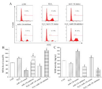
Endothelial progenitor cell (EPC) transplantation is a safe and effective method to treat acute myocardial infarction (AMI). However, oxidative stress leads to the death of a large number of EPCs in the early stage of transplantation, severely weakening the therapeutic effect. Previous studies demonstrated that microRNAs regulate the biological function of EPCs. The aim of the current study was to investigate the effect of microRNA on the biological function of EPCs under oxidative stress. Quantitative reverse transcription PCR was performed to detect the expression of miR-126, miR-508-5p, miR-150, and miR-16 in EPCs from rats, among which miR-126 showed a relatively higher expression. Treatment with H2O2 decreased miR-126 expression in EPCs in a dose-dependent manner. EPCs were further transfected with miR-126 mimics or inhibitors, followed by H2O2 treatment. Overexpression of miR-126 enhanced the proliferation, migration, and tube formation of H2O2-treated EPCs. MiR-126 overexpression also inhibited reactive oxygen species and malondialdehyde levels and enhanced superoxide dismutase levels, as well as increased angiopoietin (Ang)1 expression and decreased Ang2 expression in H2O2-treated EPCs. Moreover, miR-126 participated in the regulation of phosphatidylinositol 3-kinase (PI3K)/protein kinase B (Akt)/glycogen synthase kinase 3β (GSK3β) and extracellular signal-regulated kinase 1/2 (ERK1/2) signaling in EPCs, where both pathways were activated after miR-126 overexpression in H2O2-treated EPCs. Overall, we showed that miR-126 promoted the biological function of EPCs under H2O2-induced oxidative stress by activating the PI3K/Akt/GSK3β and ERK1/2 signaling pathway, which may serve as a new therapeutic approach to treat AMI.

Downloads

Download data is not yet available.
MicroRNA-126 enhances the biological function of endothelial progenitor cells under oxidative stress via PI3K/Akt/GSK-3β and ERK1/2 signaling pathways

Downloads

Additional Files

Published

2021-02-01

How to Cite

1.
Wu Q, Qi B, Duan X, Ming X, Yan F, He Y, Bu X, Sun S, Zhu H. MicroRNA-126 enhances the biological function of endothelial progenitor cells under oxidative stress via PI3K/Akt/GSK3β and ERK1/2 signaling pathways. Biomol Biomed [Internet]. 2021Feb.1 [cited 2023Mar.28];21(1):71-80. Available from: https://www.bjbms.org/ojs/index.php/bjbms/article/view/4493

Issue

Section

Translational and Clinical Research

INTRODUCTION

Acute myocardial infarction (AMI) is a common and critical acute cardiovascular disease. Great progress has been achieved in the treatment of AMI with drugs and surgical procedures, which partially restore blood supply to the infarcted area and prevent deterioration of cardiac function. However, the therapeutic effect is limited, and the mortality rate of AMI has not been satisfactorily controlled [1]. Recently, stem cell transplantation has increasingly gained attention in the treatment of myocardial infarction [2,3]. Among various types of stem cells, endothelial progenitor cells (EPCs) have consistently shown good efficacy in transplantation [4,5]. Many animal [6-8] and clinical [9] experiments have validated EPC transplantation as a safe and effective method to treat myocardial infarction. The procedure shows good application prospects by reducing the size of infarction, improving the wall motion of infarcted areas, increasing the left ventricular ejection fraction after infarction, and improving disease prognosis. Despite these advantages, post-AMI hypoxic-ischemic microenvironment and oxidative stress lead to the death of a large number of EPCs in the early transplantation stage, severely weakening the therapeutic effect [10,11]. We previously demonstrated that hydrogen peroxide (H2O2)-induced oxidative stress significantly affected the biological functions of EPCs such as secretion, proliferation, migration, and adhesion [12]. Similarly, augmented levels of post-AMI oxidative stress in the myocardium severely influence the biological function of transplanted EPCs [13]. Therefore, it is essential to improve the biological function of EPCs under oxidative stress to promote post-AMI myocardial repair.

MicroRNAs are small non-coding RNAs that act as post-transcriptional regulators of gene expression in animals and plants [14]. They actively participate in pathophysiological processes such as growth and development, hematopoietic processes, angiogenesis, cell proliferation, and apoptosis and are closely related to cardiovascular diseases [15,16]. Several microRNAs, such as miR-126 and miR-17, are specifically expressed in EPCs and regulate EPC proliferation, migration, angiogenesis, aging, and other EPC functions [17-19]. In addition, microRNAs enhanced the capacity of EPCs to repair heart injury [20]. However, the molecular mechanism of microRNAs underlying the regulation of EPCs is not clear.

Previous research has revealed that microRNAs influenced cellular functions by targeting phosphatidylinositol 3-kinase (PI3K)/protein kinase B (Akt) signaling [21-23]. This pathway is involved in regulating various biological processes and was shown to exert a protective effect on oxidative stress-induced EPC injury [24-26]. However, whether the microRNA-mediated regulation of EPC function is related to PI3K/Akt signaling remains unclear. In this study, based on the previous research and databases, the expression of miR-126, miR-508-5p, miR-150, and miR-16 was evaluated in EPCs [27-29]. Among them, miR-126 was detected with relatively high expression in EPCs. EPCs were stimulated with H2O2, which decreased the expression of miR-126 in a dose-dependent manner. To this end, miR-126 was overexpressed or inhibited to evaluate its regulatory effect on EPC function and PI3K/Akt signaling under oxidative stress.

MATERIALS AND METHODS

Cell extraction and culture

Four-week-old male Sprague Dawley rats were purchased from the Hubei Provincial Center for Disease Control and Prevention. Bone marrow-derived mononuclear cells were isolated from the rats through density gradient centrifugation using Ficoll-Hypaque (TBD, Tianjin, China). The harvested cells were seeded into 6-well plates pre-coated with fibronectin (5 μg/mL, Millipore, Boston, MA, USA) at 1 × 10⁶ cells/well with endothelial growth medium (EGM-2, LONZA, Basel, Switzerland). After 24 h of incubation at 37°C in an atmosphere with 5% CO2, the medium was changed and non-adherent cells were discarded. Passaging was performed when the confluence reached 70–80%, and the medium was changed every 2 days. Cell morphology was observed by fluorescence microscopy (Olympus, Japan) and spindle-shaped cells were identified as bone marrow-derived EPCs.

Characterization of EPCs

CD133 and CD34, which are expressed in bone marrow-derived EPCs at the early stage, are characteristic molecular markers of EPCs [30,31]. The percentage of harvested CD133+ and CD34+ cells was detected by flow cytometry (Beckman, Coulter, Brea, CA, USA). Cells (1 × 10⁶) were centrifuged at 1000 × g for 5 min and resuspended in 300 μL of phosphate-buffered saline [PBS] (Bioswamp, Myhalic Biotechnology Co., Ltd., Wuhan, China) containing 10% fetal bovine serum (FBS) in a 1.5 mL centrifuge tube. Then, 6 μL of CD133-allophycocyanin (Abcam, Cambridge, UK) and 6 μL of CD34-fluorescein isothiocyanate [FITC] (eBioscience, CA, USA) were added. Thereafter, the cells were incubated in the dark for 1 h at 4°C. After two washes with pre-cooled PBS and centrifugation at 300 × g for 5 min, the cells were resuspended in 300 μL of flow cytometry buffer (BD bioscience, USA). The cells were analyzed and data were acquired using a flow cytometer (Beckman Coulter, Brea, CA, USA). Furthermore, EPCs were identified by double staining of 1,1’-dioctadecyl-3,3,3’,3’-tetramethylindocarbocyanine-labeled acetylated low-density lipoprotein (Dil-AcLDL, Molecular Probes, Invitrogen, Carlsbad, CA, USA) and FITC-labeled lectin from Ulex europaeus agglutinin-1 (FITC-UEA-1, Sigma, Missouri, USA) [32]. Cells in the logarithmic phase (1 × 10⁶ cells/mL) were cultured for 3 days, and adherent cells were incubated with Dil-AcLDL (24 μg/mL) for 1 h. The cells were then fixed in 4% paraformaldehyde for 10 min and counterstained with FITC-UEA-1 (10 μg/mL) for 1 h. Fluorescent images were acquired under an inverted fluorescence microscope (Olympus, Tokyo, Japan). The expression of miR-126, miR-508-5p, miR-150, and miR-16 in EPCs was measured by quantitative reverse transcription polymerase chain reaction (qRT-PCR), with miR-126 showing relatively high expression. Therefore, the relationship between miR-126 and the biological function of EPCs was explored in the subsequent experiments.

Cell treatment

EPCs in the logarithmic phase were treated with H2O2 at different concentrations (200, 400, 600, 800, and 1000 μM). Cell viability and miR-126 expression were evaluated using cell counting kit-8 (CCK-8) assay and qRT-PCR, respectively. The optimal experimental concentration of H2O2 was selected to be 600 μM. EPCs were transfected with miR-126 mimics or inhibitors (Guangzhou RiboBio, Co., Ltd. Guangzhou, China), followed by H2O2 treatment for 6 h. The EPCs were divided into eight experimental groups based on treatment: control (CON, no treatment); H2O2 (treated with 600 μM H2O2); miR-126 mimic; miR-126 mimic negative control (mimic-NC); miR-126 inhibitor; miR-126 inhibitor negative control (inhibitor-NC); H2O2 + miR-126 mimic; and H2O2 + miR-126 inhibitor.

qRT-PCR

Total RNA was extracted using Trizol reagent (Ambion, TX, USA) and DNA in the extracted RNA was eliminated using DNase I (Fermentas, Thermo Fisher, Massachusetts, USA). cDNA was synthesized from total RNA (500 ng) using the M-MuLV kit (TAKARA, Dalian, China). qRT-PCR was carried out using the SYBR Green PCR kit according to the manufacturer’s instructions. The primer sequences are as follows: miR-126-F, 5’-GGGCATTATTACTTTT-3’, miR-126-R, 5’-AACTGGTGTCGTGGAGTCGGC-3’; miR-508-5p-F, 5’-GGGTACTCCAGAGGGC-3’, miR-508-5p-R, 5’-AACTGGTGTCGTGGAGTGGC-3’; miR-150-F, 5’-GGG TCTCCCAACCCTTG-3’, miR-150-R, 5’-AACTGGTGTCG TGGAGTCGGC-3’; miR-16-F, GGGTAGCAGCACGTA AA-3’, miR-16-R, 5’-AACTGGTGTCGTGGAGTCGGC-3’; U6-F, 5’-CTCGCTTCGGCAGCACATATACT-3’, U6-R, 5’-ACGCTTCACGAATTTGCGTGTC-3’. U6 served as an internal control. The 2−ΔΔCt method was utilized to calculate the relative expression level of miR-126 in EPCs treated with different concentrations of H2O2. The expression of miR-126, miR-508-5p, miR-150, and miR-16 in EPCs was calculated using the 2−ΔCt method [33]. All experiments were performed in triplicate.

CCK-8 assay

The CCK-8 (Bioswamp) assay was performed to evaluate cell viability according to the manufacturer’s instructions. Harvested cells were seeded into a 96-well plate at 3 × 10³ cells/well. The cells were treated with H2O2 at different concentrations and/or transfected with miR-126 mimics or inhibitors, and 10 μL of CCK-8 solution was added to each well. After 4 h of incubation, the absorbance of the wells was measured using a microplate reader (Thermo Scientific, USA) at 450 nm.

Flow cytometry

Intracellular reactive oxygen species (ROS) levels and apoptosis were assessed by flow cytometry. For the intracellular ROS assay, the harvested cells at a concentration of 1 × 10⁷ cells/mL were mixed with the diluted DCFH-DA (Bioswamp) fluoroprobes. The cells were incubated for 20 min at 37°C with gentle shaking every 4 min to ensure sufficient contact with the probes. After three washes with a serum-free culture medium, the cells were collected and measured by flow cytometry. For the apoptosis assay, the Annexin V-FITC/propidium iodide (PI) assay (Bioswamp) was performed according to the manufacturer’s protocol. The harvested cells at a concentration of 5 × 10⁵ cells were resuspended in 200 μL of binding buffer, 10 μL of Annexin V-FITC, and 10 μL of PI. The cells were incubated for 30 min in the dark and subjected to flow cytometry.

Transwell migration assay

Treated cells were cultured in serum-free EGM-2 for 24 h and digested with 0.25% trypsin. After washing with serum-free EGM-2, the cells were resuspended in EGM-2 supplemented with 1% FBS at 1 × 10⁵ cells/mL. Then, 0.5 mL of cells were added to the top Transwell chamber, while 0.75 mL of EGM-2 containing 10% FBS was added to the lower chamber. After 48 h of culture at 37°C, the cells were fixed with 4% formaldehyde for 10 min and stained with 0.5 % crystal violet for 30 min. Finally, the cells were observed under a microscope (Nikon, Japan).

Tube formation assay

After transfection and treatment with H2O2 for 6 h, the EPCs were resuspended in EGM-2 supplemented with 10% FBS and seeded in a Matrigel-coated 96-well plate (CORNING, USA) at 2 × 10⁵ cells/mL. After 4 h of incubation at 37°C, the formation of capillary-like structures was photographed under an inverted fluorescence microscope (Olympus, Tokyo, Japan).

Enzyme-linked immunosorbent assay (ELISA)

The activity of superoxidase dismutase (SOD) and level of malondialdehyde (MDA) in the supernatant of treated EPCs were determined using respective ELISA kits (Bioswamp) according to the manufacturer’s protocols.

Western blot

Total proteins were extracted from EPCs using radioimmunoprecipitation assay lysis buffer (Bioswamp) supplemented with protease and phosphatase inhibitors. The proteins were quantified using a bicinchoninic acid assay kit (Bioswamp). The obtained proteins (20 μL) were separated using sodium dodecyl sulfate-polyacrylamide gel electrophoresis and transferred to polyvinylidene fluoride membranes (Millipore). The membranes were blocked with 5% skim milk for 2 h at room temperature and incubated overnight at 4°C with the following primary antibodies: PI3K (Abcam, 1:1000), Akt (Bioswamp, 1:1000), p-Akt (Bioswamp, 1:1000), glycogen synthase kinase 3β (GSK3β, Abcam, 1:5000), p-GSK3β (Abcam, 1:1000), extracellular signal-regulated kinase 1/2 (ERK1/2, Abcam, 1:1000), p-ERK1/2 (Abcam, 1:1000); caspase 3 (Bioswamp, 1:1000), angiopoietin (Ang)1 (Abcam, 1:500), Ang 2 (Abcam, 1:5000), and glyceraldehyde 3-phosphate dehydrogenase [GAPDH] (CST, 1:1000). After washing, the membranes were incubated with a goat anti-rabbit IgG secondary antibody (Bioswamp, 1:20000) at room temperature for 1 h. Immunoreactivity was visualized by colorimetric reaction using enhanced chemiluminescence substrate buffer (Millipore) using an automatic chemiluminescence analyzer (Tanon-5200, Shanghai, China). The band gray values were measured by TANON GIS software.

Statistical analysis

Data are expressed as the mean ± standard deviation (SD). One-way analysis of variance followed by the least significant difference test was used to compare differences between groups using IBM SPSS Statistics for Windows, Version 19.0. (IBM Corp., Armonk, NY, USA). A value of p < 0.05 was considered statistically significant.

RESULTS

EPCs were successfully extracted and exhibited relatively high miR-126 expression

To verify that EPCs have been successfully extracted, we visualized the typical morphology of EPCs using microscopy (Figure 1A). Flow cytometry was carried out to measure the percentage of CD133+ and CD34+ populations in the isolated cells. These markers are expressed in the early stage of bone marrow-derived EPCs. The results showed that the percentage of CD133+ and CD34+ cells was as high as 79.99% (Figure 1B). Meanwhile, positive staining was demonstrated for both Dil-AcLDL and FITC-UEA-1 (Figure 1C), confirming that the isolated cells were differentiating EPCs [34]. Furthermore, the expression of miR-126, miR-508-5p, miR-150, and miR-16 in EPCs was measured by qRT-PCR. The threshold cycle of miR-126 was lower than that of the other microRNAs (Figure 1D), demonstrating relatively high miR-126 expression in EPCs. Thus, the relationship between miR-126 and the biological function of EPCs was explored in subsequent experiments.

FIGURE 1: Characterization of EPCs. (A) Morphology of EPCs under a bright-field microscope. (B) Identification of the percentage of CD133+ and CD34+ EPCs. (C) Dil-AcLDL and FITC-UEA-1 double staining of EPCs. (D) Expression of miR-126, miR-508-5p, miR-150, and miR-16 in EPCs. Data are expressed as the mean ± standard deviation [SD] (n = 3). EPCs: Endothelial progenitor cells; Dil-AcLDL: 1,1’-dioctadecyl-3,3,3’,3’-tetramethylindocarbocyanine-labeled acetylated low-density lipoprotein; FITC: Fluorescein isothiocyanate; UEA-1: Ulex europaeus agglutinin 1.

Selection of H2O2 treatment concentration

The isolated EPCs were treated with H2O2 at 200, 400, 600, 800, and 1000 μM to select the optimal treatment concentration. CCK-8 and qRT-PCR were performed to evaluate cell viability and miR-126 expression, respectively. Figure 2 shows that the cell viability was decreased in a H2O2 dose-dependent manner, with significant differences compared with CON group (p < 0.05). At a concentration of 600 μM, H2O2 significantly downregulated miR-126 expression compared to that in control EPCs (p < 0.05). Thus, 600 μM H2O2 was chosen for the subsequent experiments.

FIGURE 2: Selection of optimal hydrogen peroxide (H2O2) concentration. (A) Viability and (B) miR-126 expression of endothelial progenitor cells after treatment with H2O2 at different concentrations. Data are expressed as the mean ± standard deviation [SD] (n = 3), *p < 0.05 vs. control (CON).

miR-126 expression

After transfection and/or H2O2 treatment, the expression of miR-126 was measured by qRT-PCR (Figure 3). Compared to CON group, cells transfected with miR-126 mimics or inhibitors showed significantly higher or lower expression of miR-126 (p < 0.05), respectively. The expression of miR-126 among mimic-NC, inhibitor-NC, and CON groups showed no difference. Compared to H2O2 group, the expression of miR-126 in H2O2+miR-126 mimic group was upregulated (p < 0.05), while that in H2O2+miR-126 inhibitor group was downregulated (p < 0.05).

FIGURE 3: Expression of miR-126 in endothelial progenitor cells subjected to transfection and/or hydrogen peroxide (H2O2) treatment. Data are expressed as the mean ± standard deviation [SD] (n = 3), *,#,&, and @ represent p < 0.05 vs. control (CON), mimic-negative control (NC), inhibitor-NC, and H2O2 , respectively.

Effect of miR-126 on the biological function of EPCs

Evaluation of EPC function showed that H2O2 and miR-126 inhibitors significantly reduced EPC viability (p < 0.05). Compared to H2O2 group, cell viability was increased in H2O2+miR-126 mimic group (p < 0.05) but decreased in H2O2+miR-126 inhibitor group (p < 0.05; Figure 4A). As anticipated, the percentage of apoptosis showed the opposite trend as that of cell viability (Figure 4B). Moreover, the expression of the pro-apoptotic protein caspase 3 in H2O2 and miR-126 inhibitor groups was enhanced compared to that of control EPCs (p < 0.05; Figure 4C). Compared to H2O2 group, the expression of caspase 3 was reduced in H2O2+miR-126 mimic group (p < 0.05) but elevated in H2O2+miR-126 inhibitor group (p < 0.05). These results are consistent with those of apoptosis. Transwell migration and tube formation assays (Figure 4D and E) showed that H2O2 inhibited EPC migration and tube formation. MiR-126 inhibitors further contributed to this inhibition, while miR-126 mimics remarkably improved EPC migration and tube formation.

FIGURE 4: Evaluation of the biological function of EPCs. (A) EPC viability. (B) Percentage of EPC apoptosis. (C) Protein expression of caspase 3. (D) EPC migration. (E) Tube formation of EPCs. Data are expressed as the mean ± standard deviation [SD] (n = 3), * and # represent p < 0.05 vs. CON and H2O2, respectively. EPCs: Endothelial progenitor cells; GAPDH: Glyceraldehyde 3-phosphate dehydrogenase; CON: Control.

Effect of miR-126 on ROS production, MDA level, and SOD activity

As shown in Figure 5, compared with CON group, H2O2 and miR-126 inhibitor notably elevated the levels of ROS and MDA (p < 0.05), while significantly decreasing SOD activity (p < 0.05). In addition, compared to H2O2 group, ROS production and MDA activity were reduced in H2O2+miR-126 mimic group (p < 0.05), whereas SOD activity was significantly increased (p < 0.05). The H2O2+miR-126 inhibitor group showed the opposite trend (p < 0.05).

FIGURE 5: Assessment of oxidative stress-related indicators. (A) ROS production; (B) MDA level; and (C) SOD activity in EPCs. Data are expressed as the mean ± [SD] (n = 3), * and # represent p < 0.05 vs. CON and H2O2, respectively. ROS: Reactive oxygen species; MDA: Malondialdehyde; SOD: Super oxidase dismutase; EPCs: Endothelial progenitor cells; CON: Control.

Effect of miR-126 on the protein expression of Ang1 and Ang2

The protein expression of Ang1 and Ang2 was evaluated by western blot (Figure 6). Compared with CON group, H2O2 and miR-126 inhibitors decreased the expression of Ang1 (p < 0.05) and increased the expression of Ang2 (p < 0.05). Compared to H2O2 group, Ang1 was upregulated in H2O2+miR-126 mimic group (p < 0.05), whereas Ang2 was downregulated (p < 0.05). Furthermore, the expression of Ang1 was decreased in H2O2+miR-126 inhibitor group (p < 0.05) but the expression of Ang2 was increased (p < 0.05).

FIGURE 6: Expression of Ang1 and Ang2 in EPCs. Data are expressed as the mean ± standard deviation [SD] (n = 3), * and # represent p < 0.05 vs. CON and H2O2, respectively. Ang: Angiopoietin; EPCs: Endothelial progenitor cells; CON: Control; GAPDH: Glyceraldehyde 3-phosphate dehydrogenase.

Effect of miR-126 on the expression of proteins associated with PI3K/Akt/GSK3β and ERK1/2 signaling

Compared to CON group, the expression of p-Akt, p-GSK3β, and p-ERK1/2 was notably decreased by H2O2 and miR-126 inhibitors (p < 0.05; Figure 7). Compared to H2O2 group, the expression of PI3K, p-Akt, p-GSK3β, and p-ERK1/2 was increased in H2O2+miR-126 mimic group (p < 0.05) but decreased in H2O2+miR-126 inhibitor group (p < 0.05).

FIGURE 7: Investigation of signaling pathways involved in miR-126-mediated effects. Expression of proteins associated with (A) PI3K/Akt/GSK3β and (B) ERK1/2 signaling pathway. Data are expressed as the mean ± [SD] (n = 3), *and #represent p < 0.05 vs. CON and H2O2, respectively. PI3K: Phosphatidylinositol 3-kinase; Akt: Protein kinase B; GSK3β: Glycogen synthase kinase 3β; ERK1/2: Extracellular signal-regulated kinase 1/2; CON: Control; GAPDH: Glyceraldehyde 3-phosphate dehydrogenase.

DISCUSSION

The present study demonstrated that H2O2 downregulated miR-126 in EPCs and suppressed EPC viability, migration, and tube formation. However, miR-126 overexpression notably improved the biological function of H2O2-damaged EPCs. Our findings are consistent with the previous reports showing that miR-126 relieved myocardial damage after AMI [35], protected cells from apoptosis, and elevated angiogenesis to prevent myocardial injury [36]. In addition, targeted deletion of miR-126 led to partial embryonic lethality, leaky vessels, and hemorrhaging in mice. This was due to the loss of vascular integrity and impaired endothelial cell migration, proliferation, and angiogenesis [37].

MiR-126 regulates multiple genes and signaling pathways, and its overexpression attenuated vascular endothelial cell apoptosis by activating PI3K/Akt signaling [21]. Upregulation of miR-126 resulted in improved ischemic angiogenesis in mesenchymal stem cells by stimulating Akt/ERK-related signaling [38]. Our study demonstrated that overexpression of miR-126 increased the protein expression of PI3K, Akt, GSK3β, and ERK1/2. The biological function of H2O2-damaged EPCs, such as cell proliferation, migration, and angiogenesis, was notably improved. In addition, ROS and MDA levels were attenuated, whereas SOD activity was enhanced. It is widely accepted that MDA is involved in the occurrence of oxidative stress [39], while SOD alleviates it [40]. Oxidative stress severely impedes the therapeutic effect of EPC transplantation in myocardial infarction treatment, and miR-126 overexpression inhibits H2O2-induced oxidative stress in EPCs [10,11]. The PI3K/Akt/GSK3β and ERK1/2 signaling pathways are involved in regulating a variety of cellular processes, such as cell proliferation, apoptosis, and oxidative stress. GSK3β is a downstream signaling molecule of Akt [41], and activation of Akt/GSK3β signaling inhibited oxidative stress and apoptosis in rats with cerebral hypoxic-ischemic injury [42]. Inhibition of MAPK/ERK and PI3K/Akt signaling was previously suggested to suppress angiogenesis in endometrial carcinoma [43]. Additionally, the Akt/GSK3β pathway is involved in the protection against AMI, and its activation stimulated tube formation and accelerated human aortic endothelial cell migration [44]. Consistent with the previous studies, our study showed that miR-126 overexpression promoted EPC proliferation, migration, and tube formation and inhibited H2O2-stimulated oxidative stress. We hypothesize that the underlying mechanism is associated with the regulation of PI3K/Akt/GSK3β and ERK1/2 signaling.

Ang1 and Ang2 are angiopoietin subtypes that are involved in angiogenesis and vessel maturation. They mainly bind to the endothelial receptor tyrosine kinase Tie-2, which is expressed in endothelial cells and is related to microvascular sprouting and stabilization [45]. Ang1 reportedly played an important role in promoting endothelial cell migration and vessel maturation, while Ang2 is involved in accelerating vascular destabilization and regression [46]. In addition, Ang2 served as an antagonist that inhibited Ang1-induced Tie-2 phosphorylation, thus disrupting angiogenesis [47,48]. According to the previous studies, the ERK1/2 pathway is involved in angiogenesis regulation. Inhibition of ERK1/2 expression reduced angiogenesis in the synovial membrane, which was associated with a decrease in Ang1 and vascular endothelial growth factor expression [49]. Our study suggested that miR-126 overexpression led to the upregulation of Ang1 and downregulation of Ang2, in turn, promoting EPC angiogenesis. These findings are consistent with the results of biological function assays.

CONCLUSION

Overall, we demonstrated that miR-126 overexpression promoted H2O2-induced EPC proliferation, migration, and tube formation by regulating PI3K/Akt/GSK3β and ERK1/2 signaling. Our results may contribute to the development of potential therapeutic strategies against AMI. However, the study was designed and conducted only at the cellular level. Further in-depth experiments will be performed in animals to verify our conclusions. In addition, specific target genes of miR-126 will be investigated in prospective studies.

Acknowledgements

ACKNOWLEDGMENTS

This study is supported by the Wuhan Municipal Health Commission (WX17Q03).

REFERENCES

  1. , , (). Moving beyond SHOCK: New paradigms in the management of acute myocardial infarction complicated by cardiogenic shock. Can J Cardiol. https://doi.org/10.1016/j.cjca.2016.10.018
  2. , , , , , (). Stem cell therapies for myocardial infarction in clinical trials:Bioengineering and biomaterial aspects. Lab Invest. https://doi.org/10.1038/labinvest.2017.100
  3. , , (). Progress of stem cell transplantation for treating myocardial infarction. Curr Stem Cell Res Ther. https://doi.org/10.2174/1574888X12666170927114854
  4. , , , (). Endothelial progenitor cells and cardiovascular cell-based therapies. Cytotherapy. https://doi.org/10.1080/14653240802714827
  5. , (). Endothelial cells facilitate cell-based cardiac repair:Progress and challenge. Curr Stem Cell Res Ther. https://doi.org/10.2174/1574888x09666140710100143
  6. , , , (). Potential mechanism for endothelial progenitor cell therapy in acute myocardial infarction:Activation of VEGF-PI3K/Akte-NOS pathway. Ann Clin Lab Sci.
  7. , , , , , (). Transplantation of endothelial progenitor cells improves neovascularization and left ventricular function after myocardial infarction in a rat model. Basic Res Cardiol. https://doi.org/10.1007/s00395-007-0685-9
  8. , , , , , (). Stromal cell-derived factor-1alpha activation of tissue-engineered endothelial progenitor cell matrix enhances ventricular function after myocardial infarction by inducing neovasculogenesis. Circulation. https://doi.org/10.1161/CIRCULATIONAHA.109.930404
  9. , , , , (). Does mobilisation of CD34+stem cells along with VEGF, angiogenin, IL-6, IL-8, and hsCRP levels allow predicting the direction of left ventricular ejection fraction and wall motion score index changes in patients with myocardial infarction?. Kardiol Pol. https://doi.org/10.5603/KP.2013.0092
  10. , , , , , (). Safety and efficacy of autologous thymosin β4 pre-treated endothelial progenitor cell transplantation in patients with acute ST segment elevation myocardial infarction:A pilot study. Cytotherapy. https://doi.org/10.1016/j.jcyt.2016.05.006
  11. , , , , (). Suppression of oxidative stress in endothelial progenitor cells promotes angiogenesis and improves cardiac function following myocardial infarction in diabetic mice. Exp Ther Med. https://doi.org/10.3892/etm.2016.3236
  12. , , , , , (). Mechanisms underlying protective effects of trimetazidine on endothelial progenitor cells biological functions against H2O2-induced injury:Involvement of antioxidation and Akt/eNOS signaling pathways. Eur J Pharmacol. https://doi.org/10.1016/j.ejphar.2013.03.027
  13. , , , , , (). Tissue kallikrein-modified human endothelial progenitor cell implantation improves cardiac function via enhanced activation of Akt and increased angiogenesis. Lab Invest. https://doi.org/10.1038/labinvest.2013.48
  14. (). MicroRNAs:Target recognition and regulatory functions. Cell. https://doi.org/10.1016/j.cell.2009.01.002
  15. , , , (). MicroRNAs in cardiovascular disease:An introduction for clinicians. Heart. https://doi.org/10.1136/heartjnl-2013-305402
  16. , , , (). MicroRNAs in cardiovascular disease:Perspectives and reality. Cardiol Rev. https://doi.org/10.1097/CRD.0000000000000078
  17. , , , , , (). Synthetic microparticles conjugated with VEGF165 improve the survival of endothelial progenitor cells via microRNA-17 inhibition. Nat Commun. https://doi.org/10.1038/s41467-017-00746-7
  18. , , , , , (). MicroRNA 126 inhibits the transition of endothelial progenitor cells to mesenchymal cells via the PIK3R2-PI3K/Akt signalling pathway. PLoS One. https://doi.org/10.1371/journal.pone.0083294
  19. , , , , (). MiR-126 modulates angiogenic growth parameters of peripheral blood endothelial progenitor cells. Biol Chem. https://doi.org/10.1515/hsz-2014-0259
  20. , , (). Regulation of endothelial progenitor cell function by micrornas. Minerva Cardioangiol.
  21. , , , , , (). MiR-126 inhibits vascular endothelial cell apoptosis through targeting PI3K/Akt signaling. Ann Hematol. https://doi.org/10.1007/s00277-015-2567-9
  22. , , , (). MicroRNA-520a-3p inhibits cell growth and metastasis of non-small cell lung cancer through PI3K/AKT/mTOR signaling pathway. Eur Rev Med Pharmacol Sci. https://doi.org/10.26355/eurrev_201804_14822
  23. , , , , (). MiR-126 regulates proliferation and invasion in the bladder cancer BLS cell line by targeting the PIK3R2-mediated PI3K/Akt signaling pathway. Onco Targets Ther. https://doi.org/10.2147/OTT.S105198
  24. , , , , , (). Research progress on signaling pathway-associated oxidative stress in endothelial cells. Oxid Med Cell Longev. https://doi.org/10.1155/2017/7156941
  25. , , , , , (). FOXO-dependent expression of the proapoptotic protein Bim:Pivotal role for apoptosis signaling in endothelial progenitor cells. FASEB J. https://doi.org/10.1096/fj.04-2727fje
  26. , , , , , (). HMG-CoA reductase inhibitors (statins) increase endothelial progenitor cells via the PI 3-kinase/Akt pathway. J Clin Invest. https://doi.org/10.1172/JCI13152
  27. , , , , (). Expression of miR-126 and miR-508-5p in endothelial progenitor cells is associated with the prognosis of chronic heart failure patients. Int J Cardiol. https://doi.org/10.1016/j.ijcard.2013.01.160
  28. , , , , , (). The effects of microvesicles on endothelial progenitor cells are compromised in type 2 diabetic patients via downregulation of the miR-126/VEGFR2 pathway. Am J Physiol Endocrinol Metab. https://doi.org/10.1152/ajpendo.00056.2016
  29. , , , , , (). MicroRNA-16 affects key functions of human endothelial progenitor cells. J Leukoc Biol. https://doi.org/10.1189/jlb.1012511
  30. , , (). Concise review:Circulating endothelial progenitor cells for vascular medicine. Stem Cells. https://doi.org/10.1002/stem.745
  31. , (). Endothelial progenitor cells:Characterization, pathophysiology, and possible clinical relevance. J Cell Mol Med. https://doi.org/10.1111/j.1582-4934.2004.tb00474
  32. , , , , , (). High glucose impairs early and late endothelial progenitor cells by modifying nitric oxide-related but not oxidative stress-mediated mechanisms. Diabetes. https://doi.org/10.2337/db06-1103
  33. , , , , , (). Targeting tumor vascular CD99 inhibits tumor growth. Front Immunol. https://doi.org/10.3389/fimmu.2019.00651
  34. , , , , , (). Allogenic endothelial progenitor cell transplantation increases flap survival through an upregulation of eNOs and VEGF on venous flap survival in rabbits. J Plast Reconstr Aesthet Surg. https://doi.org/10.1016/j.bjps.2018.12.042
  35. , , , , (). Effects of rosuvastatin and MiR-126 on myocardial injury induced by acute myocardial infarction in rats:Role of vascular endothelial growth factor A (VEGF-A). Med Sci Monit. https://doi.org/10.12659/msm.896983
  36. , , , , , (). Exosomes from MiR-126-overexpressing adscs are therapeutic in relieving acute myocardial ischaemic injury. Cell Physiol Biochem. https://doi.org/10.1159/0004→9
  37. , , , , , (). The endothelial-specific microRNA miR-126 governs vascular integrity and angiogenesis. Dev Cell. https://doi.org/10.1016/j.devcel.2008.07.002
  38. , (). Mesenchymal stem cells overexpressing MiR-126 enhance ischemic angiogenesis via the AKT/ERK-related pathway. Cardiol J. https://doi.org/10.5603/cj.2011.0032
  39. , , , , (). Today's oxidative stress markers. Med Pr. https://doi.org/10.13075/mp.5893.00137
  40. , , , , , (). Targeting free radicals in oxidative stress-related human diseases. https://doi.org/10.1016/j.tips.2017.04.005
  41. , , , , (). Sulfuretin attenuates MPP+-induced neurotoxicity through Akt/GSK3βand ERK signaling pathways. Int J Mol Sci. https://doi.org/10.3390/ijms18122753
  42. , , (). Oxymatrine attenuates brain hypoxic-ischemic injury from apoptosis and oxidative stress:Role of p-Akt/GSK3β/HO-1/Nrf-2 signaling pathway. Metab Brain Dis. https://doi.org/10.1007/s11011-018-0293-4
  43. , , , , (). MicroRNA-29b inhibits angiogenesis by targeting VEGFA through the MAPK/ERK and PI3K/Akt signaling pathways in endometrial carcinoma. Cell Physiol Biochem. https://doi.org/10.1159/000460510
  44. , (). Electron transfer in proteins:In search of preferential pathways. FASEB J. https://doi.org/10.1096/fasebj.5.11.1868979
  45. , , , , , (). Early vessel destabilization mediated by angiopoietin-2 and subsequent vessel maturation via angiopoietin-1 induce functional neovasculature after ischemia. PLoS One. https://doi.org/10.1371/journal.pone.0061831
  46. , (). Angiopoietins in angiogenesis. Cancer Lett. https://doi.org/10.1016/j.canlet.2012.08.018
  47. , , (). Pericytes:Developmental, physiological, and pathological perspectives, problems, and promises. Dev Cell. https://doi.org/10.1016/j.devcel.2011.07.001
  48. , , , , , (). Angiopoietin-2, a natural antagonist for Tie2 that disrupts in vivo angiogenesis. Science. https://doi.org/10.1126/science.277.5322.55
  49. , , , (). Inhibition of ERK pathway decreases the synovial hyperplasia and angiogenesis of rheumatoid arthritis rats. Eur J Inflamm. https://doi.org/10.1177/205∣218794531

Conflict of interest statement: The authors declare no conflict of interests