Immunohistochemical study of dental pulp cells with 3D collagen type I gel in demineralized dentin tubules in vivo

Authors

  • Sultan Zeb Khan Department of Clinical Pathophysiology, Graduate School of Tokyo Dental College, Tokyo, Japan
  • Sana Mirza Department of Oral Pathology, Faculty of Dentistry, Ziauddin University, Karachi, Pakistan
  • Samina Karim Department of Ophthalmology, Hayatabad Medical Complex, Khyber Girls Medical College, Peshawar, Pakistan
  • Takashi Inoue Department of Clinical Pathophysiology, Graduate School of Tokyo Dental College, Tokyo, Japan
  • Mohammed S. Bin-Shuwaish Department of Restorative Dentistry, College of Dentistry, King Saud University, Riyadh, Saudi Arabia https://orcid.org/0000-0001-8127-0165
  • Laila Al Deeb Department of Restorative Dentistry, College of Dentistry, King Saud University, Riyadh, Saudi Arabia
  • Khold Al Ahdal Department of Restorative Dentistry, College of Dentistry, King Saud University, Riyadh, Saudi Arabia
  • Rana S. Al-Hamdan Department of Restorative Dentistry, College of Dentistry, King Saud University, Riyadh, Saudi Arabia
  • Ahmed M. Maawadh Department of Restorative Dentistry, College of Dentistry, King Saud University, Riyadh, Saudi Arabia
  • Fahim Vohra Department of Prosthetic Dental Sciences, College of Dentistry, King Saud University, Riyadh, Saudi Arabia https://orcid.org/0000-0002-6204-7985
  • Tariq Abduljabbar Department of Prosthetic Dental Sciences, College of Dentistry, King Saud University, Riyadh, Saudi Arabia

DOI:

https://doi.org/10.17305/bjbms.2020.4614

Keywords:

Collagen type I gel, demineralized dentin tubules, dental pulp cells, osteogenesis

Abstract

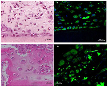
Dental pulp cells (DPCs) represent good candidates for the regeneration of dental tissue. This study aimed to evaluate the growth and differentiation potential of DPCs cultured inside demineralized dentin tubules in vivo. Six green fluorescent protein-transgenic rats (body weight 100 g each) and thirty-two Sprague-Dawley (SD) male rats (body weight 250 g each) were used for DPC collection and dentin tubules preparation and transplantation, respectively. Third-passage DPCs with or without collagen gels were loaded into demineralized dentin tubules. Both types of grafts were transplanted into the rectus abdominis muscles of SD rats and were harvested after 21 days. The expression of alkaline phosphatase (ALP), bone sialoprotein (BSP), osteopontin (OPN), nestin, and dentin sialoprotein (DSP) was analyzed by immunohistochemistry. Histological analysis showed that DPCs in the collagen gel formed an osteodentin-like hard tissue matrix after 21 days. Increased positive immunoreactivity for ALP, BSP, OPN, nestin, and DSP was observed in experimental groups compared with control. Our results demonstrate that DPCs in collagen gel inside demineralized dentin tubules show increased growth and differentiation.

Immunohistochemical study of dental pulp cells with 3D collagen type I gel in demineralized dentin tubules in vivo

Downloads

Additional Files

Published

02-11-2020

How to Cite

1.
Immunohistochemical study of dental pulp cells with 3D collagen type I gel in demineralized dentin tubules in vivo. Biomol Biomed [Internet]. 2020 Nov. 2 [cited 2024 Mar. 19];20(4):438-44. Available from: https://www.bjbms.org/ojs/index.php/bjbms/article/view/4614

Funding data