Prevalence and genotype distribution of rotaviruses in children with gastroenteritis in Rize province

Authors

  • Selim Dereci Department of Pediatrics, School of Medicine Recep Tayyip Erdogan University Rize
  • Ayşegül Çopur Çiçek Department of Medical Microbiology, School of Medicine, Recep Tayyip Erdogan University Department of Pediatrics, Rize
  • Sümeyra Savaş Acar Turkey Public Health Agency, Research and Practice Laboratory of Molecular Microbiology, Ankara
  • Zekiye Bakkaloğlu Turkey Public Health Agency, Research and Practice Laboratory of Molecular Microbiology, Ankara
  • Serdar Özkasap Department of Pediatrics, School of Medicine, Recep Tayyip Erdogan University Department of Pediatrics, Rize
  • Kadri Kanber Department of Pediatrics, School of Medicine, Recep Tayyip Erdogan University Department of Pediatrics, Rize
  • Şadan Hacisalihoğlu Department of Pediatrics, School of Medicine, Recep Tayyip Erdogan University Department of Pediatrics, Rize
  • Yücehan Albayrak Department of Pediatrics, School of Medicine, Recep Tayyip Erdogan University Department of Pediatrics, Rize
  • Rıza Durmaz Turkey Public Health Agency, Research and Practice Laboratory of Molecular Microbiology, Ankara Yildirim Beyazıt University, Medicine Faculty, Department of Medical Microbiology, Ankara

DOI:

https://doi.org/10.17305/bjbms.2015.469

Keywords:

Rotaviruses, children, genotyping

Abstract

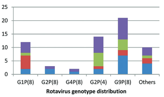
Determination of the distribution of rotavirus genotypes is essential for understanding the epidemiology of this virus responsible for nearly half a million of deaths in patients with gastroenteritis worldwide. In the present study, we aimed to genotype the rotavirus strains isolated from diarrheal stool samples in children under 5 years old. A total of 1297 fecal samples were collected, and rotavirus antigen was detected in 73 of these samples. Antigen-positive samples were transferred to the Public Health Agency of Turkey, Molecular Microbiology Research Laboratory, and were tested for determination of genotypes G and P using semi-nested multiplex polymerase chain reaction method performed with consensus- and genotype-specific primers. Twelve specimens were found to be negative for rotavirus in genotyping method. All the positive-strains were in G1-4, G8-9, P(4), P(8), and P(9) genotypes. The most frequent GP genotype combinations were found to be G9P(8) in 21 strains (34.4%), G2P(4) in 14 strains (23.0%), and G1P(8) in 12 strains (19.7%). We found 10 distinct genotypes amongst a total of 61 strains. Among the strains isolated and genotyped in our study, 90.2% (55/61) and 67.2% (41/61) have already been included in the two existing commercial vaccines. In conclusion, these findings implicate the necessity of development of region-specific vaccines after evaluation of the local genotype distribution. Further studies on the large number of rotavirus strains would contribute to this process. 

Downloads

Download data is not yet available.

References

Kim MJ, Jeong HS, Kim SG, Lee SM, Kim SH, Kee HY, et al. Diversity of rotavirus strain circulated in gwangju, republic of Korea. Osong Public Health Res Perspect 2014;5(6):364–369. doi:10.1016/j.phrp.2014.10.004.

Matthijnssens J, Otto PH, Ciarlet M. VP6-sequence-based cutoff values as a criterion for rotavirus species demarcation. Arch Virol 2012;157(6):1177–1182. doi:10.1007/s00705-012-1273-3.

CDC. Prevention of rotavirus gastroenteritis among infants and children: recommendation of the Advisory Committee on Immunization Practices (ACIP). MMWR Recomm Rep 2009; 58(2):1–25.

Durmaz R, Kalaycioglu AT, Acar S, Bakkaloglu Z, Karagoz A, Korukluoglu G, et al. Turkish Rotavirus Surveillance Network. Prevalence of Rotavirus Genotypes in Children Younger than 5 Years of Age before the Introduction of a Universal Rotavirus Vaccination Program: Report of Rotavirus Surveillance in Turkey. PLoS One 2014;9(12):e113674. doi:10.1371/journal.pone.0113674.

Iturriza-Gómara M, Dallman T, Bányai K, Böttiger B, Buesa J, Diedrich S, et al. Rotavirus genotypes co-circulating in Europe between 2006 and 2009 as determined by EuroRotaNet, a pan-European collaborative strain surveillance network. Epidemiol Infect 2011;139(6):895-909. doi:10.1017/S0950268810001810.

Simmonds MK, Armah G, Asmah R, Banerjee I, Damanka S, Esona M, et al. New oligonucleotide primers for P-typing of rotavirus strains: Strategies for typing previously untypeable strains. J Clin Virol 2008;42(4):368–373.doi :10.1016/j.jcv.2008.02.011.

Gentsch JR, Laird AR, Bielfelt B, Griffin DD, Banyai K, Ramachandran M, et al. Serotype diversity and reassortment between human and animal rotavirus strains: implications for rotavirus vaccine programs. J Infect Dis 2005;192(1):146–159.doi :10.1086/431499.

Iturriza-Gómara M., Cubitt D., Desselberger U. Amino acid substitution within the VP7 protein of G2 rotavirus strains associated with failure to serotype. J Clin Microbiol 2001;39(10):3796–3798.doi :10.1128/JCM.39.10.3796-3798.2001.

Tapisiz A, Karahan ZC, Ciftci E, Ince E, Dogru U. Changing patterns of rotavirus genotypes in Turkey. Curr Microbiol 2011;63(6):517–522.doi:10.1007/s00284-011-0014-2.

Bozdayi G, Dogan B, Dalgic B, Bostanci I, Sari S, Battaloglu NO, et al. Diversity of human rotavirus G9 among children in Turkey. J Med Virol 2008;80(4):733–740.doi:10.1002/jmv.21120.

Midgley S, Böttiger B, Jensen TG, Friis-Møller A, Person LK, Nielsen L, et al. Human group A rotavirus infections in children in Denmark; detection of reassortant G9 strains and zoonotic P(14) strains. Infect Genet Evol 2014;27:114–120.doi:10.1016/j.meegid.2014.07.008.

Anca IA, Furtunescu FL, Pleţca D, Streinu-Cercel A, Rugină S, Holl K. Hospital-based surveillance to estimate the burden of rotavirus gastroenteritis in children below five years of age in Romania. Germs 2014;4(2):30–40.doi:10.11599/germs.2014.1053.

Mullick S, Mukherjee A, Ghosh S, Pazhani GP, Sur D, Manna B, et al. Community based case-control study of rotavirus gastroenteritis among young children during 2008-2010 reveals vast genetic diversity and increased prevalence of G9 strains in Kolkata. PLoS One 2014;17;9(11):e112970. doi:10.1371/journal.pone.0112970.

Numazaki K, Ichikawa M. Clinical and virological characteristics of rotavirus gastroenteritis and prevalence of strains in Tochigi, Japan. In Vivo 2014;28(6):1141–1147.

Boula A, Waku-Kouomou D, Njiki Kinkela M, Esona MD, Kemajou G, Mekontso D, et al. Molecular surveillance of rotavirus strains circulating in Yaoundé, Cameroon, September 2007-December 2012. Infect Genet Evol 2014;28:470–475.doi:10.1016/j.meegid.2014.08.019.

Semeiko GV, Yermalovich MA, Poliakova N, Mijatovic-Rustempasic S, Kerin TK, Wasley A, et al. Rotavirus genotypes in Belarus, 2008-2012. Infect Genet Evol 2014;28:480–485.doi:10.1016/j.meegid.2014.09.007.

Cataloluk O, Iturriza M, Gray J. Molecular characterization of rotaviruses circulating in the population of Turkey. Epidemiol Infect 2005;133(4):673–678.doi:10.1017/S0950268805003882.

Altindis M, Bányai K, Kalayci R, Gulamber C, Koken R, Apan T, et al. Molecular characterization of rotaviruses in mid-western Turkey, 2006–2007. Cent Eur J Med 2010;5(5):640–645.doi :10.2478/s11536-009-0130-6.

Ruiz-Palacios GM, Pérez-Schael I, Velázquez FR, Abate H, Breuer T, Clemens SC et al. Safety and efficacy of an attenuated vaccine against severe rotavirus gastroenteritis. N Engl J Med 2006;354:11–22.doi:10.1056/NEJMoa052434.

Dulgheroff AC, Figueiredo EF, Gouvêa VS, Domingues AL. Changes in epidemiology of rotavirus in the Triângulo Mineiro region of Brazil: lack of two consecutive rotavirus seasons. Braz J Med Biol Res 2014;47(12):1091–1095.doi:10.1590/1414-431X20144156.

Prevalence and genotype distribution of rotaviruses in children with gastroenteritis in Rize province

Downloads

Additional Files

Published

2015-07-09

How to Cite

1.
Dereci S, Çopur Çiçek A, Savaş Acar S, Bakkaloğlu Z, Özkasap S, Kanber K, Hacisalihoğlu Şadan, Albayrak Y, Durmaz R. Prevalence and genotype distribution of rotaviruses in children with gastroenteritis in Rize province. Biomol Biomed [Internet]. 2015Jul.9 [cited 2023Mar.29];15(3):35-9. Available from: https://www.bjbms.org/ojs/index.php/bjbms/article/view/469

Issue

Section

Microbiology

INTRODUCTION

Rotaviruses (RVs) are the major causative agents of acute and severe diarrhea and gastroenteritis in children, particularly in those younger than 5 years. These microorganisms cause about half a million deaths per year worldwide [1].

Rotaviruses are non-enveloped RNA viruses belonging to family Reoviridae with a genome consisting of 11 segments of double-stranded (ds) RNA surrounded by a triple-layer nucleocapsid. The outer layer includes two structural viral proteins (VPs), denoted as VP4 and VP7. VP4 is a protease-cleaved protein (protein P), and VP7 is a glycoprotein (Protein G). Proteins P and G determine the serotype of the virus. Since they are targets for antibodies, these proteins are important for development of the vaccine. Rotaviruses are further subclassified into genotype G and genotype P, depending on whether their genome contains VP7 or VP4 gene, respectively [1-3].

Mapping the genotype distribution of rotavirus strains is essential for determination of the epidemiology of the virus both locally and globally, and is thus also critical to understand whether the current vaccine covers the most prevalent genotypes of a certain location.

In the present study, we aimed to determine the prevalence and the genotype distribution of rotaviruses in children admitted to Rize Training and Research Hospital in Eastern Black Sea region of Turkey. Moreover, we wanted to make a contribution to a database of Turkey Rotavirus Surveillance Network (TÜROSA) employed in process of vaccine development.

MATERIALS AND METHODS

Sample collection

Fecal samples of a total of 1297 children admitted to the outpatient clinics of pediatrics or the emergency room of Rize Training and Research Hospital in 2013 with a diagnosis of gastroenteritis were collected using a disposable container. The samples were collected within the first 48 hours of hospitalization. Patients’ demographical data as well as the clinical symptoms and findings were obtained retrospectively from the hospital records.

Antigen detection

Fecal samples were tested for the presence of rotavirus antigen using commercial immunochromatographic test (ABON Biopharm, London, U. K.) in concordance to manufacturer’s protocol. Rotavirus antigen-positive fecal samples were stored at –20 °C until the moment of genotyping testing.

Genotyping of the strains

All the antigen-positive samples (a total of 73) were transferred to the Public Health Agency of Turkey, Molecular Microbiology Research Laboratory, and were tested on determination of genotypes G and P, using semi-nested multiplex polymerase chain reaction method performed with consensus- and genotype-specific primers.

Nucleic acid extraction

RNA extraction was performed as previously described [4]. A fecal suspension of 10% (w/v) prepared in phosphate-buffered saline was vortexed and centrifuged at 3000Xg for 15 min. The supernatant was taken, and RNA was extracted using EZ1 virus Mini Kit (Qiagen GmbH, Hilden, Germany) following the manufacturer’s recommendations.

G and P Genotyping

For amplification of the VP7 gene, extracted RNA was reverse-transcribed and then amplified with the consensus forward primer VP7-F and reverse primer VP7-R, using the Superscript one-step RT-PCR kit (Invitrogen, Thermo Fisher Scientific, Waltham, MA, USA), as described before [5]. The amplification conditions were as follows: denaturation of dsRNA at 95°C for 5 min, reverse transcription at 45°C for 45 min, and then amplification of cDNA following the cycling parameters [5].

For amplification of the VP4 gene, first cDNA was synthesized with random-hexamer primer using the first strand cDNA synthesis kit (Thermo Fisher Scientific, Waltham, MA, USA). Then, the cDNA was amplified using the consensus forward primer (VP4F) and the reverse primer (VP4R), as previously described [1, 6-7]. The thermal-cycling conditions were carried out as follows: denaturation at 95°C for 3 min, 35 cycles at 95°C for 45 s, 54°C for 45 s, and 72°C for 1 min, and final extension at 72°C for 10 min [6] (Table 1).

TABLE 1: Primers used for genotyping of VP4 and VP7

Semi-nested type-specific multiplex PCR was carried out for determination of the genotypes P and G. G genotyping was performed using first-round PCR product and specific primers (targeted to G1-4, G8-10), and the VP7-R consensus primer in PCR master mix (Thermo Fisher Scientific, Waltham, MA, USA) following the thermal-cycling conditions, as described before [5]. P genotyping was carried out using the first-round PCR products along with specific primers of VP4-F, P(4), P(6), and P(8-11). Amplification was performed as 35 cycles at 95°C for 45 s, 45°C for 45 s and 72°C for 1 min after denaturation. Sequences of the primers used to identify G and P genotypes and the amplicon sizes of each genotype are shown in Table 1. The PCR products were electrophoresed on a 2% agarose gel. The G and P genotypes were determined by the sizes of the amplicons.

Sequencing

The first-round PCR products were purified using Agencourt AMPure (Beckman Coulter Company, Beverly, MA, USA), the sequencing was performed using consensus primers for the VP7 and VP4 genes. Sequence reaction contained the primers, purified amplicon, and dye terminator cycle sequencing quick start kit (Beckman Coulter Company, Beverly, MA, USA). The sequencing reaction was carried out with denaturation at 94°C for 3 min, 30 cycles of denaturation at 96°C for 20 s, annealing at 55°C for 20 s, and elongation at 60°C for 4 min. The sequence data were provided from an automated analysis and sequencing system (Beckman Coulter CEQ 8000). The obtained sequences were subjected to NCBI BLAST using the National Center for Biotechnology Information (http://blast.ncbi.nlm.nih.gov) to find identical and closely related sequences for VP7 vs VP4 genotype assignment.

Statistical analysis

Statistical analysis was performed using the software SPSS, version 15.0 (IBM Corporation, Chicago, IL, USA). Differences between the groups according to the variables were analyzed using the chi-square (χ2) test, and ANOVA test in case of more than one group comparison. P values of < 0.05 were considered statistically significant.

RESULTS

Rotavirus was detected in 5.6% (73/1297) patient samples. Twelve out of 73 (16.4%) antigen-positive samples turned out as negative in the genotyping method. All the strains were in G1-4, G8-9, P(4), P(8), and P(9) genotypes. The most seen G-P genotype combinations were found to be G9P(8) in 21 strains (34.4%), G2P(4) in 14 strains (23.0%), and G1P(8) in 12 strains (19.7%). We found a total of 10 different genotypes amongst 61 strains, while 77.0 (47/61) of them were in the three most commonly isolated genotypes (Table 2).

TABLE 2: Genotype distribution according to age, gender, and months of the year

All the rotavirus cases were reported during the “cold months”, i.e., between November and May. The highest number of cases was observed in April (16 of 61), while only one case was seen in January (Table 2). No significant difference was found among the months in terms of the genotype frequencies (p > 0.05).

All the rotavirus strains were detected in children who were between 13 and 60 months old. However, the highest rate of positivity was seen in the 5-year-old patients (49-60 months) as 36.1% of the cases (Table 2). No significant difference was found among the age groups in terms of the genotype frequencies (p > 0.05). A total of 34 (55.8%) of the patients were male, while the 27 (44.2%) were female (Table 2). No significant gender difference was found in terms of the genotype frequencies (p > 0.05).

In addition, there were only two cases of severe dehydration, while no deaths were reported in our patients. The genotype distribution of the strains according to age is presented in Figure 1.

FIGURE 1: Genotype distribution of rotaviruses detected in children according to age groups.

DISCUSSION

Understanding the epidemiology of rotavirus, responsible for about half a million deaths worldwide [3], is of critical importance for community health. Determining the distribution of rotavirus genotypes can contribute to better understanding of epidemiology, as well as for the planning of the vaccine coverage both locally and nationwide.

Rotavirus infections are predominantly reported during the cold months of the year [4]. In accordance to the literature, all the rotavirus cases in our study were observed between November and May. However, the cases didn’t follow the normal distribution; for instance, the highest number of cases was reported in April (16 of 61), while only one case was seen in January.

Rotavirus commonly infects children younger than two years of age, with a significant peak between 13 and 24 months, both in our country and throughout Europe [4, 8]. However, in the present study, the highest incidence (36.1%) was observed in patients of five years of age (49-60 months). However, this finding supports the conclusion of Durmaz et al. [4], who published the most comprehensive and updated study, stating that surveillance studies on rotavirus should broaden its age limits to include children younger then 5 years instead of taking into account only children up to 2 years of age.

It is reported that the majority of the G genotypes (more than 85%) are identified as G1-4, and G9, while of the P genotypes are identified as P(8), P(4), and P(6) [4]. Durmaz et al. [4] found that more than 97% of their strains corresponded to these genotypes. In concordance to these data, 100% of our strains were in these genotype groups (Table 2).

To date, a total of 42 different G-P genotype combinations have been reported in Turkey [4]. Tapisiz et al. [9] reported 34 different genotype combinations (n=90), while Bozdayi et al. [10] found 20 combinations (n=128). Durmaz et al. [4] identified 26 genotypes (n=1644). In our study, however, 10 distinct genotypes were found.

In the present study, 83.6% (51/61) rotavirus strains were in the five common G-P genotype combinations. Amongst those, G9P(8) was the most frequent genotype with a rate of 34.4% (n=21). The following most frequent genotypes were G2P(4) (23.0%) and G1P(8) (19.7%). In the United States of America (USA), the most prevalent genotypes were reported to be G1P(8), G2P(4), G3P(8), G4P(8), G9P(8), and G9P(6). In the USA, it is also found that in recent years, G3P(8) showed increasing trend in frequency among the gastroenteritis cases [3]. Similarly, G1P(8) was reported to be the most frequent genotype in majority of the countries in Europe, amongst more than 25 thousand isolated strains (49.9%). The exceptions are Romania and Denmark, where G9P(8) is predominant type, similarly to our study and the other published studies from Turkey. The other common genotypes isolated in Europe were: G4P(8), G2P(4), G9P(8), and G3P(8) [4-5, 11-12]. In addition, Mullick et al. [13] reported the most common genotypes as G1P(8), G9P(8), G2P(4) and G9P(4) in India, while Numazaki et al. [14] identified G1P(8), G3P(8) and G9P(8) as the most frequent in Japan. In Republic of Korea, the most common genotypes were G1P(8), G2P(4) and G9P(8) in the study published by Kim et al. [1], while Boula et al. [15] reported G9P(8), G1P(8) and G3P(6) genotypes in Cameroon. In contrast, Semeiko et al. [16] found the G4P(8) as the predominant genotype in Belarus.

In distinct locations of Turkey, G9P(8) and G1P(8) were found to be the most prevalent genotypes [4, 10,17]. Durmaz et al. [4] found the most common genotypes as G9P(8) (40.5%), G1P(8) (21.6%), G2P(8) (9.3%), and G2P(4) (6.5%); these findings were in concordance with our data, with a difference in frequency of G2P(4) (23.0% vs. 6.5%). However, Altindis et al. [18] also reported the most frequent genotype as G2P(4) with a frequency rate of 47.2%, the finding that supports the results of our study. Though Cataloluk et al. [17] reported the most prevalent genotype as G4P(8) (42.2%), we found only two strains (3.3%) belonging to that particular type. These findings implicate that the distribution of rotavirus genotypes is different between in industrialized and developing countries. However, available data are insufficient to understand the reasons for this difference.

A live, oral, human-bovine reassortant rotavirus vaccine (RotaTeq®, Merck and Company, Whitehouse Station, NJ, USA) has been recommended for routine vaccination of infants. It contains five reassortant common rotavirus types such as G1, G2, G3, G4, and P(8) (subgroup P1A). Another live vaccine Rotarix® (GlaxoSmithKline Biologicals, Rixensart, Belgium) contains the attenuated monovalent G1P(8) human rotavirus strain [3, 19]. Both of these vaccines cover the most common types throughout the world, and are currently in use in several countries. They aim to control severe diarrhea and to prevent death in children younger than 5 years of age [20]. Amongst our strains, 90.2% (55/61) were in the coverage of RotaTeq®, while 67.2% (41/61) were in coverage of Rotarix®. This finding supports the necessity of developing nation-specific vaccines in relation to the clinical outcomes. In addition, this can be critical in prevention of a selective increase in the prevalence of G9 or other emerging genotypes.

CONCLUSION

In conclusion, we found that the most common genotype was G9P(8) amongst 10 distinct genotypes that is in accordance with the data reported in studies in Turkey. In addition, 77.0% of the strains (47/61) were identified as being one of the three most common genotypes: G9P(8), G2P(4) and G1P(8). Although all the cases were seen during the cold months, the peak of infection was observed in April. In contrast to other studies, all the observed rotavirus infections occurred in children aged 13-60 months, with a peak between 49 and 60 months of age. However, 90.2% and 67.2% of our strains were covered by the two currently commercially available vaccines. These data suggest that G-P genotype combinations vary in relation to geographical location and seasons of the year. Further studies containing large number of rotavirus strains would contribute to development of nation-specific vaccines.

DECLARATION OF INTERESTS

The authors declare no conflict of interests.

REFERENCES

  1. , , , , , (). Diversity of rotavirus strain circulated in gwangju, republic of Korea. Osong Public Health Res Perspect. doi:10.1016/j.phrp.2014.10.004
  2. , , (). VP6-sequence-based cutoff values as a criterion for rotavirus species demarcation. Arch Virol. doi:10.1007/s00705-012-1273-3
  3. (). Prevention of rotavirus gastroenteritis among infants and children: recommendation of the Advisory Committee on Immunization Practices (ACIP). MMWR Recomm Rep.
  4. , , , , , (). Turkish Rotavirus Surveillance Network. Prevalence of Rotavirus Genotypes in Children Younger than 5 Years of Age before the Introduction of a Universal Rotavirus Vaccination Program: Report of Rotavirus Surveillance in Turkey. PLoS One. doi:10.1371/journal.pone.0113674
  5. , , , , , (). Rotavirus genotypes co-circulating in Europe between 2006 and 2009 as determined by EuroRotaNet, a pan-European collaborative strain surveillance network. Epidemiol Infect. doi:10.1017/S0950268810001810
  6. , , , , , (). New oligonucleotide primers for P-typing of rotavirus strains: Strategies for typing previously untypeable strains. J Clin Virol. doi:10.1016/j.jcv.2008.02.011
  7. , , , , , (). Serotype diversity and reassortment between human and animal rotavirus strains: implications for rotavirus vaccine programs. J Infect Dis. doi:10.1086/431499
  8. , , (). Amino acid substitution within the VP7 protein of G2 rotavirus strains associated with failure to serotype. J Clin Microbiol. doi:10.1128/JCM.39.10.3796-3798.2001
  9. , , , , (). Changing patterns of rotavirus genotypes in Turkey. Curr Microbiol. doi:10.1007/s00284-011-0014-2
  10. , , , , , (). Diversity of human rotavirus G9 among children in Turkey. J Med Virol. doi:10.1002/jmv.21120
  11. , , , , , (). Human group A rotavirus infections in children in Denmark;detection of reassortant G9 strains and zoonotic P(14) strains. Infect Genet Evol. doi:10.1016/j.meegid.2014.07.008
  12. , , , , , (). Hospital-based surveillance to estimate the burden of rotavirus gastroenteritis in children below five years of age in Romania. Germs. doi:10.11599/germs.2014.1053
  13. , , , , , (). Community based case-control study of rotavirus gastroenteritis among young children during 2008-2010 reveals vast genetic diversity and increased prevalence of G9 strains in Kolkata. PLoS One. doi:10.1371/journal.pone.0112970
  14. , (). Clinical and virological characteristics of rotavirus gastroenteritis and prevalence of strains in Tochigi, Japan. In Vivo.
  15. , , , , , (). Molecular surveillance of rotavirus strains circulating in Yaoundé, Cameroon, September 2007-December 2012. Infect Genet Evol. doi:10.1016/j.meegid.2014.08.019
  16. , , , , , (). Rotavirus genotypes in Belarus 2008-2012. Infect Genet Evol. doi:10.1016/j.meegid.2014.09.007
  17. , , (). Molecular characterization of rotaviruses circulating in the population of Turkey. Epidemiol Infect. doi:10.1017/S0950268805003882
  18. , , , , , (). Molecular characterization of rotaviruses in mid-western Turkey 2006–2007. Cent Eur J Med. doi:10.2478/s11536-009-0130-6
  19. , , , , , (). Safety and efficacy of an attenuated vaccine against severe rotavirus gastroenteritis. N Engl J Med. doi:10.1056/NEJMoa052434
  20. , , , (). Changes in epidemiology of rotavirus in the Triângulo Mineiro region of Brazil: lack of two consecutive rotavirus seasons. Braz J Med Biol Res. doi:10.1590/1414-431X20144156