Cardiotrophin-1: A new predictor of atrial fibrillation relapses after successful cardioversion

Authors

  • Ibrahim Altun Mugla Sitki Kocman University, Faculty of Medicine, Department of Cardiology
  • Burak Pamukcu Istanbul University, Istanbul Medical Faculty, Department of Cardiology
  • Cenk Eray Yildiz Istanbul University, Institute of Cardiology, Department of Cardiovascular Surgery
  • Selda Can Arkaya Istanbul University, Istanbul Medical Faculty, Department of Biochemistry
  • Goksel Guz Istanbul University, Istanbul Medical Faculty, Department of Cardiology
  • Akar Yilmaz Istanbul University, Istanbul Medical Faculty, Department of Cardiology
  • Ahmet Kaya Bilge Istanbul University, Istanbul Medical Faculty, Department of Cardiology
  • Umit Mutlu Turkoglu Istanbul University, Istanbul Medical Faculty, Department of Biochemistry
  • Kamil Adalet Istanbul University, Istanbul Medical Faculty, Department of Cardiology

DOI:

https://doi.org/10.17305/bjbms.2015.503

Keywords:

Atrial fibrillation, cardioversion, cardiotrophin-1

Abstract

We aimed to investigate whether or not cardiotrophin-1 (CT-1) can be used as a predictor of sinus rhythm constancy in patients with atrial fibrillation (AF) converted to sinus rhythm. Thirty two patients with AF (48-78 years), without any structural heart disease were enrolled for the study. The control group consisted of 32, age and gender matched healthy persons. Measurements of CT-1 were made after transthoracic and transesophageal echocardiography prior to cardioversion (CV). Relapses of AF were investigated by monthly electrocardiograms (ECGs) and ambulatory ECGs at 1st, 3rd, and 6th month. At the end of 6th month, measurements of CT-1 were repeated. At the beginning patients with AF had increased CT-1 levels when compared to controls (0.94 ± 0.32 pg/mL vs. 0.30 ± 0.12 pg/mL, [p < 0.001]). At the end of follow-up of the 32 patients, 17 (53%) had AF relapse. Age, initial duration of AF, left ventricle diameters, ejection fraction, left atrium appendix flow rates were similar among patients with and without AF relapse. However, basal left atrium diameter (4.24 ± 0.14 cm vs. 4.04 ± 0.22 cm, p = 0.005), pulmonary artery pressure (32.82 ± 5 vs. 28.60 ± 6.23 mmHg, p = 0.004) and CT-1 values (1.08 ± 0.37 vs. 0.82 ± 0.16 pg/mL, p = 0.02) were significantly increased in patients with AF relapse. Furthermore, patients with relapsed AF had higher CT-1 levels at 6th month when compared to those in sinus rhythm (1.00 ± 0.40 vs. 0.71 ± 0.23 pg/mL). We conclude that post-CV, AF relapses are more frequent among patients with increased baseline CT-1 levels, and CT-1 may be a potential predictor of AF relapse.

Downloads

Download data is not yet available.

References

Lip GY, Golding DJ, Nazir M, Beevers DG, Child DL, Fletcher RI. A survey of atrial fibrillation in general practice: The West Birmingham Atrial Fibrillation Project. Br J Gen Pract 1997;47(418):285-289.

Kannel WB, Wolf PA, Benjamin EJ, Levy D. Prevalence, incidence, prognosis, and predisposing conditions for atrial fibrillation: Population-based estimates. Am J Cardiol 1998;82(8A):2N-9N.

Risk factors for stroke and efficacy of antithrombotic therapy in atrial fibrillation. Analysis of pooled data from five randomized controlled trials. Arch Intern Med 1994;154(13):1449-1457.

Benjamin EJ, Levy D, Vaziri SM, D'Agostino RB, Belanger AJ, Wolf PA. Independent risk factors for atrial fibrillation in a population-based cohort. The Framingham Heart Study. JAMA 1994 16;271(11):840-844. http://www.dx.doi.org/10.1001/jama.271.11.840

http://www.dx.doi.org/10.1001/jama.1994.03510350050036.

Petty GW, Khandheria BK, Whisnant JP, Sicks JD, O'Fallon WM, Wiebers DO. Predictors of cerebrovascular events and death among patients with valvular heart disease: A population-based study. Stroke 2000;31(11):2628-2635. http://www.dx.doi.org/10.1161/01.STR.31.11.2628.

Allessie M, Ausma J, Schotten U. Electrical, contractile and structural remodeling during atrial fibrillation. Cardiovasc Res 2002;54(2):230-246. http://www.dx.doi.org/10.1016/S0008-6363(02)00258-4.

Frustaci A, Chimenti C, Bellocci F, Morgante E, Russo MA, Maseri A. A. Histological substrate of atrial biopsies in patients with lone atrial fibrillation. Circulation 1997;96(4):1180-1184. http://www.dx.doi.org/10.1161/01.CIR.96.4.1180.

Ausma J, Litjens N, Lenders MH, Duimel H, Mast F, Wouters L, et al. Time course of atrial fibrillation-induced cellular structural remodeling in atria of the goat. J Mol Cell Cardiol 2001;33(12):2083-2094. http://www.dx.doi.org/10.1006/jmcc.2001.1472.

Boldt A, Wetzel U, Lauschke J, Weigl J, Gummert J, Hindricks G, et al. Fibrosis in left atrial tissue of patients with atrial fibrillation with and without underlying mitral valve disease. Heart 2004;90(4):400-405. http://www.dx.doi.org/10.1136/hrt.2003.015347.

Park JH, Pak HN, Choi EJ, Jang JK, Kim SK, Choi DH, et al. The relationship between endocardial voltage and regional volume in electroanatomical remodelled left atria in patients with atrial fibrillation: comparison of three-dimensional computed tomographic images and voltage mapping. J Cardiovasc Electrophysiol 2009;20(12):1349-1356. http://www.dx.doi.org/10.1111/j.1540-8167.2009.01557.x.

Pennica D, King KL, Shaw KJ, Luis E, Rullamas J, Luoh SM, et al. Expression cloning of cardiotrophin 1, a cytokine that induces cardiac myocyte hypertrophy. Proc Natl Acad Sci U S A 1995;92(4):1142-1146. http://www.dx.doi.org/10.1073/pnas.92.4.1142.

Sheng Z, Pennica D, Wood WI, Chien KR. Cardiotrophin-1 displays early expression in the murine heart tube and promotes cardiac myocyte survival. Development 1996;122(2):419-428.

Yoshida K, Taga T, Saito M, Suematsu S, Kumanogoh A, Tanaka T, et al. Targeted disruption of gp130, a common signal transducer for the interleukin 6 family of cytokines, leads to myocardial and hematological disorders. Proc Natl Acad Sci U S A 1996;93(1):407-411. http://www.dx.doi.org/10.1073/pnas.93.1.407.

Liao Z, Brar BK, Cai Q, Stephanou A, O'Leary RM, Pennica D, et al. Cardiotrophin-1 (CT-1) can protect the adult heart from injury when added both prior to ischaemia and at reperfusion. Cardiovasc Res 2002;53(4):902-910. http://www.dx.doi.org/10.1016/S0008-6363(01)00531-4.

Hausenloy DJ, Yellon DM. Cardioprotective growth factors. Cardiovasc Res 2009;83(2):179-194. http://www.dx.doi.org/10.1093/cvr/cvp062.

Hongya H, Yujie Z, Congya B, Zhe F, Zhenxian Y, Hanying M, et al. Role of cardiotrophin-1 in a canine model of atrial fibrillation and the effect of irbesartan on cardiotrophin-1. Heart 2011;97 Suppl 3:A25. http://www.dx.doi.org/10.1136/heartjnl-2011-300867.71.

Ruiz-Hurtado G, Gómez-Hurtado N, Fernández-Velasco M, Calderón E, Smani T, Ordoñez A, et al. Cardiotrophin-1 induces sarcoplasmic reticulum Ca(2) leak and arrhythmogenesis in adult rat ventricular myocytes. Cardiovasc Res 2012;96(1):81-89. http://www.dx.doi.org/10.1093/cvr/cvs234.

Tsai CT, Lin JL, Lai LP, Lin CS, Huang SK. Membrane translocation of small GTPase Rac1 and activation of STAT1 and STAT3 in pacing-induced sustained atrial fibrillation. Heart Rhythm 2008;5(9):1285-1293. http://www.dx.doi.org/10.1016/j.hrthm.2008.05.012.

Calabrò P, Limongelli G, Riegler L, Maddaloni V, Palmieri R, Golia E, et al. Novel insights into the role of cardiotrophin-1 in cardiovascular diseases. J Mol Cell Cardiol 2009;46(2):142-148. http://www.dx.doi.org/10.1016/j.yjmcc.2008.11.002.

López B, González A, Lasarte JJ, Sarobe P, Borrás F, Díaz A, et al. Is plasma cardiotrophin-1 a marker of hypertensive heart disease? J Hypertens 2005;23(3):625-632. http://www.dx.doi.org/10.1097/01.hjh.0000160221.09468.d3.

Talwar S, Squire IB, Downie PF, Davies JE, Ng LL. Plasma N terminal pro-brain natriuretic peptide and cardiotrophin 1 are raised in unstable angina. Heart 2000;84(4):421-424. http://www.dx.doi.org/10.1136/heart.84.4.421.

Talwar S, Squire IB, O'brien RJ, Downie PF, Davies JE, Ng LL. Plasma cardiotrophin-1 following acute myocardial infarction: relationship with left ventricular systolic dysfunction. Clin Sci (Lond) 2002;102(1):9-14. http://www.dx.doi.org/10.1042/CS20010105.

Tsutamoto T, Asai S, Tanaka T, Sakai H, Nishiyama K, Fujii M, et al. Plasma level of cardiotrophin-1 as a prognostic predictor in patients with chronic heart failure. Eur J Heart Fail 2007;9(10):1032-1037. http://www.dx.doi.org/10.1016/j.ejheart.2007.07.015.

Konii H, Sato K, Kikuchi S, Okiyama H, Watanabe R, Hasegawa A, et al. Stimulatory effects of cardiotrophin 1 on atherosclerosis. Hypertension 2013;62(5):942-950. http://www.dx.doi.org/10.1161/HYPERTENSIONAHA.113.01653.

Mittal S, Ayati S, Stein KM, Schwartzman D, Cavlovich D, Tchou PJ, et al. Transthoracic cardioversion of atrial fibrillation: comparison of rectilinear biphasic versus damped sine wave monophasic shocks. Circulation 2000;101(11):1282-1287. DOI: 10.1161/01.CIR.101.11.1282.

Capucci A, Villani GQ, Aschieri D, Rosi A, Piepoli MF. Oral amiodarone increases the efficacy of direct-current cardioversion in restoration of sinus rhythm in patients with chronic atrial fibrillation. Eur Heart J 2000;21(1):66-73. http://www.dx.doi.org/10.1053/euhj.1999.1734.

Boriani G, Diemberger I, Biffi M, Domenichini G, Martignani C, Valzania C, et al. Electrical cardioversion for persistent atrial fibrillation or atrial flutter in clinical practice: predictors of long-term outcome. Int J Clin Pract 2007;61(5):748-756. http://www.dx.doi.org/10.1111/j.1742-1241.2007.01298.x.

García-Cenador MB, Lopez-Novoa JM, Díez J, García-Criado FJ. Effects and mechanism of organ protection by cardiotrophin-1. Curr Med Chem 2013;20(2):246-256. http://www.dx.doi.org/10.2174/0929867311320020005.

http://www.dx.doi.org/10.2174/092986713804806702.

Khan SQ, Kelly D, Quinn P, Davies JE, Ng LL. Cardiotrophin-1 predicts death or heart failure following acute myocardial infarction. J Card Fail 2006;12(8):635-640. http://www.dx.doi.org/10.1016/j.cardfail.2006.06.470.
Cardiotrophin-1: A new predictor of atrial fibrillation relapses after successful cardioversion

Downloads

Additional Files

Published

2015-07-23

How to Cite

1.
Altun I, Pamukcu B, Yildiz CE, Arkaya SC, Guz G, Yilmaz A, Bilge AK, Turkoglu UM, Adalet K. Cardiotrophin-1: A new predictor of atrial fibrillation relapses after successful cardioversion. Biomol Biomed [Internet]. 2015Jul.23 [cited 2023Mar.25];15(3):68-73. Available from: https://www.bjbms.org/ojs/index.php/bjbms/article/view/503

Issue

Section

Translational and Clinical Research

INTRODUCTION

Atrial fibrillation (AF) is the most common sustained cardiac arrhythmia [1]. The prevalence of AF doubles with each advancing decade of age, from 0.5% at age 50-59 years to almost 9% at age 80-89 years [2]. The annual risk for ischemic stroke is estimated around 5% in patients with AF without anti-thrombotic therapy. Ageing, history of diabetes mellitus, hypertension, cardiac failure, prior stroke or transient ischemic attack, female gender, and vascular diseases are risk factors for stroke in AF while some of these factors are also involved in the development and persistence of AF [3-5].

Electrical remodeling that can be defined as shortening of atrial refractoriness develops within the first days of AF, stabilizes AF and contributes to its persistence. Atrial contractile remodeling, that can be defined as the loss of atrial contractility, leads to a reduced atrial transport function after cardioversion (CV) of AF. Reduced atrial contractility may also enhance atrial dilatation and contribute to the persistence of AF. In the long-term, persistent tachycardia causes myolysis and loss of atrial contractile force. Fibrosis of the atrial tissue leads intra-atrial conduction disturbances. Other underlying cardiovascular diseases also contribute to remodeling [6]. Thus, recurrent fibrillatory activity promotes progressive electrical and tissue structural remodeling, [6-8] and reduction of left atrial (LA) endocardial voltage through fibrosis [9]. These remodeling mechanisms affect LA volumes, endocardial voltages and conduction velocities [10].

Cardiotrophin-1 (CT-1) is a cytokine belonging to interleukin-6 (IL-6) family that plays pivotal roles including maturation of cardiac myocytes, promotion of cell survival in cardiomyocytes via anti-apoptotic pathways mediated by mitogen-activated protein kinase, extracellular signal-regulated protein kinases 1 and 2, and induction of hypertrophy in neonatal ventricular cardiomyocytes [11,12]. Recent studies established that CT-1 acts via transmembrane signaling protein glycoprotein 130 (gp130), and mice lacking gp130 exhibit severe ventricular hypoplasia [13]. Of note, experimental studies indicated that CT-1 can limit myocardial injury even when administered at the onset of reperfusion [14]. Increased levels of CT-1 has been determined in patients with stable or unstable angina, acute myocardial infarction, aortic stenosis, mitral regurgitation, left ventricular (LV) hypertrophy, and LV systolic dysfunction [15]. A recent study investigated the role of CT-1 on atrial myocardial fibrosis in a canine model, and established that expression of CT-1 significantly increased in atrial fibrosis and irbesartan could decrease its expression [16]. The association between the arrhythmogenic potential of CT-1 has been established in another rat ventricle model in which cardiomyocytes treated with CT-1 have presented high spontaneous calcium release during diastole, a proposed mechanism that links to arrhythmogenicity in the pathologic heart [17].

In this study, we aimed to investigate the role of CT-1 in AF remodeling and whether CT-1 can be used as a predictor of sinus rhythm maintenance among persistent AF patients converted to sinus rhythm via electrical CV.

MATERIALS AND METHODS

Patient selection

The study was approved by Institutional Ethics Committee, and all the patients gave written informed consent prior enrolment. Thirty two consecutive persistent AF patients (18 women (56.25%), mean age 56.47 ± 9.02 years) without any structural heart and valve disease were enrolled to the study from the outpatient clinic. The mean duration of AF was 6.3 ± 3.42 months.

Patients with severe LV hypertrophy, chronic liver disease (or over 3-fold increased serum transaminases), renal disease (serum creatinine levels of >132.6 µmol/L), hypotension, pregnancy, prominent heart valve disease (stenosis or regurgitation), hypothyroidism or hyperthyroidism, pulmonary embolism, hypoxemia, anemia, a prior history of hypersensitivity to either angiotensin converting enzyme inhibitors or angiotensin receptor blockers, an ejection fraction (EF) of <50%, a left atrium of >4.5 cm, and contraindication for oral anticoagulation were excluded. The control group consisted of 32, age and sex matched healthy people (mean age 56.66 ± 8.92 years, 55.4% women). These healthy controls enrolled to the study were randomLy chosen among both staff and patients’ families. A total of 45 patients have been excluded according to the exclusion criteria.

Study protocol

Baseline medical history and complete physical examination including weight and height measurements of the participants were recorded at the beginning. Standard echocardiographic examinations were also performed. Patients on oral anticoagulation therapy underwent direct transesophageal echocardiographic (TOE) study while patients who were not taking anticoagulants were prescribed warfarin for 3 weeks with a target international normalized ratio 2.0-3.0. These patients underwent TOE study after successful oral anticoagulation therapy. All the patients were hospitalized for electrical direct current CV (DC-CV). After 12 hours fasting propafenone infusion was started intravenously, and electrical CV was performed under anesthesia in the coronary intensive care unit. Oral propafenone and warfarin therapies were continued after successful conversion to sinus rhythm. Patients were discharged 48 hours after DC-CV. Patients were followed up with monthly physical examinations and electrocardiograms (ECGs) for 6 months (in the first month, once every week). Patients were advised to come immediately whenever they had palpitations. At the end of 1st, 3rd and 6th month 24 hours ambulatory ECGs were recorded. At 6th month, echocardiographic studies were repeated.

Blood samples were collected via superficial veins of the right arm in 10 mL ethylenediamine tetra acetic acid (EDTA) containing tubes before CV and at 6th month.

Echocardiography

All echocardiographic studies were performed by an experienced cardiologist with commercially available “Vivid 7 dimension” system (Vingmed Ultrasound, GE Healthcare, USA) using a 2.5-3.5 Mhz probe. Two-dimensional echocardiographic measurements were recorded in parasternal long axis, apical 2, 4, and 5 chambers windows according to the principles defined by the “American Society of Echocardiography.” LV septal and posterior wall thicknesses, LV diastolic and systolic dimensions were measured via M-mode echocardiography on parasternal long axis windows. LVEF was calculated via “Teichholz” method. LA diameters were measured from parasternal and apical windows’. Color Doppler echocardiography was used to determine valve function. TOEs were performed by an experienced cardiologist in the echocardiography laboratory under local anesthesia with 10% xylocain solution and sedation with 1-5 mg intravenous midazolam. Upper transesophageal, mid transesophageal and transgastric images were recorded. Left atrium appendage (LAA) was examined for potential thrombi and LAA flows were recorded.

Ambulatory ECGs

Ambulatory ECG monitorization was performed during the follow-up period at 1st, 3rd, and 6th month of the study. We have used the “Cardio Navigator 8.407” system (Del Mar Reynolds Medical, Edinburg and Hertford, UK). The results were evaluated by the investigators who are experienced in the field.

Measurement of CT-1 levels

Blood samples collected in EDTA-containing tubes were stored at –80°C. Levels of CT-1 were determined with a commercially available CT-1 “enzyme-linked immunosorbent assay” (ELISA) kit (Human CT-1 ELISA, RayBiotech, Norcross, GA) on a manual “DNM-9602 MicroPlate Reader.” The automatic washing procedure was performed with a “DNX-9620 MicroPlate Washer” (Nanjing Perlove Medical Equipment, Jiangsu, China) device. The linearity was 106% (97-113). The intra-assay and interassay variability were 6 and 8%, respectively.

Statistics

Descriptive statistics were expressed as mean ± standard deviation. Median and percentage distribution have been used to determine deviation from normality. Continuous variables having normal distribution were evaluated by Student t test. Categorical data were evaluated by Chi-square test. Mann-Whitney U test was used to evaluate continuous variables having abnormal distribution. Receiver operating characteristic (ROC) curves were used to calculate cut-off values for AF relapse in AF patients and healthy controls. Pearson’s linear regression test was used to determine the relationship between CT-1 and relapse of AF. Furthermore, logistic regression analysis was used to evaluate potential confounders to the relapse of AF. A p < 0.05 was accepted as statistically significant. All statistical analyses were prepared with commercially available SPSS V 15.0 software (Statistical Package for the Social Sciences INC., Chicago, Illinois, USA).

RESULTS

The mean basal CT-1 level was 0.30 ± 0.12 pg/mL and 0.94 ± 0.32 pg/mL among healthy controls and AF patients, respectively (p < 0.001). The ROC curve analysis indicated a cut-off of 0.47 pg/mL with 94% sensitivity and 93% specificity (Figure 1). At the end of 6 months, follow-up, of the 32 patients, 17 (53%) had AF relapse. The mean time between DC-CV and AF relapse was 25.29 ± 32.30 (2-120) days.

FIGURE 1: The receiver operating characteristic curve for cardiotrophin-1, as a predictor of atrial fibrillation.

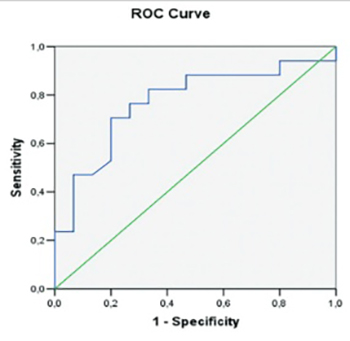
Age, initial duration of AF, LV diameters, right ventricle diameters, EF, left atrium appendix flow rates were similar among patients with and without AF relapse. However, basal left atrium diameter (4.24 ± 0.14 cm vs. 4.04 ± 0.22 cm, p = 0.005), pulmonary artery pressure (32.82 ± 5 vs. 28.60 ± 6.23 mmHg, p = 0.004) and CT-1 values (1.08 ± 0.37 vs. 0.82 ± 0.16 pg/mL, p = 0.02) were found significantly increased in patients with AF relapse. Furthermore, patients with relapsed AF had higher CT-1 levels at 6th month when compared to those in sinus rhythm (1.00 ± 0.40 vs. 0.71 ± 0.23 pg/mL). We did not determine a significant correlation between the LA diameter and CT-1 levels (r = 0.17, p = not significant). The ROC curve analysis indicated CT-1 level of 0.89 pg/mL as the cut-off value for the prediction of AF relapse (76% sensitivity and 74% specificity, p < 0.001) (Figure 2). Of note, we determined that LA diameter over 4 cm can predict AF relapse with 88% sensitivity, but only 40% specificity (Figure 3).

FIGURE 2: The receiver operating characteristic curve for cut-off value of cardiotrophin-1 for the prediction of atrial fibrillation relapse.
FIGURE 3: The receiver operating characteristic curve used for the cut-off value of left atrium diameter for the prediction of atrial fibrillation relapse.

The relationship between the echocardiographic parameters and relapse of AF are shown in Table 1. The relationship between CT-1 levels and AF relapse after CV are shown in Table 2. Patients with relapsed AF had higher CT-1 levels at 6th month when compared to those in sinus rhythm (1.00 ± 0.40 vs. 0.71 ± 0.23 pg/mL, p = 0.009). Patients without relapsed AF had higher CT-1 levels at 6th month when compared to those in sinus rhythm (1.08 ± 0.37 vs. 0.82 ± 0.16, p = 0.008).

TABLE 1: Relationship between the echocardiographic parameters and AF relapse after CV
TABLE 2: Relationship between CT-1 levels and AF relapse after CV

The logistic regression analysis indicated that a CT-1 levels were independent predictor of AF relapse and values ≥0.89 pg/mL increased ~9-fold the risk of AF relapse (Odds ratio [OR]: 8.93; 95% confidence interval [CI] 1.80-44.3).

DISCUSSION

In our study CT-1 levels over 0.47 pg/mL reflected the pathologic limit for healthy controls and CT-1 > 0.89 pg/mL was associated with approximately 9-fold increased AF relapse risk in patients converted to sinus rhythm. Although several parameters including low EF, pulmonary hypertension, increased LA diameter, duration of AF and CT-1 level were found associated with AF relapse after successful CV, our multivariate analysis indicated CT-1 level as the prominent predictor of AF relapse. We think that atrial remodeling in patients with persistent AF is associated with increased CT-1 levels, and the increase of this mediator contributes to AF relapses after successful CV.

This is the first human study indicating that CT-1 levels are increased in patients with persistent AF and increased CT-1 levels over a cut-off value can be used for the prediction of AF relapse after successful CV. Recently, a single experimental study based on pacing-induced sustained AF model on pigs, has indicated no increase in tissue levels of IL-6, leukemia inhibitory factor (LIF) and CT-1, which are known to activate signal transducers and activators of transcriptions through membrane gp130 and Janus kinase [18].

Recent studies determined that CT-1 plays an active role in promoting structural changes (hypertrophy of myocytes, collagen synthesis, and progression of remodeling) in cardiovascular diseases including hypertension, valve diseases, congestive heart failure, and coronary artery disease [19]. Two experimental studies revealed the importance of CT-1 in the pathogenesis of atrial myocardial fibrosis and developing arrhythmias over the release of high concentrations of calcium during diastole [16,17]. There are evidence that CT-1 plasma levels can be elevated in patients with hypertension [20], coronary artery disease [21,22], and heart failure [23]. A recent experimental study indicated that CT-1 accelerates the development of atherosclerotic lesions by stimulating the inflammasome, foam cell formation associated with CD36 and acyl-CoA: cholesterol acyltransferase-1 upregulation in macrophages, and migration, proliferation, and collagen-1 production in vascular smooth muscle cells [24]. Our study provided new information on increased CT-1 among persistent AF patients without structural heart disease.

The success of CV depends on several factors including, the duration of AF, LA diameter, LA appendage flow velocities, underlying organic heart disease, local factors (e.g. chest wall impedance) and technique. In general, successful conversion to sinus rhythm is achieved in about 79% (monophasic) to 94% (biphasic) of patients after CV, however, 50-60% of the patients return to AF in a few months [25,26]. A recent study indicated that sinus rhythm was restored in 89% by DC-CV but maintained only in 34% of the AF patients at a median follow-up of 930 days [27]. In our study, all the patients returned to sinus rhythm with CV, but during the follow-up (6 months) relapsed AF was determined in 17 patients (53%). It is conceivable that structural remodeling comprising progressive LA enlargement, atrial fibrosis, and loss of rate concordance might have a pivotal contribution to the relapse of AF.

CT-1 is reported to exhibit cytoprotective, pro-proliferative and profibrotic effects on the heart via a unique receptor system comprising LIF receptor (LIFRβ) and a common signal transducer, the gp130 [28]. CT-1 was also proposed as a prognostic biomarker for death and heart failure in patients with acute myocardial infarction [29] as well as increased levels of CT-1 were reported to be associated with fibrosis, remodeling and depression of myocardial contractility in patients with heart failure [23]. We think that inflammation leads structural and electrical remodeling that consequently induces AF relapse and investigation of this vicious circle may provide pivotal information for the prevention of AF.

In our study, we excluded patients with severe structural heart disease in order to decrease the effect of potential contributors to AF relapse. We also aimed to investigate more accurately the relationship between CT-1 levels and cardiac remodeling seen in AF patients. Our study has limitations including the sample size, and the duration of follow-up.

CONCLUSION

The increased CT-1 levels were associated with increased AF relapses after successful CV in persistent AF patients. We conclude that CT-1 can be a potential predictor for AF relapse among persistent AF patients who are candidates for CV.

DECLARATION OF INTERESTS

The authors declare no conflicts of interests.

Acknowledgements

ACKNOWLEDGMENT

The abstract of this manuscript was presented in ESC Congress.

REFERENCES

  1. , , , , , (). A survey of atrial fibrillation in general practice: The West Birmingham Atrial Fibrillation Project. Br J Gen Pract.
  2. , , , (). Prevalence, incidence, prognosis, and predisposing conditions for atrial fibrillation: Population-based estimates. Am J Cardiol.
  3. (). Risk factors for stroke and efficacy of antithrombotic therapy in atrial fibrillation. Analysis of pooled data from five randomized controlled trials. Arch Intern Med.
  4. , , , , , (). Independent risk factors for atrial fibrillation in a population-based cohort. The Framingham Heart Study. JAMA. http://www.dx.doi.org/10.1001/jama.271.11.840 http://www.dx.doi.org/10.1001/jama.1994.03510350050036
  5. , , , , , (). Predictors of cerebrovascular events and death among patients with valvular heart disease: A population-based study. Stroke. http://www.dx.doi.org/10.1161/01.STR.31.11.2628
  6. , , (). Electrical, contractile and structural remodeling during atrial fibrillation. Cardiovasc Res. http://www.dx.doi.org/10.1016/S0008-6363(02)00258-4
  7. , , , , , (). A Histological substrate of atrial biopsies in patients with lone atrial fibrillation. Circulation. http://www.dx.doi.org/10.1161/01.CIR.96.4.1180
  8. , , , , , (). Time course of atrial fibrillation-induced cellular structural remodeling in atria of the goat. J Mol Cell Cardiol. http://www.dx.doi.org/10.1006/jmcc.2001.1472
  9. , , , , , (). Fibrosis in left atrial tissue of patients with atrial fibrillation with and without underlying mitral valve disease. Heart. http://www.dx.doi.org/10.1136/hrt.2003.015347
  10. , , , , , (). The relationship between endocardial voltage and regional volume in electroanatomical remodelled left atria in patients with atrial fibrillation: Comparison of three-dimensional computed tomographic images and voltage mapping. J Cardiovasc Electrophysiol. http://www.dx.doi.org/10.1111/j.1540-8167.2009.01557.x
  11. , , , , , (). Expression cloning of cardiotrophin 1, a cytokine that induces cardiac myocyte hypertrophy. Proc Natl Acad Sci U S A. http://www.dx.doi.org/10.1073/pnas.92.4.1142
  12. , , , (). Cardiotrophin-1 displays early expression in the murine heart tube and promotes cardiac myocyte survival. Development.
  13. , , , , , (). Targeted disruption of gp130, a common signal transducer for the interleukin 6 family of cytokines, leads to myocardial and hematological disorders. Proc Natl Acad Sci U S A. http://www.dx.doi.org/10.1073/pnas.93.1.407
  14. , , , , , (). Cardiotrophin-1 (CT-1) can protect the adult heart from injury when added both prior to ischaemia and at reperfusion. Cardiovasc Res. http://www.dx.doi.org/10.1016/S0008-6363(01)00531-4
  15. , (). Cardioprotective growth factors. Cardiovasc Res. http://www.dx.doi.org/10.1093/cvr/cvp062
  16. , , , , , (). Role of cardiotrophin-1 in a canine model of atrial fibrillation and the effect of irbesartan on cardiotrophin-1. Heart. http://www.dx.doi.org/10.1136/heartjnl-2011-300867.71
  17. , , , , , (). Cardiotrophin-1 induces sarcoplasmic reticulum Ca(2) leak and arrhythmogenesis in adult rat ventricular myocytes. Cardiovasc Res. http://www.dx.doi.org/10.1093/cvr/cvs234
  18. , , , , (). Membrane translocation of small GTPase Rac1 and activation of STAT1 and STAT3 in pacing-induced sustained atrial fibrillation. Heart Rhythm. http://www.dx.doi.org/10.1016/j.hrthm.2008.05.012
  19. , , , , , (). Novel insights into the role of cardiotrophin-1 in cardiovascular diseases. J Mol Cell Cardiol. http://www.dx.doi.org/10.1016/j.yjmcc.2008.11.002
  20. , , , , , (). Is plasma cardiotrophin-1 a marker of hypertensive heart disease?. J Hypertens. http://www.dx.doi.org/10.1097/01.hjh.0000160221.09468.d3
  21. , , , , (). Plasma N terminal pro-brain natriuretic peptide and cardiotrophin 1 are raised in unstable angina. Heart. http://www.dx.doi.org/10.1136/heart.84.4.421
  22. , , , , , (). Plasma cardiotrophin-1 following acute myocardial infarction: relationship with left ventricular systolic dysfunction. Clin Sci (Lond). http://www.dx.doi.org/10.1042/CS20010105
  23. , , , , , (). Plasma level of cardiotrophin-1 as a prognostic predictor in patients with chronic heart failure. Eur J Heart Fail. http://www.dx.doi.org/10.1016/j.ejheart.2007.07.015
  24. , , , , , (). Stimulatory effects of cardiotrophin 1 on atherosclerosis. Hypertension. http://www.dx.doi.org/10.1161/HYPERTENSIONAHA.113.01653
  25. , , , , , (). Transthoracic cardioversion of atrial fibrillation: comparison of rectilinear biphasic versus damped sine wave monophasic shocks. Circulation. doi:10.1161/01.CIR.101.11.1282
  26. , , , , (). Oral amiodarone increases the efficacy of direct-current cardioversion in restoration of sinus rhythm in patients with chronic atrial fibrillation. Eur Heart J. http://www.dx.doi.org/10.1053/euhj.1999.1734
  27. , , , , , (). Electrical cardioversion for persistent atrial fibrillation or atrial flutter in clinical practice: predictors of long-term outcome. Int J Clin Pract. http://www.dx.doi.org/10.1111/j.1742-1241.2007.01298.x
  28. , , , (). Effects and mechanism of organ protection by cardiotrophin-1. Curr Med Chem. http://www.dx.doi.org/10.2174/0929867311320020005 http://www.dx.doi.org/10.2174/092986713804806702
  29. , , , , (). Cardiotrophin-1 predicts death or heart failure following acute myocardial infarction. J Card Fail. http://www.dx.doi.org/10.1016/j.cardfail.2006.06.470