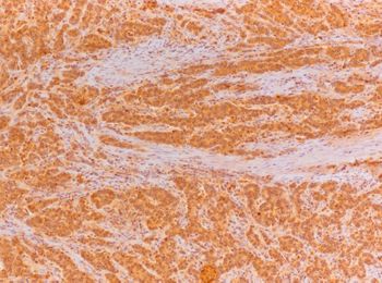
Expression of NEDD9 and connexin-43 in neoplastic and stromal cells of gastric adenocarcinoma
DOI:
https://doi.org/10.17305/bjbms.2020.5379Keywords:
Gastric cancer, NEDD9, conexin-43, microenvironment, metastasesAbstract
Gastric cancer is related to high mortality rates and advanced disease stage at the time of diagnosis. Its carcinogenesis is extensively studied and is associated with genetic and epigenetic changes, changed the interaction between tumor and adjacent stromal cells, and changes in the microenvironment molecule status. Neural precursor cell-expressed developmentally down-regulated 9 (NEDD9) affects different signaling proteins and pathways, apoptosis, adhesion, cell migration, and invasiveness. Connexin-43 (Cx43) also assists in intercellular communications and has several channel-independent functions. Aberrant expression of those two gap junction proteins plays an essential role in metastatic processes. Our scope was to detect the expression of Cx43 and NEDD9 in epithelial and stromal gastric cancer compartments and its relation to tumor progression and lymph node metastases. Cancer tissue from 53 cases of node-negative and 55 cases of node-positive primary gastric carcinoma patients was analyzed for Cx43 and NEDD9 expression by immunohistochemical assay, and the results were correlated with the remaining clinical and pathological findings and survival. In our cohort of patients with lymph node metastases, we detected higher expression of epithelial Cx43 in the primary tumor and stromal Cx43 expression correlated with both epithelial NEDD9 (rho = 0.453) and stromal NEDD9 (rho = 0.484). Higher epithelial Cx43 and NEDD9 expression were associated with higher mortality (HR 1.54, 95% CI 1.01-2.37, p = 0.048). Epithelial Cx43 expression, both epithelial and stromal NEDD9 expression, T and N status were all independently associated with shorter survival. In summary, our findings suggest that increased expression of both epithelial and stromal NEDD9 and epithelial Cx43 could potentially be used as prognostic gastric cancer biomarkers.
Citations
Downloads
Downloads
Additional Files
Published
How to Cite
Accepted 2021-01-08
Published 2021-10-01


