Breast MRI, digital mammography and breast tomosynthesis: comparison of three methods for early detection of breast cancer
DOI:
https://doi.org/10.17305/bjbms.2015.616Keywords:
breast, breast MRI, breast tomosynthesis, digital mammographyAbstract
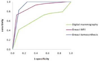
Breast cancer is the most common malignancy in women and early detection is important for its successful treatment. The aim of this study was to investigate the sensitivity and specificity of three methods for early detection of breast cancer: breast magnetic resonance imaging (MRI), digital mammography, and breast tomosynthesis in comparison to histopathology, as well as to investigate the intraindividual variability between these modalities. We included 57 breast lesions, each detected by three diagnostic modalities: digital mammography, breast MRI, and breast tomosynthesis, and subsequently confirmed by histopathology. Breast Imaging-Reporting and Data System (BI-RADS) was used for characterizing the lesions. One experienced radiologist interpreted all three diagnostic modalities. Twenty-nine of the breast lesions were malignant while 28 were benign. The sensitivity for digital mammography, breast MRI, and breast tomosynthesis, was 72.4%, 93.1%, and 100%, respectively; while the specificity was 46.4%, 60.7%, and 75%, respectively. Receiver operating characteristics (ROC) curve analysis showed an overall diagnostic advantage of breast tomosynthesis over both breast MRI and digital mammography. The difference in performance between breast tomosynthesis and digital mammography was significant (p < 0.001), while the difference between breast tomosynthesis and breast MRI was not significant (p = 0.20).
Citations
Downloads
References
American College of Radiology. ACR practice parameter for the performance of screening and diagnostic mammography. American College of Radiology; 2014.
Chan H-P, Wei J, Sahiner B, et al. Computer-aided detection system for breast masses on digital tomosynthesis mammograms: preliminary experience. Radiology 2005;237(3):1075-1080.
Andersson I, Ikeda D, Zackrisson S, et al. Breast tomosynthesis and digital mammography: a comparison of breast cancer visibility and BIRADS classification in a population of cancer with subtle mammographic findings. Eur Radiol 2008;18:2817-2825.
Teertstra H, Loo C, van den Bosch M, et al. Breast tomosynthesis in clinical practice: initial results. Eur Radiol 2010;20(1):16-24.
Kontos D, Bakic PR, Carton AK, Troxel AB, Conant EF, Maidment A. Parenchymal texture analysis in digital breast tomosynthesis for breast cancer risk estimation: a preliminary study. Acad Radiol 2009;16(3):283-298.
Helvie MA. Digital Mammography Imaging: Breast Tomosynthesis and Advanced Applications. Radiol Clin North Am. 2010;48(5):917-929.
American College of Radiology. ACR practice parameter for the performance of contrast-enhanced magnetic resonance imaging (MRI) of the breast. American College of Radiology; 2014.
Orel SG, Schnall MD. MR Imaging of the breast for the detection, diagnosis, and staging of breast cancer. Radiology 2001;220(1):13-30.
Esserman L, Wolverton D, Hylton N. Magnetic resonance imaging for primary breast cancer management: current role and new applications. Endocrine-Related Cancer 2002;9:141-153.
Van Goethem M, Schelfout K, Dijckmans L, et al. MR mammography in the pre-operative staging of breast cancer in patients with dense breast tissue: comparison with mammography and ultrasound. Eur Radiol 2004;14:809-816.
Park SH, Goo JM, Jo CH. Receiver Operating Characteristic (ROC) curve: Practical Review for Radiologists. Korean J Radiol 2004;5(1):11-18
Altman DG. Practical statistics for medical research. London: Chapman and Hall; 1991.
Uematsu T, Kasami M, Watanabe J. Does the degree of background enhancement in breast MRI affect the detection and staging of breast cancer. Eur Radiol 2011;21:2261-2267
Poplack S, Tosteson T, Kogel C, Nagy H. Digital breast tomosynthesis: Initial experience in 98 women with abnormal digital screening mammography. AJR 2007;189:616-623
Majid AS, de Paredes ES, Doherty RD, Sharma N, Salvador X. Missed breast carcinoma: pitfalls and pearls. Radiographics 2003;23:881-895
Macura KJ, Ouwerkerk R, Jacobs MA, Bluemke DA. Patterns of enhancement on breast MR images: interpretation and imaging pitfalls. Radiographics 2006;26:1719-1734.
Djilas-Ivanovic D, Schultze-Haack H, Pavic D, Semelka RC: Breast. In: Semelka RC (ed.). Abdominal-Pelvic MRI, 3rd edn. New Jersey: Wiley-Blackwell; 2010, pp. 1687-1766.
Mann RM, Kuhl CK, Kinkel K, Boetes C. Breast MRI: guidelines from the European Society of breast imaging. Eur Radiol 2008:1307-1318.
Cheng L, Li X. Breast magnetic resonance imaging: kinetic curve assessment. Gland Surg 2013;2(1):50-53.

Downloads
Additional Files
Published
Issue
Section
Categories
How to Cite
Accepted 2015-08-24
Published 2015-11-16


