G-protein-coupled estrogen receptor-30 gene polymorphisms are associated with uterine leiomyoma risk

Authors

  • Burcu Kasap Department of Obstetrics and Gynecology, School of Medicine, Mugla Sitki Kocman University, Mugla, Turkey
  • Nilgün Öztürk Turhan Department of Obstetrics and Gynecology, School of Medicine, Mugla Sitki Kocman University, Mugla, Turkey
  • Tuba Edgünlü School of Health Sciences, Mugla Sitki Kocman University, Mugla, Turkey.
  • Müzeyyen Duran Department of Obstetrics and Gynecology, School of Medicine, Mugla Sitki Kocman University, Mugla, Turkey
  • Eren Akbaba Department of Obstetrics and Gynecology, School of Medicine, Mugla Sitki Kocman University, Mugla, Turkey
  • Gökalp Öner Department of Obstetrics and Gynecology, School of Medicine, Mugla Sitki Kocman University, Mugla, Turkey

DOI:

https://doi.org/10.17305/bjbms.2016.683

Keywords:

uterine leiomyoma, GPR30 (GPER1), gene, polymorphism, estrogen receptor

Abstract

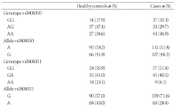
The G-protein-coupled estrogen receptor (GPR30, GPER-1) is a member of the G-protein-coupled receptor 1 family and is expressed significantly in uterine leiomyomas. To understand the relationship between GPR30 single nucleotide polymorphisms and the risk of leiomyoma, we measured the follicle-stimulating hormone (FSH) and estradiol (E2) levels of 78 perimenopausal healthy women and 111 perimenopausal women with leiomyomas. The participants’ leiomyoma number and volume were recorded. DNA was extracted from whole blood with a GeneJET Genomic DNA Purification Kit. An amplification-refractory mutation system polymerase chain reaction approach was used for genotyping of the GPR30 gene (rs3808350, rs3808351, and rs11544331). The differences in genotype and allele frequencies between the leiomyoma and control groups were calculated using the chi-square (χ2) and Fischer’s exact test. The median FSH level was higher in controls (63 vs. 10 IU/L, p=0.000), whereas the median E2 level was higher in the leiomyoma group (84 vs. 9.1 pg/mL, p=0.000). The G allele of rs3808351 and the GG genotype of both the rs3808350 and rs3808351 polymorphisms and the GGC haplotype increased the risk of developing leiomyoma. There was no significant difference in genotype frequencies or leiomyoma volume. However, the GG genotype of the GPR30 rs3808351 polymorphism and G allele of the GPR30 rs3808351 polymorphism were associated with the risk of having a single leiomyoma. Our results suggest that the presence of the GG genotype of the GPR30 rs3808351 polymorphism and the G allele of the GPR30 rs3808351 polymorphism affect the characteristics and development of leiomyomas in the Turkish population.


Downloads

Download data is not yet available.

Author Biography

Burcu Kasap, Department of Obstetrics and Gynecology, School of Medicine, Mugla Sitki Kocman University, Mugla, Turkey

Obstetrics and Gynecology

References

Baird DD, Dunson DB, Hill MC, Cousins D, Schectman JM. High cumulative incidence of uterine leiomyoma in black and white women: ultrasound evidence. Am J Obstet Gynecol. 2003;188(1):100-107.

http://dx.doi.org/10.1067/mob.2003.99

Ekin M, Cengiz H, Öztürk E, Kaya C, Yasar L, Savan K. Genitourinary symptoms and their effects on quality of life in women with uterine myomas. Int Urogynecol J. 2014;25(6):807-810

http://dx.doi.org/10.1007/s00192-013-2295-4

Flake GP, Andersen J, Dixon D. Etiology and pathogenesis of uterine leiomyomas: a review. Environ Health Perspect. 2003;111(8):1037-1054.

http://dx.doi.org/10.1289/ehp.5787

Grandien K, Berkenstam A, Gustafsson JA. The estrogen receptor gene: promoter organization and expression. Int J Biochem Cell Biol. 1997;29(12):1343-1369

http://dx.doi.org/10.1016/S1357-2725(97)89967-0

Pedram A, Razandi M, Levin ER. Nature of functional estrogen receptors at the plasma membrane. Mol Endocrinol. 2006;20(9):1996-2009.

http://dx.doi.org/10.1210/me.2005-0525

Revankar CM, Cimino DF, Sklar LA, Arterburn JB, Prossnitz ER. A transmembrane intracellular estrogen receptor mediates rapid cell signaling. Science. 2005;307(5715):1625-1630.

http://dx.doi.org/10.1126/science.1106943

Otto C, Rohde-Schulz B, Schwarz G, Fuchs I, Klewer M, Brittain D,et al. G protein-coupled receptor 30 localizes to the endoplasmic reticulum and is not activated by estradiol. Endocrinology. 2008;149(10):4846-4856.

http://dx.doi.org/10.1210/en.2008-0269

Hewitt SC, Harrell JC, Korach KS. Lessons in estrogen biology from knockout and transgenic animals. Annu Rev Physiol. 2005;67:285-308.

http://dx.doi.org/10.1146/annurev.physiol.67.040403.115914

Dennis MK, Burai R, Ramesh C, Petrie WK, Alcon SN, Nayak TK, et al. In vivo effects of a GPR30 antagonist. Nat Chem Biol. 2009;5(6):421-427.

http://dx.doi.org/10.1038/nchembio.168

Smith HO, Leslie KK, Singh M, Qualls CR, Revankar CM, Joste NE, et al. GPR30: a novel indicator of poor survival for endometrial carcinoma. Am J Obstet Gynecol. 2007;196(4):386.e1-9; discussion 386.e9-11.

Huang GS, Gunter MJ, Arend RC, Li M, Arias-Pulido H, Prossnitz ER, et al. Co-expression of GPR30 and ERbeta and their association with disease progression in uterine carcinosarcoma. Am J Obstet Gynecol. 2010;203(3):242.e1-5.

http://dx.doi.org/10.1016/j.ajog.2010.04.046

Plante BJ, Lessey BA, Taylor RN, Wang W, Bagchi MK, Yuan L, et al. G protein-coupled estrogen receptor (GPER) expression in normal and abnormal endometrium. Reprod Sci. 2012;19(7):684-693.

http://dx.doi.org/10.1177/1933719111431000

Filardo EJ, Graeber CT, Quinn JA, Resnick MB, Giri D, DeLellis RA, et al. Distribution of GPR30, a seven membrane-spanning estrogen receptor, in primary breast cancer and its association with clinicopathologic determinants of tumor progression. Clin Cancer Res. 2006;12(21):6359-6366.

http://dx.doi.org/10.1158/1078-0432.CCR-06-0860

He YY, Cai B, Yang YX, Liu XL, Wan XP. Estrogenic G protein-coupled receptor 30 signaling is involved in regulation of endometrial carcinoma by promoting proliferation, invasion potential, and interleukin-6 secretion via the MEK/ERK mitogen-activated protein kinase pathway. Cancer Sci. 2009;100(6):1051-1061.

http://dx.doi.org/10.1111/j.1349-7006.2009.01148.x

Tica AA, Dun EC, Tica OS, Gao X, Arterburn JB, Brailoiu GC, et al. G protein-coupled estrogen receptor 1-mediated effects in the rat myometrium. Am J Physiol Cell Physiol. 2011;301(5):C1262-1269.

http://dx.doi.org/10.1152/ajpcell.00501.2010

Welsh T, Johnson M, Yi L, Tan H, Rahman R, Merlino A, et al. Estrogen receptor (ER) expression and function in the pregnant human myometrium: estradiol via ERα activates ERK1/2 signaling in term myometrium. J Endocrinol. 2012;212(2):227-238.

http://dx.doi.org/10.1530/JOE-11-0358

Hermon TL, Moore AB, Yu L, Kissling GE, Castora FJ, Dixon D. Virchows Arch. Estrogen receptor alpha (ERalpha) phospho-serine-118 is highly expressed in human uterine leiomyomas compared to matched myometrium. 2008;453(6):557-569.

Jakimiuk AJ, Bogusiewicz M, Tarkowski R, Dziduch P, Adamiak A, Wróbel A, et al. Estrogen receptor alpha and beta expression in uterine leiomyomas from premenopausal women. Fertil Steril. 2004;82:1244-1249.

http://dx.doi.org/10.1016/j.fertnstert.2004.02.130

Tian R, Wang Z, Shi Z, Li D, Wang Y, Zhu Y, et al. Differential expression of G-protein-coupled estrogen receptor-30 in human myometrial and uterine leiomyoma smooth muscle. Fertil Steril. 2013;99(1):256-263.

http://dx.doi.org/10.1016/j.fertnstert.2012.09.011

Giess M, Lattrich C, Springwald A, Goerse R, Ortmann O, Treeck O. GPR30 gene polymorphisms are associated with progesterone receptor status and histopathological characteristics of breast cancer patients. J Steroid Biochem Mol Biol. 2010;118(1-2):7-12.

http://dx.doi.org/10.1016/j.jsbmb.2009.09.001

Andersen J. Growth factors and cytokines in uterine leiomyomas. Semin Reprod Endocrinol. 1996;14(3):269-282.

http://dx.doi.org/10.1055/s-2007-1016336

Olde B, Leeb-Lundberg LM. GPR30/GPER1: searching for a role in estrogen physiology. Trends Endocrinol Metab. 2009;20(8):409-416.

http://dx.doi.org/10.1016/j.tem.2009.04.006

Haas E, Meyer MR, Schurr U, Bhattacharya I, Minotti R, Nguyen HH, et al. Differential effects of 17beta-estradiol on function and expression of estrogen receptor alpha, estrogen receptor beta, and GPR30 in arteries and veins of patients with atherosclerosis. Hypertension. 2007;49(6):1358-1363.

http://dx.doi.org/10.1161/HYPERTENSIONAHA.107.089995

Smith HO, Arias-Pulido H, Kuo DY, Howard T, Qualls CR, Lee SJ, et al. GPR30 predicts poor survival for ovarian cancer. Oncol. 2009;114(3):465-471.

http://dx.doi.org/10.1016/j.ygyno.2009.05.015

Sirianni R, Chimento A, Ruggiero C, De Luca A, Lappano R, Andò S, et al. Endocrinology. The novel estrogen receptor, G protein-coupled receptor 30, mediates the proliferative effects induced by 17beta-estradiol on mouse spermatogonial GC-1 cell line. 2008;149(10):5043-5051.

Kleuser B, Malek D, Gust R, Pertz HH, Potteck H. 17-Beta-estradiol inhibits transforming growth factor-beta signaling and function in breast cancer cells via activation of extracellular signal-regulated kinase through the G protein-coupled receptor 30. Mol Pharmacol. 2008;74(6):1533-1543.

http://dx.doi.org/10.1124/mol.108.046854

Chevalier N, Paul-Bellon R, Camparo P, Michiels JF, Chevallier D, Fénichel P. Genetic variants of GPER/GPR30, a novel estrogen-related G protein receptor, are associated with human seminoma. Int J Mol Sci. 2014;15(1):1574-1589.

http://dx.doi.org/10.3390/ijms15011574

Korkmaz HA, Edgünlü T, Eren E, Demir K, Çakir ED, Çelik SK, et al. GPR30 Gene Polymorphisms Are Associated with Gynecomastia Risk in Adolescents. Horm Res Paediatr. 2015;83(3):177-182.

http://dx.doi.org/10.1159/000369013

Snieder H, MacGregor AJ, Spector TD. Genes control the cessation of a woman's reproductive life: a twin study of hysterectomy and age at menopause. J Clin Endocrinol Metab. 1998;83(6):1875-1880.

http://dx.doi.org/10.1210/jc.83.6.1875

Luoto R, Kaprio J, Rutanen EM, Taipale P, Perola M, Koskenvuo M. Heritability and risk factors of uterine fibroids--the Finnish Twin Cohort study. Maturitas. 2000;37(1):15-26.

http://dx.doi.org/10.1016/S0378-5122(00)00160-2

Cha PC, Takahashi A, Hosono N, Low SK, Kamatani N, Kubo M, et al. A genome-wide association study identifies three loci associated with susceptibility to uterine fibroids. Nat Genet. 2011;43(5):447-450.

http://dx.doi.org/10.1038/ng.805

Edwards TL, Hartmann KE, Velez Edwards DR. Variants in BET1L and TNRC6B associate with increasing fibroid volume and fibroid type among European Americans. Hum Genet. 2013;132(12):1361-1369.

http://dx.doi.org/10.1007/s00439-013-1340-1

Aissani B, Zhang K, Wiener H.Follow-up to genome-wide linkage and admixture mapping studies implicates components of the extracellular matrix in susceptibility to and size of uterine fibroids. Fertil Steril. 2015;103(2):528-534.e13.

http://dx.doi.org/10.1016/j.fertnstert.2014.10.025

Morosova EB, Chukhlovin AB, Kulagina NV, Kipich NV, Totolian AA. Functional gene polymorphism of matrix metalloproteinase-1 is associated with benign hyperplasia of myo- and endometrium in the Russian population. Genet Test Mol Biomarkers. 2012;16(9):1032-1037.

http://dx.doi.org/10.1089/gtmb.2011.0376

Attar R, Atasoy H, İnal-Gültekin G, Timirci-Kahraman Ö, Güleç-Yilmaz S, Dalan AB, et al. The effects of PON1 gene Q192R variant on the development of uterine leiomyoma in Turkish patients. In Vivo. 2015;29(2):243-246.

Gultekin GI, Yilmaz SG, Kahraman OT, Atasoy H, Dalan AB, Attar R, et al. Lack of influence of the ACE1 gene I/D polymorphism on the formation and growth of benign uterine leiomyoma in Turkish patients. Asian Pac J Cancer Prev. 2015;16(3):1123-1127.

http://dx.doi.org/10.7314/APJCP.2015.16.3.1123

Shen Y, Ren ML, Xu J, Xu Q, Ding YQ, Wu ZC, et al. A multicenter case-control study on screening of single nucleotide polymorphisms in estrogen-metabolizing enzymes and susceptibility to uterine leiomyoma in han chinese. Gynecol Obstet Invest. 2014;77(4):224-230.

http://dx.doi.org/10.1159/000360083

Ates O, Demirturk F, Toprak M, Sezer S. Polymorphism of catechol-o-methyltransferase and uterine leiomyoma. Mol Cell Biochem. 2013;375(1-2):179-183.

Alleyne AT, Austin S, Williams A. Distribution of CYP17α polymorphism and selected physiochemical factors of uterine leiomyoma in Barbados. Meta Gene. 2014; 9(2):358-365.

http://dx.doi.org/10.1016/j.mgene.2014.03.006

G-protein-coupled estrogen receptor-30 gene polymorphisms are associated with uterine leiomyoma risk

Downloads

Additional Files

Published

2016-01-06

How to Cite

1.
Kasap B, Öztürk Turhan N, Edgünlü T, Duran M, Akbaba E, Öner G. G-protein-coupled estrogen receptor-30 gene polymorphisms are associated with uterine leiomyoma risk. Biomol Biomed [Internet]. 2016Jan.6 [cited 2023Dec.1];16(1):39-45. Available from: https://www.bjbms.org/ojs/index.php/bjbms/article/view/683

Issue

Section

Molecular Biology

INTRODUCTION

Uterine leiomyoma is the most common benign tumor originating from uterine smooth muscle cells of the myometrium [1]. Leiomyomas impair the quality of life and reproductive health of women, causing abnormal uterine bleeding, pelvic pain, preterm labor, recurrent pregnancy loss, and infertility [2]. Besides ethnicity, nulliparity, obesity, early menarche, and age as predisposing factors, estrogen is regarded as the main promoter of leiomyomas [3]. Estrogens, mainly estradiol (E2), exert their classic slow biological effects via activation of and interaction with the nuclear alpha (ERα) and beta (ERβ) estrogen receptors (ERs) [4]. Rapid non-genomic responses to estrogen are mediated by either ERs at the plasma membrane [5] or the extranuclear seven-transmembrane domain receptor (GPR30) [6]. GPR30 is a member of the G-protein-coupled receptor 1 family and encodes a multi-pass membrane protein that localizes to the endoplasmic reticulum. GPR30 protein binds estrogen, resulting in intracellular calcium mobilization and the synthesis of phosphatidylinositol 3,4,5-trisphosphate in the nucleus. Therefore, this protein plays a role in the rapid non-genomic signaling events widely observed following the stimulation of cells and tissues with estrogen [7]. ERα plays a major role in estrogen-mediated responses, such as the proliferation and induction of gene expression in the uterus [8]. In addition to ERα, GPR30 is expressed in human endometrium and promotes human uterine epithelial proliferation [9]. Elevated GPR30 is associated with high-grade, poor-prognosis endometrial cancer; uterine carcinosarcoma [10,11]; and endometriosis [12]. GPR30 expression is also strongly associated with tumor progression in breast cancer [13]. The proliferative effect of GPR30 acts via activation of the mitogen-activated protein kinase pathway [14]. GPR30 is expressed and functional in rat myometrium, and its activation is associated with increased contractility in rat myometrial cells [15]. Subsequently, GPR30 was detected in the human myometrium at term in pregnancy, but its contribution to mediating estrogen action in the myometrium during labor was less than that of ERα [16]. ERα is also more highly expressed in leiomyoma than in the myometrium [17], whereas ERβ is expressed in both myometrium and leiomyoma tissues [18]. Recently, GPR30 was also found to be more highly expressed in leiomyoma than in matched myometrium [19]. More than 1,000 single nucleotide polymorphisms (SNPs) of the GPR30 gene have been identified (http//www.ncbi.nlm.nih.gov/snp/). We selected three nonsynonymous SNPs of the GPR30 gene as genotyping targets based on the HapMap project database (http://hapmap.ncbi.nlm.nih.gov/): rs3808350, rs3808351, and rs11544331. Functionally, SNPs are nonsynonymous polymorphisms. The rs3808350 and rs3808351polymorphisms are located in the 5’-region of the GPR30 gene; rs11544331 is located in exon 3 and leads to a Pro16Leu amino acid substitution [20]. We hypothesized that GPR30 gene SNPs might affect susceptibility to leiomyoma development and leiomyoma characteristics because cumulative estrogen exposure contributes to the development of leiomyoma [21]. The first genome-wide association study of leiomyoma was conducted in a Japanese population and showed that three loci on chromosomes 10q24.33, 22q13.1, and 11p15.5 had significant genome-wide associations with leiomyomas. However, GPR30 polymorphisms have not been investigated in human leiomyoma. We investigated the association between GPR30 gene polymorphisms and leiomyoma for the first time. We compared the genotype, haplotype, and allele frequencies of three GPR30 SNPs in women with leiomyoma and healthy perimenopausal women as well as within leiomyoma subgroups based on leiomyoma number and size. These comparisons were performed to determine whether GPR30 SNPs are related to the risk and characteristics of leiomyoma and to evaluate the early diagnostic potential of those SNPs.

MATERIALS AND METHODS

Study population

This study was conducted in the Department of Obstetrics and Gynecology, Muğla Sıtkı Koçman University, School of Medicine. The study population comprised 78 perimenopausal healthy women (age, 40–55 years) who visited our hospital for routine gynecological examinations and 111 perimenopausal women (age, 40–55 years) with leiomyoma seen from January 2015 to April 2015. All participants underwent transvaginal or pelvic ultrasonography to detect leiomyoma. The volume of each leiomyoma was calculated using the formula 0.521 × D1 × D2 × D3, where D1, D2, and D3 are three perpendicular dimensions (length, width, and depth). In patients with more than one leiomyoma, the volume was the sum of all leiomyoma measurements. Age, body mass index (BMI), and E2 and follicle-stimulating hormone (FSH) levels were recorded. Patients with endocrinological disorders, on hormone therapy, or with a history of myomectomy were excluded from the study. The patients with leiomyoma were subcategorized according to leiomyoma number (single or multiple leiomyomas) and leiomyoma volume (<20 mL, 20–100 mL, and >100 mL).

Ethics

The study protocol was approved by Muğla Sıtkı Koçman University Medical Sciences Ethics Committee (2015-7), and informed consent was obtained from all participants. This study was performed in conformity with the Declaration of Helsinki, as revised in 2000.

Hormone evaluation

Blood for measuring the FSH and E2 levels was drawn from an antecubital vein and analyzed using an electrochemiluminescence immunoassay.

DNA extraction and genotyping

Venous blood samples (2 mL) were collected in Vacutainer tubes containing sodium/potassium EDTA. DNA was extracted with a GeneJET Genomic DNA Purification Kit (Thermo K0772; Thermo Fisher Scientific, Waltham, MA, USA). We used the amplification refractory mutation system polymerase chain reaction (PCR) approach for genotyping SNPs of the GPR30 gene. PCR was performed in a 25-µL volume with 100 ng of DNA, 100 µm dNTPs, 20 pmol of each primer, 1.5 mM MgCl2, 1× PCR buffer with (NH4)2SO4, and 2 U of Taq DNA polymerase (Thermo Scientific EP0401), using an automated thermal cycler (Techne Flexigene, Cambridge, UK). The primer sequences and PCR conditions are listed in Table 1. The PCR products were separated at 120 V for 40–50 minutes on 2.5% agarose gels containing 0.5 mg/mL ethidium bromide. We used a 100-bp DNA ladder (Fermentas Vilnius, Lithuania) as the size standard for each gel lane. The gel was visualized under ultraviolet light using a gel electrophoresis visualizing system (Vilber Lourmat Deutschland, Germany).

TABLE 1: Oligonucleotides and PCR conditions used for SNPs of GPR30 genes

Statistical analysis

All of the frequency and statistical calculations were performed using SPSS, ver. 20.0 (IBM Corporation, Armonk, NY, USA). Calculated continuous variables are presented as median and range, and categorical variables are presented as proportions. The independent sample t-test and the Mann–Whitney U-test were used to compare continuous variables between two groups. Chi-square analysis and Fisher’s exact test were used to compare frequencies of the GPR30 ­genotypes, haplotypes, and alleles between the patients in the leiomyoma and control groups and within the leiomyoma subgroups, with p<0.05 considered statistically significant.

RESULTS

The patient cohort characteristics are summarized in Table 2. Briefly, the study included 111 patients with leiomyoma and 78 controls. There were no significant differences in median age, hemoglobin level, or BMI between the leiomyoma and control patients (p=0.530, p=0.183, and p>0.05, respectively). The median FSH level was higher in the control group (63 vs. 10 IU/L, respectively; p=0.000), while the median E2 level was higher in the leiomyoma group (84.0 vs. 9.1 pg/mL, respectively; p=0.000), as shown in Table 2.

TABLE 2: The demographic and clinical characteristics of patients with uterine leiomyoma and controls

The genotype and allele frequencies of the GPR30 gene polymorphisms (rs3808350, rs3808351, and rs11544331) are shown in Table 3. All of the observed genotype frequencies were at Hardy–Weinberg equilibrium in the controls (p>0.05). The respective frequencies of the GG, AG, and AA genotypes for the rs3808350 GPR30 polymorphism were 17.9%, 47.4%, and 34.6% in the control group and 33.3%, 29.7%, and 36.9% in the leiomyoma group. The relative risk for developing leiomyoma in individuals with the rs3808350 GPR30 GG genotype was higher than with the AG genotype (p=0.017) (Table 3). The GG, GA, and AA genotype distributions of the rs3808351 GPR30 polymorphism were 28%, 32%, and 18% for the controls and 57%, 45%, and 9% for the leiomyoma patients. The relative risk for individuals with the GG genotype of the rs3808351 GPR30 polymorphism was higher than with the AA genotype (p=0.08) (Table 3). In the allele frequency analysis, individuals with the rs3808351 GPR30 polymorphism G allele had a higher risk of developing leiomyoma than did individuals with the A allele (p=0.004) (Table 3). There were no significant associations between the allele distributions for the rs3808350 GPR30 polymorphism (p=0.250) or between the genotype and allele distributions for the rs11544331 GPR30 polymorphism among the control and leiomyoma patients (p=0.346 and p=0.208, respectively). Haplotype analysis of the rs3808350, rs3808351, and rs11544331 GPR30 polymorphisms revealed that the GGC haplotype frequency was 19.8% in leiomyoma patients and 6.3% in the controls, and the difference was significant (OR=3.00; 95% CI, 1.395–6.450) (Table 4). Thus, the GGC haplotype increased the risk of development of leiomyoma by three times.

TABLE 3: Distributions of GPR30 genotypes and allele frequencies and the risk of uterine leiomyoma development.
TABLE 4: GPR30 rs3808350/rs3808351/rs11544331 haplotypes and risk of uterine leiomyoma

The distributions of the GPR30 rs3808350, rs3808351, and rs11544331 genotypes and alleles according to leiomyoma number and size were also examined. There were no significant differences between the GPR30 rs3808350, rs3808351, and rs11544331 polymorphism genotype (p=0.170, p=0.104, and p=0.596, respectively) or allele (p=0.915, p=0.195, and p=0.228, respectively) frequencies and leiomyoma range in size (data not shown). In relation to leiomyoma number, the GG genotype of the GPR30 rs3808351 polymorphism and G allele of the GPR30 rs3808351 polymorphism increased the risk for having only one leiomyoma (p=0.028 and p=0.011, respectively) (Table 5).

TABLE 5: Genotype and allele frequencies of GPR30 in patients categorized according to uterine leiomyoma number

DISCUSSION

We hypothesized that GPR30 gene SNPs might affect the susceptibility to leiomyoma development and leiomyoma characteristics because cumulative estrogen exposure contributes to the development of leiomyoma [21]. Consequently, we studied three SNPs that might alter the GPR30 expression levels or GPR30 protein structure and function. For the first time, we showed that the rs3808351 polymorphism of the GPR30 gene was associated with both the risk and number of leiomyomas, whereas the rs3808350 polymorphism was associated only with the risk of leiomyoma development in the Turkish population.

GPR30, has been detected in many tissues, including the uterus [22]. GPR30 expression was discovered in the vascular smooth muscle cells of patients with atherosclerosis [23]. Subsequently, it was found that GPR30 expression mediated the proliferation of ovarian [24], endometrial [10], breast [13], and sperm cell [25] tumors. Tian et al. [19] investigated the potential proliferative effect of estrogen-driven GPR30 signaling on uterine smooth muscle cells in samples of leiomyoma and adjacent myometrium. They reported that GPR30 was more intensely expressed in the smooth muscle cells of leiomyoma than in matched myometrium and speculated that the up-regulation of GPR30 by E2 might affect intracellular signaling in leiomyoma smooth muscle cells [19]. ICI 182.780 is an ERα–ERβ antagonist that is thought to be involved in GPR30 signaling [26]. Tian et al. [19] confirmed this for leiomyoma cells and demonstrated that the treatment of leiomyoma cells with ICI 182.780 stimulated E2-induced expression of GPR30. We found that the GG genotypes of both the rs3808350 and rs3808351 polymorphisms of the GPR30 gene showed an increased risk for developing leiomyoma; thus, these findings might lead to the development of new leiomyoma treatment modalities customized according to the SNP status of the GPR30 gene.

Although associations between SNPs of GPR30 and several diseases, such as breast carcinoma [20], seminoma [27], and gynecomastia [28], have been investigated, this is the first study to investigate the association between leiomyoma development and GPR30 SNPs. Giess et al. [20] evaluated the association between GPR30 SNPs and breast carcinoma and reported no significant differences in the allele, genotype, or haplotype frequencies of SNPs between breast carcinoma and healthy individuals, whereas SNPs of the GPR30 gene were strongly associated with lymph node involvement, tumor size, histological grade, and progesterone receptor status. Chevalier et al. [27] demonstrated that the GG genotype in both the rs3808350 and rs3808351 polymorphisms protected against seminoma in Caucasian males. Finally, Korkmaz et al. [28] reported that the GG genotype of rs3808350, AA genotype of rs3808351, G allele of rs3808350, and A allele of rs3808351 increased the risk of gynecomastia in a Turkish population.

In our study, the G allele of rs3808351 was frequently seen in leiomyoma patients. The GG genotype of both the rs3808350 and rs3808351 polymorphisms and GGC haplotype increased the risk of developing leiomyoma. Our findings are consistent with those of Korkmaz et al. [28] and with the SNP location, which might explain the changes in GPR30 expression described by Tian et al. [19].

Hereditability studies conducted in several European countries showed that genetic factors were responsible for 26% to 69% of leiomyoma [29,30]. Consequently, new gene interactions strongly related to the pathways of leiomyoma etiology were hypothesized to be involved in this complex disease. The first genome-wide association study of leiomyoma was subsequently conducted in a Japanese population and revealed three SNPs that have strong associations with leiomyoma [31]. Edwards et al. [32] replicated these findings and tested related SNPs for associations with leiomyoma characteristics; they observed a strong association between SNPs of BET1L and intramural leiomyoma and between SNPs of TNRC6B and larger leiomyomas in a European–American population. New data from a genome-wide association study suggested that SNPs of the COL6A3 and COL13A genes affect the susceptibility to leiomyoma development and characteristics in African–American and European–American populations [33]. Supporting this finding, a polymorphism of the MMP-1 gene affecting collagen remodeling in the myometrium was suggested to be a potential risk factor for developing leiomyoma in a Russian population [34]. In a Turkish population, the effects of the PON1 gene Q192 variant [35] and ACE1 gene I/D polymorphism were investigated in leiomyoma patients, and an association between the PON1 gene Q192 variant and risk of developing leiomyoma was found [36]. In these two studies conducted in Turkish populations, the PON1 gene Q192 variant and ACE1 gene I/D polymorphism were found to be unrelated to tumor size or number [35,36]. We showed that the rs3808351 polymorphism of the GPR30 gene was associated with the risk and number of leiomyoma, whereas the rs3808350 polymorphism was associated only with the risk of developing leiomyoma in a Turkish population. Because the number of patients in the leiomyoma subgroups was insufficient to perform an adequate statistical analysis, we were unable to analyze the association between myoma localization (intramural, subserosal, and submucosal) and SNPs of GPR30.

Regarding estrogen as the main promoter of leiomyoma development, genetic polymorphisms of genes for estrogen-metabolizing enzymes, such as COMT, CYP1A1, and CYP1B1, were studied in a Chinese Han population, which found that SNPs of these genes were associated with the risk of developing leiomyoma [37]. By contrast, Ateş et al. [38] found that the COMT Val158Met polymorphism was not related to the risk of leiomyoma development in Turkey, although larger leiomyomas were associated with this polymorphism. Similarly, an SNP of the CYP17 gene, which encodes an enzyme for estrogen biosynthesis, did not increase the risk of developing leiomyoma in black women in Barbados, whereas estrogen levels and BMI were found to be contributing risk factors [39]. Our findings are consistent with the literature in terms of an association between estrogen levels and leiomyoma development, but our findings did not support an association between BMI and leiomyoma development or between the rs3808350, rs3808351, and rs11544331 genotype frequencies and leiomyoma volume.

CONCLUSION

The findings of this study support the contribution of increased E2 levels and genetic factors to the pathogenesis of leiomyoma.

Acknowledgements

ACKNOWLEDGEMENTS

We thank Sevim Karakaş Çelik for helping with the statistical analysis.

DECLARATION OF INTERESTS

The authors declare no competing interests.

REFERENCES

  1. , , , , (). High cumulative incidence of uterine leiomyoma in black and white women:Ultrasound evidence. Am J Obstet Gynecol. http://dx.doi.org/10.1067/mob.2003.99
  2. , , , , , (). Genitourinary symptoms and their effects on quality of life in women with uterine myomas. Int Urogynecol J. http://dx.doi.org/10.1007/s00192-013-2295-4
  3. , , (). Etiology and pathogenesis of uterine leiomyomas:A review. Environ Health Perspect. http://dx.doi.org/10.1289/ehp.5787
  4. , , (). The estrogen receptor gene:Promoter organization and expression. Int J Biochem Cell Biol. http://dx.doi.org/10.1016/S1357-2725(97)89967-0
  5. , , (). Nature of functional estrogen receptors at the plasma membrane. Mol Endocrinol. http://dx.doi.org/10.1210/me.2005-0525
  6. , , , , (). A transmembrane intracellular estrogen receptor mediates rapid cell signaling. Science. http://dx.doi.org/10.1126/science.1106943
  7. , , , , , (). G protein-coupled receptor 30 localizes to the endoplasmic reticulum and is not activated by estradiol. Endocrinology. http://dx.doi.org/10.1210/en.2008-0269
  8. , , (). Lessons in estrogen biology from knockout and transgenic animals. Annu Rev Physiol. http://dx.doi.org/10.1146/annurev.physiol.67.040403.115914
  9. , , , , , (). In vivo effects of a GPR30 antagonist. Nat Chem Biol. http://dx.doi.org/10.1038/nchembio.168
  10. , , , , , (). GPR30:A novel indicator of poor survival for endometrial carcinoma. Am J Obstet Gynecol. discussion 386.e9-11
  11. , , , , , (). Co-expression of GPR30 and ERbeta and their association with disease progression in uterine carcinosarcoma. Am J Obstet Gynecol. http://dx.doi.org/10.1016/j.ajog.2010.04.046
  12. , , , , , (). G protein-coupled estrogen receptor (GPER) expression in normal and abnormal endometrium. Reprod Sci. http://dx.doi.org/10.1177/1933719111431000
  13. , , , , , (). Distribution of GPR30, a seven membrane-spanning estrogen receptor, in primary breast cancer and its association with clinicopathologic determinants of tumor progression. Clin Cancer Res. http://dx.doi.org/10.1158/1078-0432.CCR-06-0860
  14. , , , , (). Estrogenic G protein-coupled receptor 30 signaling is involved in regulation of endometrial carcinoma by promoting proliferation, invasion potential, and interleukin-6 secretion via the MEK/ERK mitogen-activated protein kinase pathway. Cancer Sci. http://dx.doi.org/10.1111/j.1349-7006.2009.01148.x
  15. , , , , , (). G protein-coupled estrogen receptor 1-mediated effects in the rat myometrium. Am J Physiol Cell Physiol. http://dx.doi.org/10.1152/ajpcell.00501.2010
  16. , , , , , (). Estrogen receptor (ER) expression and function in the pregnant human myometrium:Estradiol via ERα activates ERK1/2 signaling in term myometrium. J Endocrinol. http://dx.doi.org/10.1530/JOE-11-0358
  17. , , , , , (). Virchows Arch. Estrogen receptor alpha (ERalpha) phospho-serine-118 is highly expressed in human uterine leiomyomas compared to matched myometrium.
  18. , , , , , (). Estrogen receptor alpha and beta expression in uterine leiomyomas from premenopausal women. Fertil Steril. http://dx.doi.org/10.1016/j.fertnstert.2004.02.130
  19. , , , , , (). Differential expression of G-protein-coupled estrogen receptor-30 in human myometrial and uterine leiomyoma smooth muscle. Fertil Steril. http://dx.doi.org/10.1016/j.fertnstert.2012.09.011
  20. , , , , , (). GPR30 gene polymorphisms are associated with progesterone receptor status and histopathological characteristics of breast cancer patients. J Steroid Biochem Mol Biol. http://dx.doi.org/10.1016/j.jsbmb.2009.09.001
  21. (). Growth factors and cytokines in uterine leiomyomas. Semin Reprod Endocrinol. http://dx.doi.org/10.1055/s-2007-1016336
  22. , (). GPR30/GPER1:Searching for a role in estrogen physiology. Trends Endocrinol Metab. http://dx.doi.org/10.1016/j.tem.2009.04.006
  23. , , , , , (). Differential effects of 17beta-estradiol on function and expression of estrogen receptor alpha, estrogen receptor beta, and GPR30 in arteries and veins of patients with atherosclerosis. Hypertension. http://dx.doi.org/10.1161/HYPERTENSIONAHA.107.089995
  24. , , , , , (). GPR30 predicts poor survival for ovarian cancer. Oncol. http://dx.doi.org/10.1016/j.ygy.no.2009.05.015
  25. , , , , , (). Endocrinology. The novel estrogen receptor, G protein-coupled receptor 30, mediates the proliferative effects induced by 17beta-estradiol on mouse spermatogonial GC-1 cell line.
  26. , , , , (). 17-Beta-estradiol inhibits transforming growth factor-beta signaling and function in breast cancer cells via activation of extracellular signal-regulated kinase through the G protein-coupled receptor 30. Mol Pharmacol. http://dx.doi.org/10.1124/mol.108.046854
  27. , , , , , (). Genetic variants of GPER/GPR30, a novel estrogen-related G protein receptor, are associated with human seminoma. Int J Mol Sci. http://dx.doi.org/10.3390/ijms15011574
  28. , , , , , (). GPR30 Gene Polymorphisms Are Associated with Gynecomastia Risk in Adolescents. Horm Res Paediatr. http://dx.doi.org/10.1159/000369013
  29. , , (). Genes control the cessation of a woman’s reproductive life:A twin study of hysterectomy and age at menopause. J Clin Endocrinol Metab. http://dx.doi.org/10.1210/jc.83.6.1875
  30. , , , , , (). Heritability and risk factors of uterine fibroids--the Finnish Twin Cohort study. Maturitas. http://dx.doi.org/10.1016/S0378-5122(00)00160-2
  31. , , , , , (). A genome-wide association study identifies three loci associated with susceptibility to uterine fibroids. Nat Genet. http://dx.doi.org/10.1038/ng.805
  32. , , (). Variants in BET1L and TNRC6B associate with increasing fibroid volume and fibroid type among European Americans. Hum Genet. http://dx.doi.org/10.1007/s00439-013-1340-1
  33. , , (). Follow-up to genome-wide linkage and admixture mapping studies implicates components of the extracellular matrix in susceptibility to and size of uterine fibroids. Fertil Steril. http://dx.doi.org/10.1016/j.fertnstert.2014.10.025
  34. , , , , (). Functional gene polymorphism of matrix metalloproteinase-1 is associated with benign hyperplasia of myo- and endometrium in the Russian population. Genet Test Mol Biomarkers. http://dx.doi.org/10.1089/gtmb.2011.0376
  35. , , , , , (). The effects of PON1 gene Q192R variant on the development of uterine leiomyoma in Turkish patients. In Vivo.
  36. , , , , , (). Lack of influence of the ACE1 gene I/D polymorphism on the formation and growth of benign uterine leiomyoma in Turkish patients. Asian Pac J Cancer Prev. http://dx.doi.org/10.7314/APJCP.2015.16.3.1123
  37. , , , , , (). A multicenter case-control study on screening of single nucleotide polymorphisms in estrogen-metabolizing enzymes and susceptibility to uterine leiomyoma in han chinese. Gynecol Obstet Invest. http://dx.doi.org/10.1159/000360083
  38. , , , (). Polymorphism of catechol-o-methyltransferase and uterine leiomyoma. Mol Cell Biochem.
  39. , , (). Distribution of CYP17α polymorphism and selected physiochemical factors of uterine leiomyoma in Barbados. Meta Gene. http://dx.doi.org/10.1016/j.mgene.2014.03.006