Olanzapine decreased osteocyte maturation and Wnt/β-catenin signaling during loading of the alveolar bone in rats

Authors

  • Saranda Disha-Ibrahimi Department of Orthodontics, Faculty of Medicine, University of Ljubljana, Ljubljana, Slovenia; Department of Periodontology and Oral Medicine, Faculty of Medicine, University of Prishtina, Prishtina, Kosovo https://orcid.org/0000-0002-5493-0698
  • Borut Furlani Institute of Pharmacology and Experimental Toxicology, Faculty of Medicine, University of Ljubljana, Ljubljana, Slovenia https://orcid.org/0000-0003-0870-3467
  • Gorazd Drevenšek Institute of Pharmacology and Experimental Toxicology, Faculty of Medicine, University of Ljubljana, Ljubljana, Slovenia https://orcid.org/0000-0002-9254-4931
  • Samo Hudoklin Institute of Cell Biology, Faculty of Medicine, University of Ljubljana, Ljubljana, Slovenia https://orcid.org/0000-0002-2144-292X
  • Janja Marc Department of Clinical Biochemistry, Faculty of Pharmacy, University of Ljubljana, Ljubljana, Slovenia
  • Irena Prodan Žitnik Department of Clinical Biochemistry, Faculty of Pharmacy, University of Ljubljana, Ljubljana, Slovenia https://orcid.org/0000-0002-6843-8709
  • Jakob Sajovic Institute of Pharmacology and Experimental Toxicology, Faculty of Medicine, University of Ljubljana, Ljubljana, Slovenia; Department of Orthodontics, University Medical Centre Ljubljana, Ljubljana, Slovenia https://orcid.org/0000-0003-1633-5983
  • Martina Drevenšek Department of Orthodontics, Faculty of Medicine, University of Ljubljana, Ljubljana, Slovenia; Department of Orthodontics, University Medical Centre Ljubljana, Ljubljana, Slovenia https://orcid.org/0000-0003-3366-5706

DOI:

https://doi.org/10.17305/bjbms.2022.7523

Keywords:

Olanzapine, osteocyte maturation, bone turnover, bone modeling, Wnt/β-catenin signaling pathwa, orthodontic force

Abstract

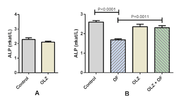
Several studies indicate the influence of olanzapine on bone metabolism; however, the results are contradictory. We evaluated the effects of olanzapine on the Wnt/β-catenin signaling pathway, physiological alveolar bone turnover, and alveolar bone modeling due to an applied orthodontic force. Adult male rats (n=48) were treated with either olanzapine or a vehicle for 21 days; then 8 rats from each group were sacrificed and the rest were divided into 4 groups: control, appliance-only, olanzapine-only, and olanzapine-appliance. The rats in the appliance groups were mounted with a superelastic closed coil spring that maintained constant orthodontic force between molars and incisors. We studied the effects of olanzapine on physiological alveolar bone turnover on day 21 of the experiment, and on alveolar bone modeling due to orthodontic force on day 56. We determined tooth movement, alveolar bone volume, activity of bone-specific cells, serum alkaline phosphatase (ALP) activity, and gene expression levels of Wnt/β-catenin signaling target genes. During forced bone modeling, olanzapine increased osteoblast volume (P<0.0001) and ALP activity (P=0.0011) and decreased osteoclast volume (P<0.0001) and gene expression of the Wnt/β-catenin signaling target genes Fosl1, Axin2, and Dkk1(P=0.001, P=0.0076, and P=0.036, respectively), and the osteocyte markers Sost and Dmp1 (P=0.0432 and P=0.0021, respectively). Similar results were obtained during physiological alveolar bone turnover on day 21, when olanzapine downregulated the gene expression of osteocyte markers and Wnt/β-catenin signaling target genes. We concluded that olanzapine attenuated osteocyte maturation during forced bone modeling and physiological alveolar bone turnover, potentially through downregulation of the Wnt/β-catenin signaling pathway.

Downloads

Download data is not yet available.
Olanzapine decreased osteocyte maturation and Wnt/β-catenin signaling during loading of the alveolar bone in rats

Downloads

Additional Files

Published

2023-01-06

How to Cite

1.
Disha-Ibrahimi S, Furlani B, Drevenšek G, Hudoklin S, Marc J, Prodan Žitnik I, Sajovic J, Drevenšek M. Olanzapine decreased osteocyte maturation and Wnt/β-catenin signaling during loading of the alveolar bone in rats. Biomol Biomed [Internet]. 2023Jan.6 [cited 2023Mar.27];23(1):114-25. Available from: https://www.bjbms.org/ojs/index.php/bjbms/article/view/7523

Issue

Section

Molecular Biology

Introduction

Schizophrenia and manic episodes in bipolar disorder are treated by prescribing antipsychotic drugs, among them olanzapine as an atypical antipsychotic [12]. Adverse effects of the long-term use of such antipsychotic medication may lead to osteoporosis in response to hyperprolactinemia and to a decrease in bone mineral density (BMD) [3]. Due to possible effects on prolactin levels, it is generally accepted that antipsychotics are categorized as either prolactin-raising or prolactin-sparing [4–6]. Antipsychotics, besides affecting prolactin release, may influence bone metabolism through various mechanisms in the presence or absence of secondary hypogonadism, the modulation of serotonergic and adrenergic signaling, the involvement of sympathetic nervous system activity, and muscular function [7]. The effects of olanzapine on BMD have previously been studied, but the results are inconclusive [8–10]. The prolactin-sparing activity of olanzapine is favored due to the minimal loss of BMD [811]. Some authors found no correlation between olanzapine and BMD [12], but found that schizophrenia itself, rather than long-term use of olanzapine, correlates with BMD loss and osteoporosis in schizophrenia patients. Some studies mention that atypical antipsychotics exhibit their pharmacological effects at least partially through the modulation of the Wnt/β-catenin signaling pathway [13], which has been shown to play a crucial role in bone remodeling by regulating osteogenic differentiation [1415]. Wnt/β-catenin signaling was shown to activate in osteocytes when mechanical loading was applied to the bone, and can be assumed to constitute a major signaling pathway in bone [16]. Interestingly, the Wnt pathway has been implicated in the homeostatic control of the periodontal ligament (PDL) and the alveolar bone [17], and shown to force induced bone modeling during orthodontic tooth movement (OTM) [18–20]. There is some evidence that the protein kinase b/glycogen synthase kinase-3 and Wnt/β-catenin signaling pathways may play a pivotal role in the progression of schizophrenia and in the molecular mechanisms of the action of antipsychotics [21–23] and their metabolic side effects [24]. In a study on osteoblast cultures it was shown that atypical antipsychotics, including olanzapine, could cause osteoblast apoptosis by downregulation of the Wnt/β-catenin signaling pathway [25]. However, to our knowledge, no such research has been conducted in vivo, nor have the effects of olanzapine on the Wnt/β-catenin signaling pathway been studied in an example of forced bone modeling. Although many studies have indicated the effects of olanzapine on bone metabolism, none have investigated its effect on bone modeling due to orthodontic force. In the present study, we evaluated the effects of olanzapine on tooth movement, alveolar bone volume, the activity of bone specific cells, and Wnt/β-catenin signaling in the alveolar bone during physiological bone turnover and bone modeling due to orthodontic force.

Figure 1.: The protocol of the study. On the 21st day after the beginning of the experiment, the first sampling was carried out. The material needed for gene expression analysis and determining serum alkaline phosphatase activity was collected. The second sampling was carried out on the 56th day after the beginning of the experiment, the materials needed for bone histomorphometry, gene expression analysis, and serum ALP activity were obtained. OF: Appliance-only group; OLZ: Olanzapine-only group; OLZ+OF: Olanazapine-appliance group.

Materials and methods

Animals

Male Wistar rats (n = 48, 260–280 g) were used in the study, between 13 and 15 weeks of age. To avoid potential dysmetabolic changes, male rats were selected over female ones [25]. The animals were housed under standard laboratory conditions at a constant temperature (23 C–25 C) and humidity. A 12-h circadian cycle was established and maintained throughout the study. The rats were fed a laboratory rat chow diet (Teklad Global Rodent Diet 2016, Harlan Laboratories, An Venray, The Netherlands) and water ad libitum. The rat chow was available in its dry form or pre-soaked in water to enable easier food intake, as it was partially impaired due to the mounted orthodontic appliance [2627].

Study protocol and olanzapine administration

At the beginning of the experiment 24 animals were treated with olanzapine 2 mg/kg daily per os for 3 weeks (Krka d.d, Novo mesto, Slovenia) and 24 animals received a vehicle for the same period of time. On the 21st day, 16 rats (OLZ21; n = 8 and control 21 group; n = 8) were sacrificed and the remaining (n = 32) were divided into 4 groups: control 56 group (n = 8), olanzapine-only group (OLZ56; n = 8), appliance-only group (orthodontic force – OF56; n = 8), and olanzapine-appliance group (OLZ+OF56; n = 8). The animals in both appliance groups (the OF56 and OLZ+OF56 groups) were mounted with an orthodontic appliance and continued to receive olanzapine (OLZ+OF56 group) or a vehicle (OF56 group) daily for the next five weeks (Figure 1). The entire period of olanzapine administration lasted for 8 weeks (56 days). No orthodontic appliance was placed in the animals in the control groups. All the groups were weight-matched prior to the start of the olanzapine administration. The doses were selected to reflect therapeutic concentrations and had previously shown metabolic side effects [2829].

Orthodontic appliance

The rats from the OF56 and the OLZ-OF56 groups were equipped with an orthodontic appliance. To mount the appliance, the rats were placed under general anesthesia. Anesthesia was induced by 3 mg/kg body weight of thiopental (Tiopental, Pliva, Zagreb, Croatia), 67 mg/kg body weight of medetomidine hydrochloride (Domitor, Pfizer, Brooklyn, NY, USA), and 50 mg/kg body weight of ketamine (Bioketan, Vetoquinol Biowet, Gorzów Wielkopolski, Poland). The anesthetic mixture was injected intraperitoneally. A super-elastic closed coil spring (GAC Dentsply International, York, PA, USA), 0.15 mm in diameter and exerting a constant force of 25 cN, was placed between the first and second left maxillary molar and the incisors. A stainless-steel ligature was used to affix the spring in both groups [27]. A steel thread, to which the closed coil was attached, was placed around the first and second molars of one side of the rats’ jaw and through a hole drilled in the upper incisors of the other side. Great care was taken when drilling the hole laterally through the incisors to leave the pulp of the teeth intact, as previously described [27]. The orthodontic appliances were readjusted weekly to enable a constant amount and direction of force (Figure 2).

Figure 2.: A schematic view of the orthodontic appliance and examples of histomorphometric and tooth movement measurements carried out.Top of the figure: an illustration of the orthodontic appliance. This consisted of a superelastic closed coil, situated between the upper first and second molar of the left side and fixed there by a stainless-steel ligature. The coil was fixed to the incisors with surgical steel wire, placed through a hole drilled in the incisors, to enhance the fixation. The hole was drilled with round, a stainless-steel bur, just above the lateral gingival margin. Middle of the figure: in the three dashed-line circles, tissue samples are presented under 10-fold (left) and 40-fold (right) magnification. The areas of interest in the maxilla, with 3 molars (M1–3), are shown under 10-fold magnification. The dashed rhomboid area encompassing the M2 and M1 in the 10-fold magnification circle shows the area where the alveolar bone volume was determined, and the black arrow shows the direction of tooth movement. The dot-and-dash line areas, denoted by the green arrows within the larger rhomboid surface show the regions along the mesial and distal roots of the M2, where the osteoblast and osteoclast volumes were determined. The dashed-line circles pointed to by the green arrows show a 40-fold magnification examples of osteoclasts and osteoblasts. Bottom right of the figure: a sketch of the upper jaw of the rat, with denoted distance that was measured to assess tooth movement. Al.b: Alveolar bone; Ob: Osteoblast; Oc: Osteoclast; PDL: Periodontal ligament; T: Tooth.

Measurement of tooth movement

The measurements of tooth movement were made with a digital caliper (Wilson and Wolpert, Utrecht, The Netherlands) on the 21st and 56th days of the study, while the animals were under anesthesia [27]. The accuracy of the caliper used was ±0.01 mm. Tooth movement was measured through the distance between the mesial-most point of the left first molar of the maxilla, and the palatal-most point of the ipsilateral incisor. This measurement was done at the gingival level, in all the animal groups. Tooth movement was calculated by subtracting the distance between the teeth measured on the 56th day from the distance between the teeth measured on the 21st day. To ensure the reliability of the measurements, two independent investigators took two measurements each within a few minutes.

Bone histomorphometry

On the 21st day after beginning of olanzapine administration, 8 rats from the OZ21 and 8 from the control 21 group were sacrificed and their right maxillary bones prepared and used for the determination of the level of gene expression. All the remaining animals (n = 32) were sacrificed on the 56th day, and their maxillary bones with 3 molars obtained. The left maxillary bones of the control 56 group were prepared for histomorphometric analysis. In the group of rats with the fitted orthodontic appliances, only the left maxillary bones were collected, as these had the appliances installed. Eight were collected for histomorphometric analysis.

To determine the osteoclast and osteoblast volume density, bone histomorphometry was used in all four groups. The tissue specimens were prepared by fixing them with a 4% paraformaldehyde in a phosphate-buffered saline solution with a pH between 7.2 and 7.4, kept at 14 C for 24 h. Next, decalcification of the samples was carried out, by first submerging them in a solution of water and ethylenediamine tetraacetic acid (EDTA), at 14 C for 12 days. The samples were then dehydrated using progressively higher solutions of ethanol, at 50%, 70%, 90%, and 100% concentration, and then with xylol. Afterward, the samples were embedded in paraffin. Finally, sections were deparaffinized and stained with hematoxylin and eosin [30].

To carry out histomorphometry, a point-counting method was used. A cycloid grid system integral to the ocular of a light microscope (BX-60, Olympus, Tokyo, Japan) was used to this end [27]. At 10-fold magnification, the area of the alveolar bone was determined, expressed as the percentage of alveolar bone area versus the tissue area consisting of tooth. PDL, connective tissue, and bone marrow spaces were determined at 10-fold magnification as well. Additionally, the osteoclast and osteoblast areas were determined, using 40-fold magnification. The areas were defined as the alveolar bone area covered with osteoblasts or osteoclasts in comparison to the total alveolar bone area, where the cells were counted in the alveolar bone alongside the mesial and distal roots of the second molar [30]. As previously described [27], the alveolar bone area, osteoblast area, and osteoclast area determined in the 20 samples of each specimen were extrapolated to assess alveolar bone volume, osteoblast volume, and osteoclast volume (Figure 2).

Alkaline phosphatase measurement

Blood samples were obtained from the abdominal vena cava immediately before the euthanasia of the rats took place. The samples were then centrifuged and the serum isolated. The alkaline phosphatase (ALP) in the rat serum was measured using the IFCC method [31] with the commercially available kit ALP – AMP (Biosystems S.A., Spain), following the manufacturer’s instructions. Briefly, ALP catalyzes the transfer of a phosphate group from 4-nitrophenylphosphate to 2-amino-2-methyl-1-propanol (AMP), producing AMP-phosphate and 4-nitrophenol. The catalytic concentration of ALP was determined from the rate of 4-nitrophenol formation, measured at 405 nm, using a Beckman Coulter DU 730 spectrophotometer, and was referenced to a 154 mmol sodium chloride solution.

RNA isolation and semi-quantitative RT-PCR

The maxillary bones with all three molars and their PDLs were excised and immediately frozen in liquid nitrogen. To additionally prevent the RNA from degrading, RNaseZap reagent was used during the entire procedure. The specimens were mechanically powdered using a pestle and mortar while in liquid nitrogen, to make sure the samples remained frozen throughout the procedure. After powdering, each separate specimen was transferred into a 1.5 ml tube that contained 1 ml of Trizol (TRIzol Plus RNA Purification System; Life Technologies, Carlsbad, CA, USA) to prevent the RNA from degrading. The Trizol and powdered specimen suspension was further homogenized by the use of an ultrasound homogenizer, to improve the release of RNA from the cells of the specimens. Chloroform extraction and a PureLink RNA Mini Kit (Invitrogen, Carlsbad, CA, USA) were used to isolate the RNA. Once the RNA was isolated, its quantity and quality were assessed. For the quality check, two methods were used: spectrophotometry with a NanoDrop ND-1000 Spectrophotometer (Thermo Scientific, Waltham, Mass), and capillary electrophoresis (carried out with a Bioanalyzer 2100, Agilent Technologies, Santa Clara, CA, USA). The quantity of the RNA was measured via spectrophotometry, with the NanoDrop ND-1000. The criteria for the samples to be used in our study were a suitable concentration (greater than 100 ng/µL) and an integration number in excess of 5, as determined with the Bioanalyzer 2100.

Complementary DNA was synthesized using a total of 50 ng of RNA, with the Transcriptor High Fidelity complementary DNA synthesis kit, to which the RNase was added (Life Technologies). The reaction was carried out in accordance with the manufacturers’ recommendations, with a final volume of 50 l. Predesigned assays were used for the determination of cathepsin K, osteocalcin, Dmp1, Fosl1, Dkk1, Sost, and Axin2 gene expressions (TaqMan Gene Expression Assays, Applied Biosystems, Rn00580723_m1, Rn01455285_g1, Rn01450122_m1, Rn00564119_m1, Rn01501537_m1, Rn00577971_m1, and Rn00577441_m1, respectively). The cDNA was amplified and quantified using the Lightcycler 480 (Roche Diagnostics GmbH, Mannheim, Germany). Thermal cycling consisted of the following steps: initial cycling at 50 C for 2 min and at 95 C for 10 min, then 40 cycles at 95 C for 15 s and at 60 C for 1 min. Serial dilutions of a mix of a few samples were used to construct a standard curve. The data on cathepsin K, osteocalcin, Dmp1, Fosl1, Dkk1, Sost, and Axin2 were normalized to the GAPDH expression, used as an internal housekeeping gene, to exclude variations due to different inputs of the total mRNA to the reaction. The GAPDH expression was determined by using the forward primer 5’TGATTCTACCCACGGCAAGTT3’, reverse primer 5’TGATGGGTTTCCCATTGATGA3’ and HOT FIREPol EvaGreen qPCR Mix Plus (no ROX) (Solis BioDyne, Tartu, Estonia), following the master mix protocol. A melting curve analysis was performed to verify the specificity of the amplification. All the reactions were carried out in duplicate, for standard samples and for the samples obtained from the four groups, with the values obtained for each reaction averaged afterwards. After normalization to the expression of GAPDH, a time-course-dependent consensus profile of gene expression was observed for all tested transcripts.

The osteocalcin gene expression level was used to determine osteoblast activity; the cathepsin K gene expression level as a marker of osteoclast activity; and the dentin matrix acidic phosphoprotein 1 (Dmp1) and sclerostin (Sost) gene expressions were used as osteocyte maturation markers [16]. The Wnt/β-catenin signaling pathway was evaluated by measuring the gene expression levels of the following β-catenin target genes: axis inhibition protein 2 (Axin2), dickkopf-related protein 1 (Dkk1), and fos-related antigen 1 (Fosl1) [32–36].

Ethical statement

All the animal procedures and the study protocol were approved by the Ethics Committee for Animal Experiments of the Administration of the Republic of Slovenia for Food Safety, Veterinary Sector and Plant Protection (No. 34401-62/2008/20) and complied with the guiding principles in “The Care and Use of Animals.”

Statistical analysis

Descriptive statistics [mean and standard error of the mean (SEM)] were calculated for each parameter (amount of tooth movement, alveolar bone volume, osteoblast and osteoclast volume, serum ALP activity, and gene expression levels) for all the animals in all the groups. Interexaminer reliability for tooth movement measurements was tested by the intraclass correlation coefficient (ICC), which was used to assess systematic bias. Within and between group comparisons were made for all measurements using the analysis of variance (ANOVA) and Tukey’s post hoc test in GraphPad Prism (GraphPad Software, San Diego, CA, USA). Comparisons between the OLZ21 and the control 21 group for all measured variables on day 21 of the experiment were performed using the unpaired t-test. Values of P < 0.05 were considered statistically significant. In the results, not all the groups contained the initial number of rats (n = 8 per group); due to a few troubled experiments some of the samples had to be excluded. A minimum of five samples per group were included in the final statistical analysis.

Results

Tooth movement

Rat molars physiologically migrate in the distal direction continuously during the animal’s lifetime—a phenomenon called distal drift, which results in an enlarged distance between molars and incisors [37]. The physiological distal drift of the molars, in the absence of OTM, was more pronounced in the OLZ56 group, but the difference was not statistically significant (P = 0.056). Moreover, there was no significant difference in the amount of OTM between the OF56 group and the OLZ+OF56 group (Figure 3). The overall mean value of the ICC for the measurement of the tooth movement was 0.935.

Figure 3.: Tooth movement on the 56th day of the experiment. No significant differences in distal drift were found between the studied groups. The histograms depict group means, while the error bars show the group SEM. Statistical testing was carried out with the use of one-way ANOVA and Tukey’s post hoc tests (n = 5–8 animals per group). OF: Appliance-only group; OLZ: Olanzapine-only group; OLZ+OF: Olanazapine-appliance group; SEM: Standard error of the mean.

Histomorphometric analysis

The histomorphometric analysis after 56 days of the experiment showed that there were no significant differences between the groups in alveolar bone volume. In the OLZ+OF56 group, a significantly higher osteoblast volume was observed than in the OLZ56 group and the OF56 group (both P < 0.0001). Osteoclast volume was significantly higher in the OF56 group compared to the OLZ+OF56 and the control group (both P < 0.0001) (Figure 4).

Figure 4.: The results of the histomorphometric analysis of the maxillary bone specimens. (A) No significant difference between the groups was found on the 56th day of the experiment. (B) Osteoblast volume was significantly greater in the group receiving olanzapine and the orthodontic appliance (OLZ-OF56, OLZ-OF in figure) (P < 0.0001), when compared to the appliance-only group (OF56, OF in figure). (C) The control 56 group (control in figure, P < 0.0001) and the group receiving olanzapine and the orthodontic appliance (OLZ-OF, P < 0.0001) exhibited significantly lower osteoclast volume on the 56th day of the experiment. The data is presented as group means, with the error bars showing the SEM of the group. n = 5–8 animals per group. SEM: Standard error of the mean.

ALP activity in serum

No significant difference in the ALP activity in the rat serum was observed between the OLZ21 group and its control on the 21st day. However, on the 56th day, ALP activity was significantly higher in the OLZ+OF56 group than in the OF56 group (P = 0.0011), and in the control 56 group compared to the OF56 group (P < 0.0001) (Figure 5).

Figure 5.: The results of the measurements of ALP activity in the serum of the rats. (A) No significant difference between the control 21 group (control in figure) and the olanzapine-only group (OLZ21, OLZ in figure) was observed on the 21st day of the experiment. (B) On the 56th day of the experiment, the olanzapine-appliance group 56 (OLZ+OF56, OLZ+OF in figure) exhibited significantly higher ALP activity than the appliance only group (OF56, OF in figure, P < 0.0011). The appliance-only group also exhibited significantly lower ALP activity levels when compared to the control group (P < 0.0001). The histograms depict group means, while the error bars show the group SEM. Statistical testing was carried out with the use of the unpaired t-test (A) and one-way ANOVA with Tukey’s post hoc tests (B) (n = 5–8 animals in each group). ALP: Alkaline phosphatase; SEM: Standard error of the mean.

Gene expression levels on day 21 of the experiment

The results of the analysis of the gene expression levels of osteocalcin, cathepsin K, DMP1, and SOST on day 21 of the experiment are presented in Figure 6A. We observed significantly downregulated expression levels of Dmp1 and Sost in the OLZ21 compared to the control 21 group (P = 0.0003 and P = 0.0228, respectively), but no significant differences were observed in the expression levels of the other two bone markers (Figure 6A). Figure 6B shows the measurements of the gene expression levels of the Wnt/β-catenin signaling target genes. The gene expression levels of Dkk1, Axin2, and Fosl1 were all decreased in the OLZ21 group in comparison to the control 21 group (P = 0.0024, P = 0.0011, and P < 0.0001, respectively).

Figure 6.: The levels of gene expression on the 21st day of the experiment. (A) Gene expression levels of bone cell activity markers. The osteocyte markers Dmp1 and Sost were significantly downregulated in the olanzapine-only group (OLZ21, OLZ in figure) compared to the control 21 group (control in figure, P = 0.0003 and P = 0.0228, respectively). No significant differences were observed in the markers of osteoblast and osteoclast activity. (B) Gene expression levels of the Wnt/β-catenin signaling target genes. Gene expression levels of Dkk1, Axin2, and Fosl1 were all significantly lower in the olanzapine-only group (OLZ21) compared to the control 21 group (P = 0.0024, P = 0.0011, and P < 0.0001). The data are presented as mean ± SEM and analyzed by the unpaired t-test (n = 5–8 animals in each group). DMP1: Dentin matrix acidic phosphoprotein 1; SOST: Sclerostin; DKK1: Dickkopf-related protein 1; AXIN2: Axis inhibition protein 2; FOSL1: Fos-related antigen 1; SEM: Standard error of the mean.

Gene expression levels on day 56 of the experiment

The gene expression levels of osteoblast, osteoclast, and osteocyte markers are presented in Figure 7. On day 56 of the experiment, a higher gene expression level of Cathepsin K in the OLZ+OF56 group than in the OF56 group was observed (P = 0.0497). The gene expression levels of both osteocyte markers, Dmp1 and Sost, were significantly downregulated in the OLZ+OF56 group compared to the OF56 group (P = 0.0021 and P = 0.0432, respectively). However, there were no significant differences in the gene expression levels of osteocalcin between the groups (Figure 7). On day 56 of the experiment, the gene expression levels of the Wnt/β-catenin target genes, Axin2 and Fosl1, were significantly higher in the OF56 group compared to the control group, indicating the upregulation of Wnt/β-catenin signaling under the influence of the orthodontic force (P = 0.0215 and P = 0.0009, respectively). However, the gene expression levels of Dkk1, Axin2, and Fosl1 were significantly downregulated in the OLZ+OF56 group compared to the appliance-only OF56 group (P = 0.036, P = 0.0076, and P = 0.001, respectively) (Figure 8).

Figure 7.: Osteoblast, osteoclast, and osteocyte gene expression marker levels on the 56th day of the experiment. (A) No significant differences in the osteocalcin expression level were found between the groups. (B) The expression level of cathepsin K was higher in the olanzapine appliance group (OLZ + OF56, OLZ+OF in figure) when compared to the appliance-only group (OF56, OF in figure) (P = 0.0497). (C) The Dmp1 gene expression level was significantly downregulated in the olanzapine-appliance group (OLZ+OF56) compared to the appliance-only (OF56) group (P = 0.0021). (D) The SOST gene expression level was significantly downregulated in the olanzapine-appliance group (OLZ+OF56) compared to the appliance-only group (OF56) (P = 0.0432). The data are presented as mean ± SEM and analyzed by the one-way ANOVA and Tukey’s post hoc tests (n = 5–8 animals in each group). DMP1: Dentin matrix acidic phosphoprotein 1; SOST: Sclerostin; SEM: Standard error of the mean.
Figure 8.: Gene expression levels of the Wnt/β-catenin target genes on the 56th day of the experiment. The gene expression levels of Dkk1 (A), Axin2 (B), and Fosl1 (C) were all significantly downregulated in the olanzapine-appliance group (OLZ+OF56, OLZ+OF in figure) in comparison to the control group (P = 0.036, P = 0.0076, P = 0.001, respectively). Interestingly, the gene expression levels of Axin2 and Fosl1 were significantly upregulated in the appliance-only group (OF56, OF in figure) compared to the control (P = 0.0215 and P = 0.0009). This result indicates that under forced-bone modeling, the upregulation of the Wnt/β-catenin pathway represents an important biological mechanism. The data are presented as mean ± SEM and analyzed by the one-way ANOVA and Tukey’s post hoc tests (n = 5–8 animals in each group). DKK1: Dickkopf-related protein 1; AXIN2: Axis inhibition protein 2; FOSL1: fos-related antigen 1; SEM: Standard error of the mean.

Discussion

The results of the present study indicate that during forced bone modeling and physiological turnover of the alveolar bone, olanzapine downregulated the Wnt/β-catenin signaling pathway and osteocyte maturation. Moreover, under conditions of forced bone modeling olanzapine induced higher osteoblast volume and serum ALP activity, and lower osteoclast volume. But despite these histological and molecular effects, olanzapine did not cause significant changes in OTM, physiological distal drift, or alveolar bone volume. To study the role of olanzapine in forced bone modeling, we used an animal model of OTM established previously [2730]. OTM comprises of three stages [38], the first two lasting less than three weeks [39], and the third, late phase which represents the beginning of bone modeling and the end of the phase of necrotic tissue degradation. This process is mainly mediated by osteoblasts and osteoclasts [40]. During OTM, the process of the resorption of the alveolar bone by osteoclasts takes place. This creates space for tooth movement, while on the other side of the tooth activated osteoblasts form new bone that fills the space created by tooth movement [40]. In the late phase of OTM, which begins around 2–4 weeks after the force has been applied, tooth movement proceeds linearly through highly coordinated bone resorption and bone formation [40], and therefore presents an appropriate model to study forced bone modeling. The gene expression level for several biological molecules is quite constant in this phase of OTM, and it can be modulated by several drugs that are used clinically [27]. Selection of the time duration of the experiment must exceed four weeks to enable constant gene expression. Despite constant gene expression, changes in bone metabolism might not be significantly expressed in histological samples.

The Wnt/β-catenin signaling pathway is a key modulator of the homeostasis of the bone [41]. Wnt/β-catenin signaling has been implicated in alveolar bone homeostasis [17] and forced bone modeling during OTM [1920]. In the present study, olanzapine significantly downregulated the gene expression levels of the Wnt/β-catenin signaling direct target genes, Axin2, Fosl1, and Dkk1, during physiological bone turnover (day 21) and during bone modeling due to orthodontic force (day 56). The inactivation of the β-catenin in mesenchymal progenitors and osteoblast precursors in the early stages of osteoblastogenesis prevents osteoblast differentiation, which results in a lack of mature osteoblasts and in the absence of bone formation [41]. But in the later stages, Wnt/β-catenin signaling inactivation in mature osteoblasts and osteocytes does not seem to affect osteoblast differentiation or bone formation, but to modulate osteoclastogenesis [164142]. In a study by Tu et al., the activation of Wnt/β-catenin signaling in osteocytes stimulated osteoblast-to-osteocyte differentiation, upregulated the expression of osteocytic markers (DMP1 and SOST), and increased the number of osteoclasts [41]. Furthermore, Wnt/β-catenin signaling has been shown to be a major pathway necessary for bone modeling, as it is activated in osteocytes when a mechanical load is applied [1643]. This is confirmed in the example of mice which exhibit a haploinsufficiency of β-catenin in osteocytes, where a defective anabolic response to bone loading can be observed [44]. We observed significant upregulation of Axin2 and Fosl1 gene expression levels in the appliance-only group compared to the control group. This indicates that the Wnt/β-catenin signaling pathway was upregulated during forced bone modeling of the alveolar bone, which represents an important physiological response to mechanical loading [1945]. However, we did not observe the same anabolic response to orthodontic force in groups treated with olanzapine. Olanzapine significantly decreased the gene expression levels of the Wnt/β-catenin target genes in the olanzapine-only group versus the control group (day 21) and the olanzapine-appliance group versus the appliance-only group (day 56). These observations reveal that olanzapine downregulated Wnt/β-catenin signaling in the alveolar bone during physiological alveolar bone turnover and forced bone modeling (Figure 9).

Figure 9.: Proposed mechanism for the effects of olanzapine on forced bone modeling. Osteocyte maturation occurs in several stages, including osteoid osteocyte, early osteocyte, and mature osteocyte (68). During this process cells express different molecular markers. (A) Mechanical loading of the alveolar bone due to orthodontic force resulted in the upregulation of the Wnt/β-catenin signaling and gene expression of osteocyte marker Dmp1. (B) Olanzapine attenuated osteocyte maturation, indicated by downregulated Dmp1 and Sost gene expression levels, and Wnt/β-catenin signaling during forced bone modeling in the alveolar bone. Olanzapine also increased osteoblast volume and serum ALP activity, potentially due to a slower osteoblast-to-osteocyte transition. DMP1: Dentin matrix acidic phosphoprotein 1; SOST: Sclerostin; ALP: Alkaline phosphatase.

Olanzapine attenuated osteocyte maturation, evidenced by the downregulated gene expression levels of the osteocytic markers, Dmp1 and Sost, during physiological bone turnover and forced modeling of the alveolar bone. SOST is strongly expressed by alveolar osteocytes [4647], and its expression is limited to mature (terminally differentiated) osteocytes [4849]. The gene expression of DMP1 normally increases as osteocyte differentiation progresses to more mature osteocytes, and it is considered a marker of both early and mature osteocytes [50]. Mechanical loading has been shown to stimulate DMP1 gene expression in the osteocytes of the alveolar bone [51], and a similar observation was made in the present study, where orthodontic force significantly upregulated the Dmp1 gene expression level. Further, DMP1 is necessary for proper osteocyte maturation, as indicated by mice lacking DMP1, which exhibited increased osteoblastic marker expression levels, e.g., ALP and osteocalcin, and low osteocytic Sost expression [52]. We found increased serum ALP activity and osteoblast volume, but downregulated gene expression levels of osteocytic markers in the olanzapine-appliance group compared to the appliance-only group. Increased osteoblast volume and maintained osteoblast activity, along with significantly downregulated gene expression levels of the osteocytic markers, suggest that olanzapine attenuated osteocyte maturation, without negatively affecting osteoblast proliferation or differentiation during forced bone modeling (Figure 9). The increased osteoblast volume is related to increased osteoblast proliferation and presence of early immature osteoblasts when ALP is also normally expressed/produced [53]. However, the osteocalcin gene is mainly expressed in mature osteoblasts that are involved in the last step of bone remodeling, i.e., collagen mineralization. Our results show that olanzapine stimulated the osteoblast proliferation but had no effect on mature osteoblasts and/or on collagen mineralization.

Lower osteoclast volume in the olanzapine-appliance group compared to the appliance-only group could be a consequence of an osteocytic maturational defect. Osteocytes produce two factors that promote the proliferation and survival of osteoclasts: receptor activator of nuclear factor kappa-b ligand (RANKL) and macrophage colony-stimulating factor (M-CSF) [5455]. Mice lacking M-CSF in osteocytes/late osteoblasts showed impaired bone remodeling with decreased bone formation and resorption [56]. Osteocytes are thus an important source of M-CSF, which maintains osteocyte mediated bone remodeling [56]. Moreover, some studies mention that an increase in RANKL depends on SOST [41], which was substantially downregulated in the olanzapine-treated rats in our study. Therefore, it is reasonable to speculate that, due to an osteocytic maturational defect under olanzapine treatment, the osteocytes produced less RANKL and/or MCSF, ultimately resulting in a lower osteoclast volume, which was observed in the olanzapine appliance group.

The discrepancy between osteoclast volume and cathepsin K gene expression is similar to discrepancies between osteoblast volume and osteocalcin gene expression. Similar to osteocalcin in osteoblasts, cathepsin K in osteoclasts represents an effective, but not regulative protein. It is directly involved in bone tissue degradation/resorption and is expressed in mature osteoclasts. Therefore, olanzapine can exert effects on cell proliferation, expressed as decreased volume of osteoclasts, but has no effect on mature cells. Most probably olanzapine does not influence the specific gene expression of cathepsin K in mature osteoclasts. From our results, we can conclude that olanzapine possibly exerts an effect on osteoclast proliferation, but has no effect on cathepsin K gene expression in mature cells [57].

It is also possible that olanzapine influenced bone metabolism through direct modulation of its target receptors on osteoblasts and osteoclasts [58]. Olanzapine is a potent antagonist of serotonin 5HT2A and 5HT6 receptors [5960], and it has been reported that pharmacological modulation of serotonin receptors affected osteoblastogenesis and osteoclastogenesis [61].

In addition, olanzapine is a partial histamine H1 receptor antagonist [62], and in our previous study, we reported that cetirizine, an H1 receptor antagonist, downregulated osteoclast volume during OTM [63]. Olanzapine could therefore partially contribute to the observed phenomena through antagonistic effects on serotonin and histamine receptors expressed on bone-specific cells. However, due to olanzapine’s numerous pharmacological targets it is difficult to pinpoint which molecular mechanism was predominant.

ALP measurement as a marker of bone formation is less specific than osteocalcin or procollagen 1 pro-peptides (P1NP). Bone mass and bone strength cannot be directly related to these biomarkers and the results cannot be interpolated to systemic changes in the skeleton. In humans, biochemical bone turnover markers should be used with caution and should always be supplemented by dual energy X-ray absorptiometry (DEXA) [64]. However, the data on both is only a surrogate marker of bone quality. The best information on bone quality is given by bone tissue structure analyses obtained by quantitative computer tomography (qCT) or histomorphometry, so animal models have an important advantage and offer trustworthy bone quality examination. In the present study, the results of bone histomorphometry are shown, and additional bone markers in the serum of animals would not substantially improve the reflection of the bone remodeling process. ALP measured in serum is a bone marker that is the most frequently used in animals, regardless of N-terminal propeptide of type I procollagen (PINP) and C-terminal telopeptide of type I collagen (CTX) being suggested by the International Federation of Clinical Chemistry and Institute of Biomedical Science in humans [64].

In the present study, osteoblast volume and serum ALP activity were increased more in the olanzapine-appliance group than in the appliance-only group, which initially does not seem to be consistent with downregulated Wnt/β-catenin signaling in the olanzapine-appliance group. However, it is probable that olanzapine attenuated Wnt/β-catenin signaling in the later stages of bone formation, during osteoblast-to-osteocyte transition and osteocyte maturation. Osteocytes are much more abundant cells than osteoblasts [50], thus having a greater contribution to the overall gene expression levels of the Wnt/β-catenin target genes. During osteoblast-to-osteocyte transition, only some osteoblasts become osteocytes [65]. It is therefore possible that due to downregulated Wnt/β-catenin signaling during osteoblast-to-osteocyte transition and osteocyte maturation, fewer osteoblast cells adopted an osteocyte fate, resulting in increased osteoblast volume and serum ALP activity. This is consistent with the significantly decreased expression of the osteocyte maturation markers, Sost and Dmp1, in the alveolar bone (Figure 9). Conclusively, in the present study, we have shown that olanzapine downregulated the Wnt/β-catenin signaling pathway, as well as interfered with osteocyte maturation during physiological bone turnover and forced bone modeling in the alveolar bone.

Conclusion

Olanzapine downregulated the gene expression levels of the osteocyte markers, Dmp1 and Sost, and the Wnt/β-catenin signaling target genes, Fosl1, Axin2, and Dkk1, in the alveolar bone. Treatment with olanzapine could have attenuated osteocyte maturation, potentially through downregulation of Wnt/β-catenin signaling, during both physiological alveolar bone turnover and forced bone modeling of the alveolar bone in rats.

Ackknowledgments

We would like to thank Manja Cedilink and Nada Pavlica Dubaric for their assistance in carrying out the laboratory measurements. This work was supported by the P3-0293 research program financed by the Slovenian Research Agency and Ad futura—Slovenian human resources development and scholarship fund.

Conflicts of interest: Authors declare no conflicts of interest.

Funding: This work was supported by the P3-0293 research program financed by the Slovenian Research Agency and Ad futura—a Slovenian human resources development and scholarship fund.

References

  1. , , , , , (). Comparative efficacy and tolerability of 32 oral antipsychotics for the acute treatment of adults with multi-episode schizophrenia: a systematic review and network meta-analysis.. Lancet. https://doi.org/10.1016/S0140-6736(19)31135-3
  2. , , , , (). New antipsychotic medications in the last decade.. Curr Psychiatry Rep. https://doi.org/10.1007/s11920-021-01298-w
  3. , , (). Hyperprolactinemia and bone mineral density: the potential impact of antipsychotic agents.. Psychoneuroendocrinology. https://doi.org/10.1016/s0306-4530(02)00129-4
  4. , , (). Effects of antipsychotics on bone mineral density in patients with schizophrenia: gender differences.. Clin Psychopharmacol Neurosci. https://doi.org/10.9758/cpn.2016.14.3.238
  5. , , , , , (). Effects of long-term prolactin-raising antipsychotic medication on bone mineral density in patients with schizophrenia.. Br J Psychiatry. https://doi.org/10.1192/bjp.184.6.503
  6. , , , , , (). Bone mineral density in schizophrenia: an update of current meta-analysis and literature review under guideline of PRISMA.. Medicine (Baltimore). https://doi.org/10.1097/MD.0000000000001967
  7. , , , , , (). Possible mechanisms for the skeletal effects of antipsychotics in children and adolescents.. Ther Adv Psychopharmacol. https://doi.org/10.1177/2045125313487548
  8. , , , , , (). Risperidone, but not olanzapine, decreases bone mineral density in female premenopausal schizophrenia patients.. J Clin Psychiatry. https://doi.org/10.4088/jcp.v64n0704
  9. , (). Bone mineral density changes over a year in young females with schizophrenia: relationship to medication and endocrine variables.. Schizophr Res. https://doi.org/10.1016/j.schres.2007.01.0
  10. , (). The role of neurotrophic factors in adult hippocampal neurogenesis, antidepressant treatments and animal models of depressive-like behavior.. Behav Pharmacol. https://doi.org/10.1097/FBP.0b013e3282ee2aa8
  11. , , , , , (). The atypical anti-psychotic clozapine decreases bone mass in rats in vivo.. Schizophr Res. https://doi.org/10.1016/j.schres.2010.11.024
  12. , , , , , (). Effects of olanzapine on bone mineral density, glucose, and lipid metabolism in schizophrenia patients.. Int J Endocrinol. https://doi.org/10.1155/2019/1312804
  13. , , , , (). Study of music effect on mental stress relief based on heart rate variability.. In: 2018 IEEE Asia Pacific Conference on Circuits and Systems, APCCAS 2018. Institute of Electrical and Electronics Engineers Inc.. https://doi.org/10.1109/APCCAS.2018.8605674
  14. , (). Mechanotransduction in bone repair and regeneration.. FASEB J.. https://doi.org/10.1096/fj.10-157370
  15. , (). New insights into Wnt-Lrp5/6-β-catenin signaling in mechanotransduction.. Front Endocrinol (Lausanne). https://doi.org/10.3389/fendo.2014.00246
  16. , , , , , (). Osteocyte Wnt/β-catenin signaling is required for normal bone homeostasis.. Mol Cell Biol. https://doi.org/10.1128/MCB.01428-09
  17. , , , , (). Alveolar bone turnover and periodontal ligament width are controlled by Wnt.. J Periodontol. https://doi.org/10.1902/jop.2014.140286
  18. , , , , , (). Biological events in periodontal ligament and alveolar bone associated with application of orthodontic forces.. Sci World J. https://doi.org/10.1155/2015/876509
  19. , , , , , (). Wnt5a mediated canonical Wnt signaling pathway activation in orthodontic tooth movement: possible role in the tension force-induced bone formation.. J Mol Histol. https://doi.org/10.1007/s10735-016-9687-y
  20. , , , , (). Expression of Wnt3a, Wnt10b, β-catenin and DKK1 in periodontium during orthodontic tooth movement in rats.. Acta Odontol Scand. https://doi.org/10.3109/00016357.2015.1090011
  21. , , (). Roles of the Akt/GSK-3 and Wnt signaling pathways in schizophrenia and antipsychotic drug action.. Am J Psychiatry. https://doi.org/10.1176/appi.ajp.2009.08121873
  22. , , , (). Antipsychotics alter the protein expression levels of β-catenin and GSK-3 in the rat medial prefrontal cortex and striatum.. Biol Psychiatry. https://doi.org/10.1016/j.biopsych.2004.11.036
  23. , , (). An emerging role for Wnt and GSK3 signaling pathways in schizophrenia.. Clin Genet. https://doi.org/10.1111/cge.12111
  24. , , , , , (). The Wnt signaling pathway effector TCF7L2 mediates olanzapine-induced weight gain and insulin resistance.. Front Pharmacol. https://doi.org/10.3389/fphar.2018.00379
  25. , , , , , (). Atypical antipsychotics induce human osteoblasts apoptosis via Wnt/β-catenin signaling.. BMC Pharmacol Toxicol. https://doi.org/10.1186/s40360-019-0287-9
  26. , , , (). Effects of endothelin antagonist tezosentan on orthodontic tooth movement in rats.. Am J Orthod Dentofac Orthop. https://doi.org/10.1016/j.ajodo.2005.12.016
  27. , , , , (). The endothelin system mediates bone modeling in the late stage of orthodontic tooth movement in rats.. Bone. https://doi.org/10.1016/j.bone.2008.06.012
  28. , , , , (). A parametric analysis of olanzapine-induced weight gain in female rats.. Psychopharmacology (Berl).. https://doi.org/10.1007/s00213-005-2224-4
  29. , , , (). Antipsychotic dosing in preclinical models is often unrepresentative of the clinical condition: a suggested solution based on in vivo occupancy.. J Pharmacol Exp Ther. https://doi.org/10.1124/jpet.102.046987
  30. , , , , , (). Bone remodeling during orthodontic tooth movement in rats with type 2 diabetes.. Am J Orthod Dentofac Orthop. https://doi.org/10.1016/j.ajodo.2015.05.031
  31. , , , , , (). IFCC primary reference procedures for the measurement of catalytic activity concentrations of enzymes at 37 C. Part 9: reference procedure for the measurement of catalytic concentration of alkaline phosphatase International Federation of Clinical Chemistry and Laboratory Medicine (IFCC) Scientific Division, Committee on Reference Systems of Enzymes (C-RSE) 1.. Clin Chem Lab Med. https://doi.org/10.1515/CCLM.2002.124
  32. , , , , , (). FGF-20 and DKK1 are transcriptional targets of β-catenin and FGF-20 is implicated in cancer and development.. EMBO J. https://doi.org/10.1038/sj.emboj.7600460
  33. , , , , , (). The Wnt antagonist DICKKOPF-1 gene is a downstream target of β-catenin/TCF and is downregulated in human colon cancer.. Oncogene. https://doi.org/10.1038/sj.onc.1208303
  34. , , , , , (). Wnt/β-Catenin/Tcf signaling induces the transcription of axin2, a negative regulator of the signaling pathway.. Mol Cell Biol. https://doi.org/10.1128/MCB.22.4.1172-1183.2002
  35. , , , , , (). Negative feedback loop of Wnt signaling through upregulation of conductin/axin2 in colorectal and liver tumors.. Langenbeck’s Arch Surg. https://doi.org/10.1128/MCB.22.4.1184-1193.2002
  36. , , , , , (). Target genes of β-catenin-T cell-factor/lymphoid-enhancer-factor signaling in human colorectal carcinomas.. Proc Natl Acad Sci USA. https://doi.org/10.1073/pnas.96.4.1603
  37. , , , , , (). Root resorption and ensuing cementum repair by Wnt/β-catenin dependent mechanism.. Am J Orthod Dentofacial Orthop. https://doi.org/10.1016/j.ajodo.2019.06.021
  38. , , (). The rat as a model for orthodontic tooth movement—a critical review and a proposed solution.. Eur J Orthod. https://doi.org/10.1093/ejo/26.5.483
  39. , , , (). Focal hyalinization during experimental tooth movement in beagle dogs.. Am J Orthod Dentofac Orthop. https://doi.org/10.1016/j.ajodo.2003.08.023
  40. , (). Cellular, molecular, and tissue-level reactions to orthodontic force.. Am J Orthod Dentofac Orthop. https://doi.org/10.1016/j.ajodo.2005.10.007
  41. , , , , , (). Osteocytes mediate the anabolic actions of canonical Wnt/β-catenin signaling in bone.. Proc Natl Acad Sci USA. https://doi.org/10.1073/pnas.1409857112
  42. , , , , , (). Canonical Wnt signaling in differentiated osteoblasts controls osteoclast differentiation.. Dev Cell. https://doi.org/10.1016/j.devcel.2005.02.017
  43. , (). Osteocytes, mechanosensing and Wnt signaling.. Bone. https://doi.org/10.1016/j.bone.2007.12.224
  44. , , , , , (). Deletion of a single β-catenin allele in osteocytes abolishes the bone anabolic response to loading.. J Bone Miner Res. https://doi.org/10.1002/jbmr.2064
  45. , , , , , (). Wnt/β-catenin signaling is a normal physiological response to mechanical loading in bone.. J Biol Chem. https://doi.org/10.1074/jbc.M602308200
  46. , , , , , (). Sclerostin and DKK1 inhibition preserves and augments alveolar bone volume and architecture in rats with alveolar bone loss.. J Dent Res. https://doi.org/10.1177/0022034518766874
  47. , , , , , (). Role of osteocyte-PDL crosstalk in tooth movement via SOST/cclerostin.. J Dent Res. https://doi.org/10.1177/0022034518771331
  48. , , , , , (). Sclerostin is a delayed secreted product of osteocytes that inhibits bone formation.. FASEB J. https://doi.org/10.1096/fj.05-4221fje
  49. , (). Intercellular cross-talk among bone cells: new factors and pathways.. Curr Osteoporos Rep. https://doi.org/10.1007/s11914-012-0096-1
  50. , (). Osteocytic signalling pathways as therapeutic targets for bone fragility.. Nat Rev Endocrinol. https://doi.org/10.1038/nrendo.2016.71
  51. , , , , , (). Mechanical loading stimulates dentin matrix protein 1 (DMP1) expression in osteocytes in vivo.. J Bone Miner Res. https://doi.org/10.1359/jbmr.2003.18.5.807
  52. , , , , , (). Loss of DMP1 causes rickets and osteomalacia and identifies a role for osteocytes in mineral metabolism.. Nat Genet. https://doi.org/10.1038/ng1905
  53. , , (). Regulation of gene expression in osteoblasts.. BioFactors. https://doi.org/10.1002/biof.72
  54. , , , (). Regulation of osteoclast function.. J Bone Miner Res. https://doi.org/10.1359/jbmr.1997.12.6.869
  55. , , , , , (). Matrix-embedded cells control osteoclast formation.. Nat Med. https://doi.org/10.1038/nm.2448
  56. , , , , , (). CSF-1 in osteocytes and late osteoblasts controls major aspects of bone remodeling.. J Bone Miner Res. https://doi.org/10.1002/jbm4.10080
  57. (). The regulation of cathepsin K gene expression.. Ann NY Acad Sci. https://doi.org/10.1196/annals.1346.018
  58. , , , , , (). Constitutively elevated blood serotonin is associated with bone loss and type 2 diabetes in rats.. PLoS One.. https://doi.org/10.1371/journal.pone.0150102
  59. , (). The in vivo effects of olanzapine and other antipsychotic agents on receptor occupancy and antagonism of dopamine D1, D2, D3, 5HT2A and muscarinic receptors.. Psychopharmacol. https://doi.org/10.1007/s002130050834
  60. , , , , , (). Potent antagonism of 5-HT(3) and 5-HT(6) receptors by olanzapine.. Eur J Pharmacol. https://doi.org/10.1016/s0014-2999(01)01399-1
  61. , , , , , (). Decreased osteoclastogenesis in serotonin-deficient mice.. Proc Natl Acad Sci USA. https://doi.org/10.1073/pnas.1117792109
  62. , , (). The role of histaminergic H1 and H3 receptors in food intake: a mechanism for atypical antipsychotic-induced weight gain?. Prog Neuro-Psychopharmacology Biol Psychiatry. https://doi.org/10.1016/j.pnpbp.2009.11.009
  63. , , , , , (). Effect of cetirizine, a histamine (H(1)) receptor antagonist, on bone modeling during orthodontic tooth movement in rats.. Am J Orthod Dentofac Orthop. https://doi.org/10.1016/j.ajodo.2009.11.013
  64. , , , , (). The laboratory rat as an animal model for osteoporosis research.. Comp Med.
  65. , , , (). . Skeletal tissue mechanics. Vol. 190.