Tumor suppressor maspin as a modulator of host immune response to cancer

Authors

  • Sijana H. Dzinic 1Department of Pathology, Wayne State University School of Medicine, Detroit, Michigan
  • Maria M. Bernardo Department of Pathology, Wayne State University School of Medicine, Detroit, Michigan, Tumor Biology and Microenvironment Program, Barbara Ann Karmanos Cancer Institute, Detroit, Michigan
  • Daniel S.M. Oliveira Department of Pathology, Wayne State University School of Medicine, Detroit, Michigan, Tumor Biology and Microenvironment Program, Barbara Ann Karmanos Cancer Institute, Detroit, Michigan, USA, Department of Urology, Wayne State University School of Medicine, Detroit, Michigan
  • Marian Wahba Department of Internal Medicine, Sinai Grace Hospital, Detroit Medical Center, Detroit, Michigan, USA
  • Wael Sakr Department of Pathology, Wayne State University School of Medicine, Detroit, Michigan, Tumor Biology and Microenvironment Program, Barbara Ann Karmanos Cancer Institute, Detroit, Michigan
  • Shijie Sheng Department of Pathology, Wayne State University School of Medicine, Detroit, Michigan, Tumor Biology and Microenvironment Program, Barbara Ann Karmanos Cancer Institute, Detroit, Michigan

DOI:

https://doi.org/10.17305/bjbms.2015.783

Keywords:

Anticancer immunity, immunosuppression, immunogenicity, cancer immunoediting, innate immunity, adaptive immunity, serine protease inhibitors, tumor suppressor, maspin, autoantigen, differentiation, epigenetic, cancer stem cells, cellular plasticity, tumor

Abstract

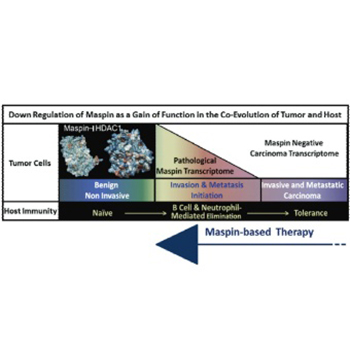
Despite the promising clinical outcome, the primary challenge of the curative cancer immunotherapy is to overcome the dichotomy of the immune response: tumor-evoked immunostimulatory versus tumor-induced immunosuppressive. The goal needs to be two-fold, to re-establish sustainable antitumor-cancer immunity and to eliminate immunosuppression. The successful elimination of cancer cells by immunosurveillance requires the antigenic presentation of the tumor cells or tumor-associated antigens and the expression of immunostimulatory cytokines and chemokines by cancer and immune cells. Tumors are heterogeneous and as such, some of the tumor cells are thought to have stem cell characteristics that enable them to suppress or desensitize the host immunity due to acquired epigenetic changes. A central mechanism underlying tumor epigenetic instability is the increased histone deacetylase (HDAC)-mediated repression of HDAC-target genes regulating homeostasis and differentiation. It was noted that pharmacological HDAC inhibitors are not effective in eliminating tumor cells partly because they may induce immunosuppression. We have shown that epithelial-specific tumor suppressor maspin, an ovalbumin-like non-inhibitory serine protease inhibitor, reprograms tumor cells toward better differentiated phenotypes by inhibiting HDAC1. Recently, we uncovered a novel function of maspin in directing host immunity towards tumor elimination. In this review, we discuss the maspin and maspin/HDAC1 interplay in tumor biology and immunology. We propose that maspin based therapies may eradicate cancer.

Downloads

Download data is not yet available.

References

Schreiber RD, Old LJ, Smyth MJ. Cancer immunoediting: Integrating immunity's roles in cancer suppression and promotion. Science 2011;331(6024):1565-70. http://dx.doi.org/10.1126/science.1203486.

Dunn GP, Old LJ, Schreiber RD. The three Es of cancer immunoediting. Annu Rev Immunol 2004;22:329-60. http://dx.doi.org/10.1146/annurev.immunol.22.012703.104803.

Mittal D, Gubin MM, Schreiber RD, Smyth MJ. New insights into cancer immunoediting and its three component phases – Elimination, equilibrium and escape. Curr Opin Immunol 2014;27:16-25. http://dx.doi.org/10.1016/j.coi.2014.01.004.

Tu E, Chia PZ, Chen W. TGFß in T cell biology and tumor immunity: Angel or devil? Cytokine Growth Factor Rev 2014;25(4):423-35. http://dx.doi.org/10.1016/j.cytogfr.2014.07.014.

Fridlender ZG, Sun J, Kim S, Kapoor V, Cheng G, Ling L, et al. Polarization of tumor-associated neutrophil phenotype by TGF-beta: "N1" versus "N2" TAN. Cancer Cell 2009;16(3):183-94. http://dx.doi.org/10.1016/j.ccr.2009.06.017.

De Smet C, Lurquin C, De Plaen E, Brasseur F, Zarour H, De Backer O, et al. Genes coding for melanoma antigens recognised by cytolytic T lymphocytes. Eye (Lond) 1997;11:243-8. http://dx.doi.org/10.1038/eye.1997.59.

Law RH, Zhang Q, McGowan S, Buckle AM, Silverman GA, Wong W, et al. An overview of the serpin superfamily. Genome Biol 2006;7(5):216. http://dx.doi.org/10.1186/gb-2006-7-5-216.

Gatto M, Iaccarino L, Ghirardello A, Bassi N, Pontisso P, Punzi L, et al. Serpins, immunity and autoimmunity: Old molecules, new functions. Clin Rev Allergy Immunol 2013;45(2):267-80. http://dx.doi.org/10.1007/s12016-013-8353-3.

Li Y, Wang LX, Pang P, Twitty C, Fox BA, Aung S, et al. Cross-presentation of tumor associated antigens through tumor-derived autophagosomes. Autophagy 2009;5(4):576-7. http://dx.doi.org/10.4161/auto.5.4.8366.

Vidalino L, Doria A, Quarta SM, Crescenzi M, Ruvoletto M, Frezzato F, et al. SERPINB3 expression on B-cell surface in autoimmune diseases and hepatitis C virus-related chronic liver infection. Exp Biol Med (Maywood) 2012;237(7):793-802. http://dx.doi.org/10.1258/ebm.2012.012024.

El-Rachkidy RG, Young HS, Griffiths CE, Camp RD. Humoral autoimmune responses to the squamous cell carcinoma antigen protein family in psoriasis. J Invest Dermatol 2008;128(9):2219-24. http://dx.doi.org/10.1038/jid.2008.71.

Chokshi NY, Sicherer SH. Molecular diagnosis of egg allergy: An update. Expert Rev Mol Diagn 2015;15(7):895-906. http://dx.doi.org/10.1586/14737159.2015.1041927.

Caubet JC, Kondo Y, Urisu A, Nowak-Wegrzyn A. Molecular diagnosis of egg allergy. Curr Opin Allergy Clin Immunol 2011;11(3):210-5. http://dx.doi.org/10.1097/ACI.0b013e3283464d1b.

Scambia G, Benedetti Panici P, Foti E, Amoroso M, Salerno G, Ferrandina G, et al. Squamous cell carcinoma antigen: Prognostic significance and role in the monitoring of neoadjuvant chemotherapy response in cervical cancer. J Clin Oncol 1994;12(11):2309-16.

Turato C, Simonato D, Quarta S, Gatta A, Pontisso P. MicroRNAs and SerpinB3 in hepatocellular carcinoma. Life Sci 2014;100(1):9-17. http://dx.doi.org/10.1016/j.lfs.2014.01.073.

Cacoub P, Frémeaux-Bacchi V, De Lacroix I, Guillien F, Kahn MF, Kazatchkine MD, et al. A new type of acquired C1 inhibitor deficiency associated with systemic lupus erythematosus. Arthritis Rheum 2001;44(8):1836-40. http://dx.doi.org/10.1002/1529-0131(200108)44:8<1836::AID-ART321>3.0.CO;2-Y.

Frazer JK, Jackson DG, Gaillard JP, Lutter M, Liu YJ, Banchereau J, et al. Identification of centerin: A novel human germinal center B cell-restricted serpin. Eur J Immunol 2000;30(10):3039-48. http://dx.doi.org/10.1002/1521-4141(200010)30:10<3039::AID-IMMU3039>3.0.CO;2-H.

Dzinic SH, Chen K, Thakur A, Kaplun A, Bonfil RD, Li X, et al. Maspin expression in prostate tumor elicits host anti-tumor immunity. Oncotarget 2014;5(22):11225-36. http://dx.doi.org/10.18632/oncotarget.2615.

Li X, Yin S, Meng Y, Sakr W, Sheng S. Endogenous inhibition of histone deacetylase 1 by tumor-suppressive maspin. Cancer Res 2006;66(18):9323-9. http://dx.doi.org/10.1158/0008-5472.CAN-06-1578.

Li X, Kaplun A, Lonardo F, Heath E, Sarkar FH, Irish J, et al. HDAC1 inhibition by maspin abrogates epigenetic silencing of glutathione S-transferase pi in prostate carcinoma cells. Mol Cancer Res 2011;9(6):733-45. http://dx.doi.org/10.1158/1541-7786.MCR-10-0505.

Bodenstine TM, Seftor RE, Khalkhali-Ellis Z, Seftor EA, Pemberton PA, Hendrix MJ. Maspin: Molecular mechanisms and therapeutic implications. Cancer Metastasis Rev 2012;31(3-4):529-51. http://dx.doi.org/10.1007/s10555-012-9361-0.

Cher ML, Biliran HR Jr, Bhagat S, Meng Y, Che M, Lockett J, et al. Maspin expression inhibits osteolysis, tumor growth, and angiogenesis in a model of prostate cancer bone metastasis. Proc Natl Acad Sci U S A 2003;100(13):7847-52. http://dx.doi.org/10.1073/pnas.1331360100.

Luo JL, Tan W, Ricono JM, Korchynskyi O, Zhang M, Gonias SL, et al. Nuclear cytokine-activated IKKalpha controls prostate cancer metastasis by repressing Maspin. Nature 2007;446(7136):690-4. http://dx.doi.org/10.1038/nature05656.

Li X, Chen D, Yin S, Meng Y, Yang H, Landis-Piwowar KR, et al. Maspin augments proteasome inhibitor-induced apoptosis in prostate cancer cells. J Cell Physiol 2007;212(2):298-306. http://dx.doi.org/10.1002/jcp.21102.

Jiang N, Meng Y, Zhang S, Mensah-Osman E, Sheng S. Maspin sensitizes breast carcinoma cells to induced apoptosis. Oncogene 2002;21(26):4089-98. http://dx.doi.org/10.1038/sj.onc.1205507.

Bernardo MM, Kaplun A, Dzinic SH, Li X, Irish J, Mujagic A, et al. Maspin expression in prostate tumor cells averts stemness and stratifies drug sensitivity. Cancer Res 2015;75(18):3970-9. http://dx.doi.org/10.1158/0008-5472.CAN-15-0234.

Bernardo MM, Meng Y, Lockett J, Dyson G, Dombkowski A, Kaplun A, et al. Maspin reprograms the gene expression profile of prostate carcinoma cells for differentiation. Genes Cancer 2011;2(11):1009-22. http://dx.doi.org/10.1177/1947601912440170.

Ning X, Shu J, Du Y, Ben Q, Li Z. Therapeutic strategies targeting cancer stem cells. Cancer Biol Ther 2013;14(4):295-303. http://dx.doi.org/10.4161/cbt.23622.

Machtens S, Serth J, Bokemeyer C, Bathke W, Minssen A, Kollmannsberger C, et al. Expression of the p53 and Maspin protein in primary prostate cancer: Correlation with clinical features. Int J Cancer 2001;95(5):337-42. http://dx.doi.org/10.1002/1097-0215(20010920)95:5<337::AID-IJC1059>3.0.CO;2-1.

Pierson CR, McGowen R, Grignon D, Sakr W, Dey J, Sheng S. Maspin is up-regulated in premalignant prostate epithelia. Prostate 2002;53(4):255-62. http://dx.doi.org/10.1002/pros.10107.

Lonardo F, Guan H, Dzinic S, Sheng S. Maspin expression patterns differ in the invasive versus lepidic growth pattern of pulmonary adenocarcinoma. Histopathology 2014;65(6):757-63. http://dx.doi.org/10.1111/his.12485.

Laresgoiti-Servitje E. A leading role for the immune system in the pathophysiology of preeclampsia. J Leukoc Biol 2013;94(2):247-57. http://dx.doi.org/10.1189/jlb.1112603.

Qi YH, Teng F, , Zhou Q, Liu YX, Wu JF, et al. Unmethylated-maspin DNA in maternal plasma is associated with severe preeclampsia. Acta Obstet Gynecol Scand 2015;94(9):983-8. http://dx.doi.org/10.1111/aogs.12691.

Taglauer ES, Gundogan F, Johnson KL, Scherjon SA, Bianchi DW. Chorionic plate expression patterns of the maspin tumor suppressor protein in preeclamptic and egg donor placentas. Placenta 2013;34(4):385-7. http://dx.doi.org/10.1016/j.placenta.2013.01.008.

Das UN. Cytokines, angiogenic, and antiangiogenic factors and bioactive lipids in preeclampsia. Nutrition 2015;31(9):1083-95. http://dx.doi.org/10.1016/j.nut.2015.03.013.

Besgen P, Trommler P, Vollmer S, Prinz JC. Ezrin, maspin, peroxiredoxin 2, and heat shock protein 27: Potential targets of a streptococcal-induced autoimmune response in psoriasis. J Immunol 2010;184(9):5392-402. http://dx.doi.org/10.4049/jimmunol.0903520.

Puga I, Cols M, Barra CM, He B, Cassis L, Gentile M, et al. Corrigendum: B cell-helper neutrophils stimulate the diversification and production of immunoglobulin in the marginal zone of the spleen. Nat Immunol 2014;15(2):205. http://dx.doi.org/10.1038/ni0214-205a.

Palanichamy A, Bauer JW, Yalavarthi S, Meednu N, Barnard J, Owen T, et al. Neutrophil-mediated IFN activation in the bone marrow alters B cell development in human and murine systemic lupus erythematosus. J Immunol 2014;192(3):906-18. http://dx.doi.org/10.4049/jimmunol.1302112.

Biliran H Jr, Sheng S. Pleiotrophic inhibition of pericellular urokinase-type plasminogen activator system by endogenous tumor suppressive maspin. Cancer Res 2001;61(24):8676-82.

Kaplun A, Dzinic S, Bernardo M, Sheng S. Tumor suppressor maspin as a rheostat in HDAC regulation to achieve the fine-tuning of epithelial homeostasis. Crit Rev Eukaryot Gene Expr 2012;22(3):249-58. http://dx.doi.org/10.1615/CritRevEukarGeneExpr.v22.i3.80.

Kelly TK, De Carvalho DD, Jones PA. Epigenetic modifications as therapeutic targets. Nat Biotechnol 2010;28(10):1069-78. http://dx.doi.org/10.1038/nbt.1678.

Schotterl S, Brennenstuhl H, Naumann U. Modulation of immune responses by histone deacetylase inhibitors. Crit Rev Oncog 2015;20(1-2):139-54. http://dx.doi.org/10.1615/CritRevOncog.2014012393.

Cao K, Wang G, Li W, Zhang L, Wang R, Huang Y, et al. Histone deacetylase inhibitors prevent activation-induced cell death and promote anti-tumor immunity. Oncogene 2015 http://dx.doi.org/10.1038/onc.2015.46.

Kroesen M, Gielen P, Brok IC, Armandari I, Hoogerbrugge PM, Adema GJ. HDAC inhibitors and immunotherapy; a double edged sword? Oncotarget 2014;5(16):6558-72. http://dx.doi.org/10.18632/oncotarget.2289.

Kim ES, Lee JK. Histone deacetylase inhibitors decrease the antigen presenting activity of murine bone marrow derived dendritic cells. Cell Immunol 2010;262(1):52-7. http://dx.doi.org/10.1016/j.cellimm.2009.12.007.

Tao R, de Zoeten EF, Ozkaynak E, Chen C, Wang L, Porrett PM, et al. Deacetylase inhibition promotes the generation and function of regulatory T cells. Nat Med 2007;13(11):1299-307. http://dx.doi.org/10.1038/nm1652.

Wong DJ, Rao A, Avramis E, Matsunaga DR, Komatsubara KM, Atefi MS, et al. Exposure to a histone deacetylase inhibitor has detrimental effects on human lymphocyte viability and function. Cancer Immunol Res 2014;2(5):459-68. http://dx.doi.org/10.1158/2326-6066.CIR-13-0188.

Tumor suppressor maspin as a modulator of host immune response to cancer

Downloads

Additional Files

Published

2015-10-25

How to Cite

1.
Dzinic SH, Bernardo MM, Oliveira DS, Wahba M, Sakr W, Sheng S. Tumor suppressor maspin as a modulator of host immune response to cancer. Biomol Biomed [Internet]. 2015Oct.25 [cited 2023Sep.23];15(4):1-6. Available from: https://www.bjbms.org/ojs/index.php/bjbms/article/view/783

Issue

Section

Reviews

CANCER IMMUNOTHERAPY, A DOUBLE-EDGED SWORD

According to the American Cancer Society, cancer is rapidly becoming a global pandemic and a leading cause of death worldwide (http://www.cancer.org/). It is estimated that there will be 1,658,370 new cancer cases diagnosed and 589,430 cancer deaths in the US in 2015. Recently, cancer immunotherapy is deemed to be one of the most promising avenues in cancer treatment, at least in part, by activating the natural host immune defense mechanisms. Of note, an ideal curative cancer immunotherapy should be both efficient and sustainable. During the past two decades, we have learned that the immune system in cancer plays a dual role: it can inhibit tumor growth by killing cancer cells, or it can have an immunosuppressive role thereby promoting tumor progression and metastasis [1]. In the co-evolution with a tumor, the host immunity is thought to undergo three phases: elimination, equilibrium, and escape [2]. During the elimination phase, the cancer immunoediting is directed towards antitumor immunity, and it mainly depends on the expression of tumor-associated antigens (TAAs) presented by major histocompatibility complex (MHC) class I molecules. Consequently, tumor cells are eliminated by adaptive immunity through the activation of cytotoxic CD8+ T-cells and NK-cells, and by innate immunity through the activation of antitumor macrophages (M1 subtype), antitumor neutrophils (N1 subtype), and granulocytes [3]. At the equilibrium phase, cancer cells may escape by repressing the host immunogenicity. For example, the accumulated genetic and epigenetic changes in tumor cells may result in reduction or loss of TAAs expression or MHC-I molecules thus inducing the tumor escape mechanism. Therefore, this phase may feature a net immune inactivation due to the counter effects of immunosurveillance and immunosuppression. During the escape phase, the tumor cells may become fully non-immunogenic, and finally survive the already weakened immunosurveillance.

The ever changing tumor cells are considered the dominant driving force for the progressive decrease of the host immune surveillance partly due to the production of various immune modifiers. Tumor cells and their immediate microenvironment may accumulate immunosuppressive cytokines and chemokines such as interleukin 10 (IL-10) and transforming growth factor beta (TGF-β). These immunosuppressive molecules can inhibit cytotoxic CD8+ T-cell proliferation and stimulate proliferation and activation of immunosuppressive regulatory T-cells or myeloid-derived suppressor cells (MDSCs) [4]. On the other hand, tumor cells are known to convert the antitumor innate immune cells to pro-inflammatory cells that further facilitate tumor survival and invasion [5]. For example, it has been shown that the pharmacological blockade of TGF-β (secreted by both cancer cells and immune cells) within the tumor microenvironment can induce neutrophil switching from N2, pro-tumor phenotype, to N1, antitumor phenotype [5]. The dichotomy between antitumor and pro-tumor immune reactions remains a major challenge in the development of cancer immunotherapies. The field needs to explore innovative approaches to develop sustainable immunotherapy through the combination of drugs that can block the plastic changes on the cancer side that may evoke immunosuppression.

NON-INHIBITORY SERINE PROTEASE INHIBITORS (SERPINS) IN IMMUNOMODULATION

A great deal of efforts has been made in the search for TAAs for cancer immunotherapy. Previously, the TAAs presented by MHC-I molecules were categorized into different classes based on various molecular criteria such as whether or not they are: overexpressed or exclusively expressed by the cancer cells, encoded by tumorigenic transforming viruses, commonly expressed in fetal tissues and in cancer cells, commonly expressed by cancer cells and adult reproductive tissues such as testis and placenta, cancer histotype and differentiation lineage specific, or idiotypic proteins [6]. In addition, TAAs may belong to a variety of protein families that are structurally and functionally distinct such as SERPINs [6].

SERPINs share a general frame of structure but have diverse functions that may be involved in normal biological and pathological processes such as blood coagulation, fibrinolysis, apoptosis, development, inflammation, and tumorigenesis [7]. A phylogenetic study of the SERPINs superfamily divided the eukaryotic SERPINs into 16 clades (termed A-P), 9 of which are found in humans [8]. Based on their activity or interaction with a target molecule, SERPINs may be classified into inhibitory or non-inhibitory SERPINs [8]. Inhibitory SERPINs, also known as “suicide” protease inhibitors insert part of their own reactive center loop (RCL) following cleavage by the protease, a hallmark of all inhibitory SERPINs, into the center of β-sheet A to form an extra strand thereby irreversibly inhibiting the enzyme [7]. Examples of inhibitory SERPINs include α1-antitrypsin and anti-thrombin among others [7]. Non-inhibitory SERPINs may function as storage proteins such as ovalbumin which is commonly found in chicken egg-white, hormone transporters such as cortisol-binding and thyroxine-binding globulins, and molecular chaperones such as heat shock protein 47 (HSP47).

To date, it remains unclear what turns a molecule into a TAA since it can be found intracellularly, on the cell surface bound or as a secreted protein [9]. As mentioned above, some of the members of SERPINs superfamily were previously characterized as antigens in cancer and autoimmune disease and as regulators of immune homeostasis [10-13]. For example, squamous cell carcinoma antigen-I is an inhibitory SERPIN that actively inhibits cathepsins L and V [7]. It is typically expressed by normal epithelial cells and B-cells, but it was shown to function as a TAA in hepatocarcinoma and cervical cancer [10,14,15]. In addition, it also functions as an autoantigen in two autoimmune diseases: psoriasis, a chronic inflammatory skin disease [11] and lupus erythematosus, a systemic autoimmune disease [16]. Interestingly, ovalbumin, a major storage glycoprotein in chicken’s egg, was demonstrated to be a driving antigen of egg allergies, the second most common food allergy in infants and young children [12,13]. Another SERPIN important for maintenance of immune homeostasis is centerin [17]. Centerin was shown to regulate the survival of naïve B-cells and the development of germinal centers of secondary lymphoid organs which are sites of B-cells differentiation. Finally, we have recently shown that re-expression of tumor suppressor maspin, an ovalbumin-like non-inhibitory SERPIN, in prostate tumor cells stimulates an antitumor immune response, leading to tumor elimination in vivo [18]. Moreover, maspin is an epithelial specific 42 kDa that acts as an endogenous inhibitor of histone deacetylase (HDAC1) [19,20].

THE BIOLOGICAL FUNCTIONS OF MASPIN IN TUMOR CELLS

The most reproducible biological effect of maspin in tumor progression is the inhibition of tumor invasion and metastasis [21]. For example, in a severe combined immunodeficiency (SCID-Hu) mouse model for prostate cancer bone metastasis, maspin expression in prostate cancer induced tumor cell redifferentiation and blocked tumor-induced bone remodeling [22]. The metastasis-promoting effects of IKKα in the transgenic adenocarcinoma of mouse prostate (TRAMP) model for prostate cancer were, at least in part, a result of maspin repression [23].

The tumor suppressive effects of maspin are attributed to its effect on gene expression reprogramming in favor of supporting better differentiated epithelial phenotype and increased sensitivity to drug-induced apoptosis [24,25]. Although maspin is an epithelial-specific protein, its impact on tumor cell biology may not be restricted to epithelial cells. We have shown that the effects of maspin are sensitive to changes of the tumor microenvironment and may have an important impact on the integrity of the tumor stroma [15,22]. For example, maspin expression in tumor cells increases fibrosis, blocks tumor angiogenesis, blocks extracellular matrix degradation, and bone remodeling [22,26]. In a recent study, we also showed that maspin inhibits cancer stem-like potential and str.jpgies drug sensitivity of prostate cancer cells [26,27]. Since cancer stem-like cells may be more resistant to chemotherapy and immunotherapy [28], it is intriguing to speculate that maspin or maspin-mimicking drugs in combination with conventional cancer treatments may re-sensitize cancer cells to chemotherapy due to inhibition of the cancer stem-like phenotype. In clinical settings, maspin down-regulation is correlated with the progression, mostly at the step of tumor invasion, of at least 15 different types of cancer [29]. Consistent with the molecular interaction between maspin and nuclear HDAC1, in benign prostate epithelium, maspin is localized predominantly in the nuclei of basal cells, whereas in high grade prostatic intraepithelial hyperplasia (high-grade prostatic intraepithelial neoplasia (HGPIN), maspin expression is significantly increased and is translocated to the cytoplasm. Maspin is progressively down regulated at the transition to invasive and high-grade prostate cancer [30]. In lung adenocarcinoma, nuclear maspin was shown to be a typical feature of the lepidic growth pattern, whereas increased and combined nuclear and cytoplasmic maspin followed by the loss of maspin characterized the invasive phenotype [31]. This clinical observation raises the possibility that apparent loss of maspin expression in invasive carcinoma may be a consequence of the elimination of maspin-expressing tumor cells, by host immunity.

A ROLE OF MASPIN IN MODULATION OF THE HOST IMMUNITY

Indeed, there are several recent reports that support the idea that maspin may function as a modulator of the immune system in the tumor microenvironment. Specifically, maspin expression correlates with preeclampsia [32-34], which is characterized as a pregnancy-induced autoimmune condition [35]. Maspin may function as an autoantigen in HLA-Cw6-associated T-cell-mediated psoriasis [36]. Maspin expression also correlates with the activation and proliferation of a fraction of CD8+ T-cells in a certain subset of psoriatic patients. We were the first to investigate the role of maspin in tumor-evoked host-immune response [18].

Using athymic nude mice, which are B-cell competent and support the growth of xenogeneic human prostate cancer cells, we demonstrated that maspin expressing prostate cancer cells were partially eliminated by antitumor innate and humoral immunity [18]. Our histological analysis of tumors revealed reorganization of the tumor mass in maspin expressing tumors resembling epithelial-like nodules associated with tumor necrotic foci due to increased neutrophil chemotaxis, infiltration, and cytotoxicity. Splenocyte analysis of tumor bearing mice revealed a significantly larger proportion of antitumor “N1” state (7/4 + CD11b + Ly6Ghigh) neutrophils in mice bearing maspin expressing tumors as compared to the controls. Previously, we showed that maspin regulates the transcription of multiple genes encoding for cytokines and chemokines such as IL-8, IL-24, chemokine (C-X-C m.jpg) ligand 10, tumor necrosis factor (ligand) superfamily (TNFSF) 10, and TGF-β that may be important in neutrophil activation and migration so the fact that we see neutrophil-mediated cytotoxicity of maspin expressing tumors should not be surprising [27]. In addition, we also demonstrated that the activation of neutrophils by maspin may be B-cell dependent since we showed that mice bearing maspin expressing tumors had elevated expression of tumor-cell reactive and maspin-specific immunoglobulin G (IgG). Although B-cell response is thought to be primarily dependent on CD4+ T-cell interaction, recent studies showed that neutrophils can promote the differentiation and activation of B-cells independently of CD4+ T-cells [37,38]. This was the first evidence demonstrating the function of maspin as an epithelial-specific antigen in cancer disease.

Experimental evidence suggests that maspin can also be found cell-surface associated or as a secreted protein [39]. Thus, it is possible that maspin may directly serve as a TAA. On the other hand, as an epithelial-specific HDAC1 inhibitor [19,40], the biological effects of maspin on immune cells may be indirect. It is important to note that a number of pharmacological HDAC inhibitors (HDACi) have been approved by the FDA for the treatment of solid and hematopoietic tumors [41]. Taking into consideration that HDACs are ubiquitously expressed by all cell types, including immune cells, systemic administration of HDACi would not only exert cytotoxicity to tumor cells but also affect all the cells that express HDAC1. Indeed, HDACi were shown to modulate the immune response of innate and adaptive immunity [42]. Specifically, they may either increase or decrease TAAs expression and presentation by tumor cells and may cause the activation of either antitumor cytotoxic CD8+ cells or pro-tumor regulatory T-cells [42-47]. Interestingly, depending on the molecular context, tumor type and stage of the disease and timing of drug administration, the net immunological impact of HDACi may be either stimulatory or suppressive. This diverse immunological consequence of HDACi that is not HDAC isoform- or cell type-specific underscores the biological necessity to have specific HDAC isoforms regulated by cell type-specific inhibitors such as maspin.

CONCLUSION AND FUTURE DIRECTIONS

As summarized in Figure 1, our view of the role of maspin in the regulation of epithelial homeostasis and tumor biology is extended into its broader biological consequence through immunomodulation. Specifically, maspin may increase the tumor cell antigenicity by depleting cancer cells of the stemness so they cannot camouflage in mesenchymal phenotypes, by regulating the HDAC1-dependent cytokine expression profile to reduce immune suppression, and by acting directly as an epithelial-specific TAA. Therefore, as compared to the pharmacological HDACi or immunotherapy, maspin or maspin-mimicking drugs may have an advantage of dually targeting aggressive tumor phenotype and the insufficiency of host immunity. This maspin-based therapy may constitute a significant advancement in immunocombination therapy, even for advanced diseases, since it can reverse the tumor cells to less aggressive and better differentiated phenotype while shifting the host immunity from tolerance or equilibrium to elimination mode.

FIGURE 1: An overarching hypothetical model for the regulation of epithelial transcriptome in tumor progression and the co-evolving host immune response. This model emphasizes the inhibition of histone deacetylase 1 as the underlying mechanisms for the biological functions of maspin and suggests the therapeutic potential of novel maspin-based drugs in eliminating invasive and metastatic cancer.

DECLARATION OF INTERESTS

This work was supported by NIH grants CA127735 and CA084176; Fund for Cancer Research and the Ruth Sager Memorial Fund (to Sheng, S). The authors declare no conflict of interests.

REFERENCES

  1. , , (). Cancer immunoediting: Integrating immunity’s roles in cancer suppression and promotion. Science. http://dx.doi.org/10.1126/science.1203486
  2. , , (). The three Es of cancer immunoediting. Annu Rev Immunol. http://dx.doi.org/10.1146/annurev.immunol.22.012703.104803
  3. , , , (). New insights into cancer immunoediting and its three component phases – Elimination, equilibrium and escape. Curr Opin Immunol. http://dx.doi.org/10.1016/j.coi.2014.01.004
  4. , , (). TGFß in T cell biology and tumor immunity: Angel or devil?Cytokine Growth Factor Rev. http://dx.doi.org/10.1016/j.cytogfr.2014.07.014
  5. , , , , , (). Polarization of tumor-associated neutrophil phenotype by TGF-beta: “N1” versus “N2” TAN. Cancer Cell. http://dx.doi.org/10.1016/j.ccr.2009.06.017
  6. , , , , , (). Genes coding for melanoma antigens recognised by cytolytic T lymphocytes. Eye (Lond). http://dx.doi.org/10.1038/eye.1997.59
  7. , , , , , (). An overview of the serpin superfamily. Genome Biol. http://dx.doi.org/10.1186/gb-2006-7-5-216
  8. , , , , , (). Serpins, immunity and autoimmunity: Old molecules, new functions. Clin Rev Allergy Immunol. http://dx.doi.org/10.1007/s12016-013-8353-3
  9. , , , , , (). Cross-presentation of tumor associated antigens through tumor-derived autophagosomes. Autophagy. http://dx.doi.org/10.4161/auto.5.4.8366
  10. , , , , , (). SERPINB3 expression on B-cell surface in autoimmune diseases and hepatitis C virus-related chronic liver infection. Exp Biol Med (Maywood). http://dx.doi.org/10.1258/ebm.2012.012024
  11. , , , (). Humoral autoimmune responses to the squamous cell carcinoma antigen protein family in psoriasis. J Invest Dermatol. http://dx.doi.org/10.1038/jid.2008.71
  12. , (). Molecular diagnosis of egg allergy: An update. Expert Rev Mol Diagn. http://dx.doi.org/10.1586/14737159.2015.1041927
  13. , , , (). Molecular diagnosis of egg allergy. Curr Opin Allergy Clin Immunol. http://dx.doi.org/10.1097/ACI.0b013e3283464d1b
  14. , , , , , (). Squamous cell carcinoma antigen: Prognostic significance and role in the monitoring of neoadjuvant chemotherapy response in cervical cancer. J Clin Oncol.
  15. , , , , (). MicroRNAs and SerpinB3 in hepatocellular carcinoma. Life Sci. http://dx.doi.org/10.1016/j.lfs.2014.01.073
  16. , , , , , (). A new type of acquired C1 inhibitor deficiency associated with systemic lupus erythematosus. Arthritis Rheum. http://dx.doi.org/10.1002/1529-0131(200108)44:8<1836: AID-ART321>3.0.CO;2-Y
  17. , , , , , (). Ide.jpgication of centerin: A novel human germinal center B cell-restricted serpin. Eur J Immunol. http://dx.doi.org/10.1002/1521-4141(200010)30:10<3039: AID-IMMU3039>3.0.CO;2-H
  18. , , , , , (). Maspin expression in prostate tumor elicits host anti-tumor immunity. Oncotarget. http://dx.doi.org/10.18632/oncotarget.2615
  19. , , , , (). Endogenous inhibition of histone deacetylase 1 by tumor-suppressive maspin. Cancer Res. http://dx.doi.org/10.1158/0008-5472.CAN-06-1578
  20. , , , , , (). HDAC1 inhibition by maspin abrogates epigenetic silencing of glutathione S-transferase pi in prostate carcinoma cells. Mol Cancer Res. http://dx.doi.org/10.1158/1541-7786.MCR-10-0505
  21. , , , , , (). Maspin: Molecular mechanisms and therapeutic implications. Cancer Metastasis Rev. http://dx.doi.org/10.1007/s10555-012-9361-0
  22. , , , , , (). Maspin expression inhibits osteolysis, tumor growth, and angiogenesis in a model of prostate cancer bone metastasis. Proc Natl Acad Sci U S A. http://dx.doi.org/10.1073/pnas.1331360100
  23. , , , , , (). Nuclear cytokine-activated IKKalpha controls prostate cancer metastasis by repressing Maspin. Nature. http://dx.doi.org/10.1038/nature05656
  24. , , , , , (). Maspin augments proteasome inhibitor-induced apoptosis in prostate cancer cells. J Cell Physiol. http://dx.doi.org/10.1002/jcp.21102
  25. , , , , (). Maspin sensitizes breast carcinoma cells to induced apoptosis. Oncogene. http://dx.doi.org/10.1038/sj.onc.1205507
  26. , , , , , (). Maspin expression in prostate tumor cells averts stemness and str.jpgies drug sensitivity. Cancer Res. http://dx.doi.org/10.1158/0008-5472.CAN-15-0234
  27. , , , , , (). Maspin reprograms the gene expression profile of prostate carcinoma cells for differentiation. Genes Cancer. http://dx.doi.org/10.1177/1947601912440170
  28. , , , , (). Therapeutic strategies targeting cancer stem cells. Cancer Biol Ther. http://dx.doi.org/10.4161/cbt.23622
  29. , , , , , (). Expression of the p53 and Maspin protein in primary prostate cancer: Correlation with clinical features. Int J Cancer. http://dx.doi.org/10.1002/1097-0215(20010920)95:5<337: AID-IJC1059>3.0.CO;2-1
  30. , , , , , (). Maspin is up-regulated in premalignant prostate epithelia. Prostate. http://dx.doi.org/10.1002/pros.10107
  31. , , , (). Maspin expression patterns differ in the invasive versus lepidic growth pattern of pulmonary adenocarcinoma. Histopathology. http://dx.doi.org/10.1111/his.12485
  32. (). A leading role for the immune system in the pathophysiology of preeclampsia. J Leukoc Biol. http://dx.doi.org/10.1189/jlb.1112603
  33. , , , , (). Unmethylated-maspin DNA in maternal plasma is associated with severe preeclampsia. Acta Obstet Gynecol Scand. http://dx.doi.org/10.1111/aogs.12691
  34. , , , , (). Chorionic plate expression patterns of the maspin tumor suppressor protein in preeclamptic and egg donor placentas. Placenta. http://dx.doi.org/10.1016/j.placenta.2013.01.008
  35. (). Cytokines, angiogenic, and antiangiogenic factors and bioactive lipids in preeclampsia. Nutrition. http://dx.doi.org/10.1016/j.nut.2015.03.013
  36. , , , (). Ezrin, maspin, peroxiredoxin 2, and heat shock protein 27: Potential targets of a streptococcal-induced autoimmune response in psoriasis. J Immunol. http://dx.doi.org/10.4049/jimmunol.0903520
  37. , , , , , (). Corrigendum: B cell-helper neutrophils stimulate the diversification and production of immunoglobulin in the marginal zone of the spleen. Nat Immunol. http://dx.doi.org/10.1038/ni0214-205a
  38. , , , , , (). Neutrophil-mediated IFN activation in the bone marrow alters B cell development in human and murine systemic lupus erythematosus. J Immunol. http://dx.doi.org/10.4049/jimmunol.1302112
  39. , (). Pleiotrophic inhibition of pericellular urokinase-type plasminogen activator system by endogenous tumor suppressive maspin. Cancer Res.
  40. , , , (). Tumor suppressor maspin as a rheostat in HDAC regulation to achieve the fine-tuning of epithelial homeostasis. Crit Rev Eukaryot Gene Expr. http://dx.doi.org/10.1615/CritRevEukarGeneExpr.v22.i3.80
  41. , , (). Epigenetic modifications as therapeutic targets. Nat Biotechnol. http://dx.doi.org/10.1038/nbt.1678
  42. , , (). Modulation of immune responses by histone deacetylase inhibitors. Crit Rev Oncog. http://dx.doi.org/10.1615/CritRevOncog.2014012393
  43. , , , , , (). Histone deacetylase inhibitors prevent activation-induced cell death and promote anti-tumor immunity. Oncogene. http://dx.doi.org/10.1038/onc.2015.46
  44. , , , , , (). HDAC inhibitors and immunotherapy;a double edged sword?Oncotarget. http://dx.doi.org/10.18632/oncotarget.2289
  45. , (). Histone deacetylase inhibitors decrease the antigen presenting activity of murine bone marrow derived dendritic cells. Cell Immunol. http://dx.doi.org/10.1016/j.cellimm.2009.12.007
  46. , , , , , (). Deacetylase inhibition promotes the generation and function of regulatory T cells. Nat Med. http://dx.doi.org/10.1038/nm1652
  47. , , , , , (). Exposure to a histone deacetylase inhibitor has detrimental effects on human lymphocyte viability and function. Cancer Immunol Res. http://dx.doi.org/10.1158/2326-6066.CIR-13-0188