Brachiomedian artery (arteria brachiomediana) revisited: a comprehensive review

Authors

  • David Kachlik Department of Anatomy, Second Faculty of Medicine, Charles University in Prague
  • Marek Konarik Department of Anatomy, Second Faculty of Medicine, Charles University in Prague
  • Jitka Riedlova Department of Anatomy, Third Faculty of Medicine, Charles University in Prague
  • Vaclav Baca Department of Health Care Studies, College of Polytechnics Jihlava

DOI:

https://doi.org/10.17305/bjbms.2016.801

Keywords:

Arteria brachiomediana, arteria brachiomediana superficialis, arterial variation, brachiomedian artery, superficial brachiomedian artery

Abstract

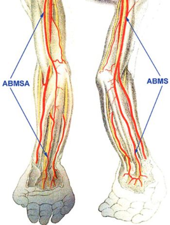
This article reviews in detail the superficial brachiomedian artery (arteria brachiomediana superficialis), a very rare variant of the main arterial trunks of the upper limb. It branches either from the axillary artery or the brachial artery, descends superficially in the arm (similar to the course of the superficial brachial artery) and continues across the cubital fossa, runs superficially in the forearm, approaches the median nerve and enters the carpal canal to reach the hand. It usually terminates in the superficial palmar arch. The first drawing was published, in 1830, and the first description was published, in 1844. Altogether, to our knowledge, only 31 cases of a true, superficial brachiomedian artery have been reported (Some cases are incorrectly reported as superficial brachioradiomedian artery or superficial brachioulnomedian artery). Based on a meta-analysis of known, available studies, the incidence is 0.23% in Caucasians and 1.48% in Mongolians. Knowing whether or not this arterial variant is present is important in clinical medicine and relevant for: The catheterization via the radial or ulnar artery; harvesting the vascular pedicle for a forearm flap based on the radial, ulnar or superficial brachiomedian arteries; the possible collateral circulation in cases of the arterial closure; and the surgical management of carpal tunnel syndrome. Its presence can elevate the danger of an injury to the superficially located variant artery or of an accidental injection.

Downloads

Download data is not yet available.

References

De Garis CF, Swartley WB. The axillary artery in white and Negro stocks. Am J Anat 1928; 41:353-97. http://dx.doi.org/10.1002/aja.1000410208.

Konarik M, Kachlik D, Baca V. A coincidental variation of the axillary artery: the brachioradial artery and the aberrant posterior humeral circumflex artery passing under the tendon of the latissimus dorsi muscle. Bosn J Basic Med Sci 2014; 14(4):239-43. http://dx.doi.org/10.17305/bjbms.2014.4.31.

Lippert H, Pabst R. Arterial Variations in Man, Munich: J.F. Bergmann; 1985. p. 71-3. http://dx.doi.org/10.1007/978-3-642-80508-0_35

http://dx.doi.org/10.1007/978-3-642-80508-0.

Trotter MI, Henderson JL, Gass H, Brua RS, Weisman S, Agress H. The origin of branches of the axillary arteries in white and American Negros. Anat Rec 1930; 46:133-7. http://dx.doi.org/10.1002/ar.1090460205.

Valsecchi O, Vassileva A, Musumeci G, Rossini R, Tespili M, Guagliumi G, et al. Failure of transradial approach during coronary interventions: Anatomic considerations. Catheter Cardiovasc Interv 2006; 67(6):870-8. http://dx.doi.org/10.1002/ccd.20732.

FCAT. Terminologia Anatomica, Stuttgart: Thieme Verlag; 1998.

Bergman RA, Afifi AK, Miyauchi R. Opus II: Cardiovascular System: Arteries: Upper Limb. In: Illustrated Encyclopedia of Human Anatomic Variation; 1992–2004. Available from: http://www.anatomyatlases.org/AnatomicVariants/Cardiovascular/Text/Arteries/Median.shtml [Last accessed on 2016 Mar 8].

Rodríguez-Niedenführ M, Sañudo JR, Vázquez T, Nearn L, Logan B, Parkin I. Median artery revisited. J Anat 1999; 195:57-63.

http://dx.doi.org/10.1046/j.1469-7580.1999.19510057.x, http://dx.doi.org/10.1017/S0021878299005075.

Natsis K, Iordache G, Gigis I, Kyriazidou A, Lazaridis N, Noussios G, et al. Persistent median artery in the carpal tunnel: Anatomy, embryology, clinical significance, and review of the literature. Folia Morphol (Warsz) 2009; 68:193-200.

Singla RK, Kaur N, Dhiraj GS. Prevalence of the persistant median artery. J Clin Diagn Res 2012; 6(9):1454-7.

Rodríguez-Niedenführ M, Vázquez T, Nearn L, Ferreira B, Parkin I, Sañudo JR. Variations of the arterial pattern in the upper limb revisited: a morphological and statistical study, with a review of the literature. J Anat 2001; 199:547-66.

http://dx.doi.org/10.1046/j.1469-7580.2001.19950547.x.

George BJ, Henneberg M. High frequency of the median artery of the forearm in South African newborns and infants. S Afr Med J 1996; 86(2):175-6.

Henneberg M, George BJ. A further study of the high incidence of the median artery of the forearm in Southern Africa. J Anat 1992; 181:151-4.

Henneberg M, George BJ. High incidence of the median artery of the forearm in a sample of recent southern African cadavers. J Anat 1992; 180:185-8.

Monro A. Outlines of the anatomy of the human body, in its sound and diseases state. Edinburgh: Archibald Constable & Co.; 1813.

Rodríguez-Niedenführ M, Burton GJ, Deu J, Sañudo JR. Development of the arterial pattern in the upper limb of staged human embryos: Normal development and anatomic variations. J Anat 2001; 199:407-17. http://dx.doi.org/10.1046/j.1469-7580.2001.19940407.x.

Green PH. An Account of the Varieties in the Arterial System of the Human Body. Dublin: James Marshal Leckie; 1830. p. 16-20.

Meckel JF. Ueber den regelwidrigen verlauf der armpulsadren [Article in German]. Dtsch Arch Physiol 1816;2:117-31.

Quain R. Anatomy of the arteries of the human body. London: Taylor and Walton; 1844. p. 326-37.

Tiedemann F. Supplementa ad tabulas arteriarum corporis humani (Supplements to tables of Arteries of the human body) [In Latin]. Heidelberg; 1846.

Gruber W. Über die neue und konstante Ellenbogenschlagader des Menschen (Arteria plicae cubiti superficialis) nebst deren Anomalien, der Arteria mediana antibrachii superficialis and ulnaris superficialis. (On new and constant elbow artery (arteria plicae cubiti superficialis) and other anomalies (arteria mediana antibrachii superficialis and ulnaris superficialis)) [In German]. Z Ges Arztezu Wien 1852; 8:481-99.

Gruber W. Über die Arteria mediana antibrachii superficialis, Arteria ulnaris antibrachii superficialis und Duplicität der Arteria ulnaris (On Arteria mediana antibrachii superficialis, arteria ulnaris antibrachii superficialis und duplication of arteria ulnaris) [In German]. Arch Anat Physiol Wissen Med 1867:668-87.

Calori L. Delle anomalie piu importanti di ossa, vasi, nervi e muscoli occorse nell'ultimo biennio facendo anatomia del corpo umano (On important anomalies of bones, vessels, nerves and muscles observed in last two years doing human anatomy) [In Italian]. Mem Acad Sci Bol 1868; 8:417-82.

Parisot L. Consideration’s sur les anomalies de i'artere humerale. Comput Rend Soc Med Nancy 1869; 1:1-28. (in French).

Giacomini C. Prematura divisione dell'arteria del braccio (sotto-varietà mediana). l’osservatore (Observation of premature division of brachial (subvariant of median artery)) [In Italian]. Gaz Clin Torino 1884; 200:129-40.

Testut L. Traité d’Anatomie Humaine [In French]. 3rd ed., Vol. 1. Fascicle 1. Paris: Octave Doin; 1895. p. 1010.

Schwalbe E. Beitrag zur kenntniss der arterienvarietäten des menschlichen armes (Contribution to knowledge of arterial variations of the human arm) [Article in German]. Morphol Arb 1898; 8:1-47.

Müller E. Beiträge zur morphologie des gefässsystems. 1. Die armarterien des menschen. Anat Hefte Abt 1903; 22:379-574. (in German).

Hoskins ER. Persistent arteriae brachii superficialis, antibrachii superficialis et mediana. Anat Rec 1914; 8:421-2.

Dubreuil-Chambardel L. Variations des artères du membre superieur. Paris: Masson et Cie; 1926. p. 180-92. (in French).

Dubreuil-Chambardel L. Variations artérielles rares (Rare arterial variaitons) [In French]. In: Bulletins et Mémoires de la Société D'anthropologie de Paris, VII. Série, tome 8; 1927. p. 86-92. http://dx.doi.org/10.3406/bmsap.1927.9087.

Schwyzer AG, De Garis CF. Three diverse patterns of the arteria brachialis superficialis in man. Anat Rec 1935; 63:405-16. http://dx.doi.org/10.1002/ar.1090630408.

Maruyama K. Seltene varietät der arterien der oberen extremität bei einem Japaner (A. brachialis superficialis lateralis inferior, A. antebrachialis superficialis mediano-ulnaris, arcus volaris superficialis vom typus mediano-ulnaris) (Rare variation of arteries of upper limb observed in a Japanese) [In German]. Folia Anat Jpn 1944; 22:551-67.

http://dx.doi.org/10.2535/ofaj1936.22.6_551.

McCormack LJ, Cauldwell EW, Anson BJ. Brachial and antebrachial arterial patterns: a study of 750 extremities. Surg Gynecol Obstet 1953; 96(1):43-54.

Mrázková O. Ontogenetic development of the median artery in man. Folia Morphol (Praha) 1975; 23(2):170-4.

Nakatani T, Izumi A, Tanaka S. Bilateral superficial median arteries. J Anat 1999;

:475-7. http://dx.doi.org/10.1046/j.1469-7580.1999.19430475.x.

Darwish HH, Khan MM, Zaher WA. Superficial median artery arises from brachioradial artery: a rare variation. Eur J Anat 2008; 12:63-6.

Jelev L, Georgiev GP. A rare case of superficial median artery of high brachial origin: anatomical and clinical considerations of the superficial brachiomedian artery. Anatomy 2010; 5:39-43.

Kachlik D, Hajek P, Konarik M, Krchov M, Baca V, et al. Coincidence of superficial brachiomedian artery and bitendinous palmaris longus: a case report. Surg Radiol Anat 2016; 38(1):147-51. http://dx.doi.org/10.1007/s00276-015-1512-x.

Poynter CW. Congenital anomalies of the arteries and veins of the human body with bibliography, Vol. 22. Lincoln: The University Studies of the University of Nebraska; 1922. p. 1-106.

Lieffring JJ. De quelques variations artérielles à la face antérieure du membre supérieur. (La coïncidence du développement de l'artère du nerf médian avec la naissance prématurée des artères cubitale et radiale) (On several arterial variations of the anterior surface of the upper limb. (The coincidence of development of the median artery and high origin of ulnar and radial arteries)) [In French]. In: Bull Mémoir Soc d'anthropol Paris, VII Série, Tome 5.; 1924. p. 67-77.

Adachi B. Das arteriensystem der Japaner, Kyoto: Maruzen; 1928. p. 285-356. (in German).

Keen JA. A study of the arterial variations in the limbs, with special reference to symmetry of vascular patterns. Am J Anat 1961; 108:245-61.

http://dx.doi.org/10.1002/aja.1001080303.

Lengele B, Dhem A. Unusual variations of the vasculonervous elements of the human axilla. Report of three cases. Arch Anat Histol Embryol 1989; 72:57-67.

Singer E. Embryological pattern persisting in arteries of the arm. Anat Rec 1933; 55:403-9. http://dx.doi.org/10.1002/ar.1090550407.

Rodríguez-Baeza A, Nebot J, Ferreira B, Reina F, Pérez J, Sañudo JR, et al. An anatomical study and ontogenetic explanation of 23 cases with variations in the main pattern of the human brachio-antebrachial arteries. J Anat 1995; 187(2):473-9.

Arey LB. Developmental anatomy. In: Development of the arteries, 6th ed. Philadelphia: W.B. Saunders Co.; 1957. p. 375-7.

Jurjus AR, Correa-De-Aruaujo R, Bohn RC. Bilateral double axillary artery: embryological basis and clinical implications. Clin Anat 1999; 12(2):135-40.

http://dx.doi.org/10.1002/(SICI)1098-2353(1999)12:2<135::AID-CA10>3.0.CO;2-M, http://dx.doi.org/10.1002/(SICI)1098-2353(1999)12:2<135::AID-CA10>3.3.CO;2-D.

Kopuz C, Gülman B, Baris S. Persistent median artery: an anatomical study in neonatal and adult cadavers. Kaibogaku Zasshi 1995; 70(6):577-80.

Jaschtschinski SN. Morphologie und topographie des Arcus volaris sublimis und profundus des Menschen (Morphology and topography of human arcus volaris sublimis and profundus) [In German]. Anat Hefte 1897; 7:161-88. http://dx.doi.org/10.1007/BF02298031.

Kachlik D, Konarik M, Horak D, Bernat I, Baca V. Anatomical difficulties of catheterization via arteria radialis. Intervent Akut kardiol 2010; 9:64-8.

Burzotta F, Trani C, De Vita M, Crea F. A new operative classification of both anatomic vascular variants and physiopathologic conditions affecting transradial cardiovascular procedures. Int J Cardiol 2010; 145(1):120-2.

http://dx.doi.org/10.1016/j.ijcard.2009.06.025.

Cohen SM. Accidental intra-arterial injection of drugs. Lancet 1948; 2(6524):409-17. http://dx.doi.org/10.1016/S0140-6736(48)90986-6.

Lister G. The hand: diagnosis and indications. Edinburgh: Churchill Livingstone; 1977. p. 96.

Devansh. Superficial ulnar artery flap. Plast Reconstr Surg 1996; 97(2):420-6. http://dx.doi.org/10.1097/00006534-199602000-00022.

Hazlett JW. The superficial ulnar artery with reference to accidental intra-arterial injection. Can Med Assoc J 1949; 61(3):289-93.

Thoma A, Young JE. The superficial ulnar artery “trap” and the free forearm flap. Ann Plast Surg 1992; 28(4):370-2. http://dx.doi.org/10.1097/00000637-199204000-00013.

Kachlik D, Baca V, Bozdechova I, Cech P, Musil V. Anatomical terminology and nomenclature: past, present and highlights. Surg Radiol Anat 2008; 30(6):459-66. http://dx.doi.org/10.1007/s00276-008-0357-y.

Kachlik D, Bozdechova I, Cech P, Musil V, Baca V. Mistakes in the usage of anatomical terminology in clinical practice. Biomed Pap Med Fac Univ Palacky Olomouc Czech Repub 2009; 153(2):157-61. http://dx.doi.org/10.5507/bp.2009.027.

Kachlik D, Musil V, Baca V. Terminologia anatomica after 17 years: inconsistencies, mistakes, and new proposals. Ann Anat 2015; 201:8-16.

http://dx.doi.org/10.1016/j.aanat.2015.04.006.

Brachiomedian artery (arteria brachiomediana) revisited: a comprehensive review

Downloads

Additional Files

Published

2016-03-10

How to Cite

1.
Kachlik D, Konarik M, Riedlova J, Baca V. Brachiomedian artery (arteria brachiomediana) revisited: a comprehensive review. Biomol Biomed [Internet]. 2016Mar.10 [cited 2023Mar.29];16(2):91-101. Available from: https://www.bjbms.org/ojs/index.php/bjbms/article/view/801

Issue

Section

Reviews

INTRODUCTION

Arteries of the upper limb belong to quite variable systems throughout the human body. The branching patterns in axilla can be more inconstant than not, reported in 10% up to 55% of cases, and in the free part of the upper limb in approximately 23% of cases [1-5]. The aim of this article is to draw attention to a very rare variant of the main forearm arterial trunks which is clinically relevant due to its interesting and important anatomical relationships.

THE VARIANTS

Arteria comitans nervi mediani (ACNM) - Median artery

The incidence of a variant artery accompanying the median nerve (NM), usually termed as the median artery Arteria comitans nervi mediani (ACNM - According to Terminologia Anatomica 1998) [6], is reported to very large extent from 0.6% to 53% (Table 1). This incidence inconsistency is based on the definition of the median artery. From the terminological point of view, it is any artery accompanying NM in the forearm and can extend into the hand via the carpal canal (canalis carpi). The older Latin anatomical nomenclatures (BNA, INA, and PNA) all used the term “arteria mediana.” But from the anatomical point of view, two main types can be classified: ACNM antebrachii (ACNMa) (antebrachial type of median artery) and ACNM manus (ACNMm) (palmar type of median artery). Both types usually originate from the proximal segment of the ulnar artery (arteria ulnaris), from the common interosseous artery (arteria interossea communis) or from the anterior interosseous artery (arteria interossea anterior).

TABLE 1: Incidence of the ACNM based on studies performed on more than 100 specimens (Natsis et al., 2009)

The first type runs only within the forearm to supply the anterior group of forearm muscles and distally fades out. Its incidence is reported in 8-12% of cases. As the ACNM does not reach the hand and supplies the forearm only, it can be denominated as the antebrachial type of the median artery (ACNMa).

The second type continues along the whole forearm and finally passes through the carpal canal and contributes to the blood supply of the palm. Its incidence is reported in approximately 4% of cases. In 35% of cases, the second type joins the trunk of the ulnar artery to form a complete superficial palmar arch (arcus palmaris superficialis) [7,8]. The second type corresponds more consistently with an embryonic structure termed the arteria mediana and that is why it is correct to use this synonym (AM) for the palmar type of the median artery (ACNMm).

As for the real incidence of both types, the data are rather unreliable because of the uncertainty of the terminology, which leads to the data being highly variable in their extent. Table 1 summarizes the most important studies of over 100 specimens (available to the authors) to see the incidence range. For other detailed data [9,10].

Bergman stated in his internet encyclopedia that the average incidence was 8% [7], while Rodríguez-Niedenführ et al., observed that the incidence of ACNMm was 18.7% in fetuses and 12% in adults, in his detailed and thorough studies on the European population. Counting every artery that runs with the NM regardless of its diameter, Rodríguez-Niedenführ et al., found the ACNMa in 53% of individuals. With such a high incidence, they considered the ACNMa to be “a normal feature rather than a variation” [8,11]. Results from our not-yet-published study show that the incidence of the ACNM in central European population is 7.4% (out of 176 limbs) with no proven relation to gender or side. Approximately half of the ACNM observed (3.4%) pass through the carpal tunnel as ACNMm, however, only 3 featured a diameter of 1 mm or larger within the canal.

The incidence varies with ethnicity and depends on age. South African newborns and infants from black communities are far more likely to have the ACNM. Henneberg and George reported the artery in 50% of forearms; over 38% of these were bilateral. In their previous work, they examined the adult population and found the incidence to be as high as 27.4% of cases, which is almost twice as high compared to the Middle European population [12-14].

The data concerning the difference in the incidence of ACNMa and ACNMm are inconsistent, the authors usually deal only with the “persistent median artery” and the amount of studied limbs is <50.

Arteria mediana superficialis (ACNMm superficialis) - Superficial median artery (Palmar type of superficial median artery)

The AM features a deep course, as does the NM. Even in the carpal canal, the AM is usually located underneath the nerve (dorsally to it). When the course of the variant artery in the midline of the forearm is superficial to the above-described one, it should be termed the superficial median artery (ACNM superficialis [ACNMS]). In that case, it can be either a branch of the radial artery or of the ulnar artery. If branching from a superficially located variant, it should belong to another terminological group. The relevant data are missing and it seems to be more of a theoretical variant. One case which fulfills the requirements of the definition is a variation mentioned by Monro in 1813. He observed a variant artery, branching from the cubital anastomosis (anastomosis cubitalis) between the superficial brachioradial artery (arteria brachioradialis superficialis) and the ulnar artery. It coursed superficially under the skin and terminated in the superficial palmar arch [15].

Arteria brachiomediana (ABM) - Brachiomedian artery

When the origin of AM is located proximally above the elbow level from the brachial artery (arteria brachialis [AB]) or from the axillary artery (arteria axillaris, AA), such a variant should be termed the brachiomedian artery (ABM), as proposed by Rodríguez-Niedenführ et al. They have summed up both the already-described and theoretically existing variants of the upper limb arterial trunks in a detailed study, in 2001, and mentioned (as the only authors) the incidence to be <0.26%, but without any detailed data [16].

ABM superficialis (ABMS) - Superficial brachiomedian artery

When the ABM courses superficially in either arm or forearm, it should be termed superficial brachiomedian artery (ABMS). It is a rare variant of the upper limb arterial trunks. As far as we know, the first indirect information comes from the book written by Green in 1830 (Figure 1). There is a depiction of the ABMS but in the text; there are no specific data to distinguish between the ABM and ABMS [17]. Already in 1816, Meckel mentioned two cases of a peculiar variation of the superficial brachioradial artery which bifurcated within the cubital fossa into a thicker lateral branch, obviously a radial artery, and a thinner medial branch, running subcutaneously and terminating in the superficial palmar arch. This variant should be termed the superficial brachioradiomedian artery (ABMS) [18]. Depictions from different authors and years are available as supplemental figures (Figure S1-S8).

FIGURE 1: The scheme of the case of the arteria brachiomediana superficialis (ABMS) artery as drawn by Green in 1830. 1 - Brachial artery (arteria brachialis), 2 - Superficial brachiomedian artery (ABMS), 3 - Radial artery (arteria radialis), 4 - Ulnar artery (arteria ulnaris).
FIGURE 1S: The scheme of two cases of the arteria brachiomediana superficialis as drawn by Gruber in 1852. Legend: ABMS – arteria brachiomediana superficialis, ABRMS – arteria brachioradiomediana superficialis, ACNM – arteria comitans nervi mediani.
FIGURE 2S: The scheme of two cases of the variant artery resembling the ABMS but passing superficially to the flexor retinaculum as drawn by Calori in 1868. Legend: X – variant artery.
FIGURE 3S: The scheme of the case of the arteria brachiomediana superficialis as drawn by Giacomini in 1884. Legend: ABMS – arteria brachiomediana superficialis.
FIGURE 4S: The scheme of two cases of a variant artery as drawn by Schwalbe in 1898. Legend: ABUMS – arteria brachioulnomediana superficialis, ACNMm – arteria comitans nervi mediani manus, AUAS – arteria ulnaris accessoria superficialis, AU – arteria ulnaris.
FIGURE 5S: The scheme of a variant artery as drawn by Hoskins in 1914. Legend: AB – arteria brachialis, X – arteria brachio-antebrachialis accessoria superficialis.
FIGURE 6S: The scheme of the case of the arteria brachioradiomediana superficialis and of the arteria brachiomediana superficialis as drawn by Dubreil-Chambardel in 1926 . Legend: ABMS – arteria brachiomediana superficialis, ABRMS – arteria brachioradiomediana superficialis, ACNMm – arteria comitans nervi mediani manus.
FIGURE 7S: The scheme of the case of the arteria brachiomediana superficialis as drawn by Adachi in 1928. Legend: ABMS – arteria brachiomediana superficialis.
FIGURE 8S: The scheme of the arteria plicae cubiti superficialis as drawn by Gruber in 1852. Legend: X – arteria plicae cubiti superficialis.

Quain in 1844 reported on two cases of ABMS out of 478 upper limbs (0.42%), one seen in Plate 45 (“vas aberrans” or “median artery”) and the other one in a very young child (no depiction) [19]. In 1843, Tiedemann depicted in his Table 46 and Figure 3, a case (No. 3) of the ABMS originating from the AB in the distal part of the arm [20]. Gruber contributed with a long-term and extensive exploration reporting on five cases out of 1,900 upper limbs (0.26%) in two subsequent articles in years 1852 and 1867, termed the “arteria mediana antebrachii superficialis.” The first case was bilateral: On the right side; the variant artery branched from the AB, and on the left side from the brachioradiomedian artery (arteria brachioradiomediana, ABM). The second case concerned the brachioradiomedian artery, and the third one was the true ABMS originating in the middle part of the AB [21,22] (Figure 2). Calori in 1868 published two cases, one concerning an absent ulnar artery replaced with a superficially located artery, branching from the AB very proximally and passing superficially to the flexor retinaculum (retinaculum musculorum flexorum) into the hand; the other branching from the AA, with a thick branch in the axilla, running superficially in the arm and forearm and passing superficially to the flexor retinaculum into the hand - both cases cannot be classified as the ABMS [23]. Parisot observed one case without stating any further details in 1869 [24]. Giacomini reported in detail in 1884 on ABMS branching from the AA [25]. Testut, in 1895, included in his anatomical textbook two cases, one originating from the AA and the other from the BA (without stating any further details) [26]. Schwalbe mentioned, in 1898, a rare and peculiar variation of a double ACNM and a double ulnar artery. The usual ACNM as well as the ulnar artery was thin (rudimentary), but superficially there were two thick arteries branching from one short stem originating in the cubital fossa to form the AB (termed “arteria mediana superficialis and arteria ulnaris superficialis”). Such a variant should be considered as a superficial brachioulnomedian artery (arteria brachioulnomediana superficialis, ABUMS - specimen No. 1961/case 22) and its branches as an accessory palmar type of median artery (ACNMm accessoria) and an accessory superficial ulnar artery (arteria ulnaris accessoria superficialis). He also observed a case of the superficial brachioulnomedian artery but without any deep counterpart (as before), however, there was a thick anastomosis with the radial artery in the middle of the forearm (specimen No. 741/case 23) [27].

FIGURE 2: The scheme of two cases, the accessory arteria brachiomediana superficialis (ABMS) joining the antebrachial type of median artery in the middle of the forearm, and the ABMS as drawn by Müller in 1903. ABMS - Superficial brachiomedian artery, ABMSA - Accessory superficial brachiomedian artery (ABMS accessoria).
FIGURE 3: The scheme of the arteria brachiomediana superficialis (ABMS) as drawn by Gruber in 1867. ABMS - superficial brachiomedian artery.

In the last century, Müller observed in a fetus (case No. 152) the “AB superficialis superior” (ABMS) branching from the AA. The NM was accompanied by the ACNMa within the proximal half of the forearm (Figure 3). He also observed a more peculiar case (case no. 153) with ABMS branching from the AA which submerged in the middle of the forearm to join the ACNMm (as a usual branch from the ulnar artery). This case can also be termed as an accessory ABMS. Finally, he observed (case No. 151) an “AB superficialis superior” (ABUMS) which bifurcated in the forearm (close to the flexor retinaculum) into the ACNM and the ulnar artery [28]. According to Müller, this variant (ABMS) is identical to the “arteria plicae cubiti superficialis” first described and denominated by Gruber, but as seen in the original articles, these are two absolutely different variants [21,28].

A case reported by Hoskins in 1914 featured a deeper course in the arm (branching form the AA and running ventrolateral to the AB proper within the brachial fascia [fascia brachii]), but running a superficial course in the forearm. The artery was immediately subjacent to the median cubital vein (vena mediana cubiti) which would predispose the vessel to an inadvertent penetration during attempts at venipuncture of the median cubital vein. No relationship between the variant artery and NM is mentioned which allows us to conclude that the reported case (originally called “arteria brachii superficialis, antebrachii superficialis et mediana”) by Hoskins should be termed as an “accessory superficial brachio-antebrachial artery” (arteria brachio-antebrachialis accessoria superficialis) to describe precisely its course [29].

In his detailed work concerning all arterial variations of the upper limb, Dubreuil-Chambardel noted his observation of a bilateral ABMS branching from the AA and featuring a cubital anastomosis with the proper AB within the cubital fossa [30]. A year later he reported a case with the ABMS branching from the AA which had a quite thick branch proximal to the cubital fossa which supplied the bellies of forearm muscles originating at the epicondyles [31]. In 1935, Schwyzer published a case report in which he described an arteria mediana (“arteria comes nervi median”) branching from the superficial brachial artery. As the ABS then continued as the radial artery, this variant should be considered as a superficial brachioradiomedian artery (arteria brachioradiomediana superficialis) [32]. Maruyama in 1944 presented a case of the brachioulnomedian artery (arteria brachioulnomediana) and reported on two similar cases published in Japan at the beginning of 20th century [33]. In 1953, McCormack reported a unique variant, namely the superficial brachioradiomediointerosseous artery (arteria brachioradiomediointerossea superficialis), depicted in his Figure 3d, which divides superficially in the cubital fossa; unfortunately giving no detailed data [34]. In 1975, Mrázková performed a study of embryonic material from surgical abortions and compared it to 75 adult specimens. She found two cases of the ABMS without stating further details [35].

Review of accessible recent literature revealed three cases: In 1999, Nakatani et al., reported on a unique case of the bilateral occurrence of ABMS (which they termed the AMS). On the right side it branched from the AA as the superficial brachioradiomedian artery and bifurcated at the level of the interepicondylar line (linea interepicondylaris) into the radial artery and ACNMS; on the left side the ABMS branched from the superficial brachial artery in the cubital fossa [36]. Darwish et al., in 2008 published a combined unique case of the ABR with the cubital anastomosis and of the ABMS branching from the ABR artery in the distal arm, which allows us to conclude that the reported case should be termed as the superficial brachioradiomedian artery to describe precisely its course and branching [37]. Last, Jelev and Georgiev described, in 2010, the ABMS branching from the initial part of the AB [38]. Our team has observed one case of the ABMS on the right side, branching from the distal segment of the AB, just above the cubital fossa [39]. The details of individual case-reports are summarized in Table 2.

TABLE 2: Details of individual case-reports

As for larger studies, consisting of the detailed description of many specimens of the upper limb arterial trunks as a whole, no relevant data can be found, except the above mentioned study made by Quain [19]. Müller studied both fetuses and adults and observed only two cases of the SBMA in 1903. Incidence stated by him is 0% in adults (0/100) and 1% in fetuses (2/200) [28]. A summary of similar variations comes from the work of Poynter (1922) who classified the variations of the BA into four groups. The Group II.1 represents the case when there are two main arterial trunks in the arm (“doubled BA”) and the variant superficial brachial artery continues as the ACNM or the common interosseous artery. No detailed data on the superficial or deep course of the ABM are stated but it includes several references (although most of them are not in context) [40]. Dubreil-Chambardel classified the origin of the ACNM into 10 types in 1926, the types VIII-X including the origin form the BA (2% out of all cases) but the real ABM can be related only to type IX [30]. Lieffring in 1924 only summarized from the above-stated case reports, and himself reported only the ACNM [41]. Adachi termed this variant as “arteria antebrachialis superficialis mediana” and summarized 8 cases (Nr. 6-14) from 410 dissected limbs of Japanese, all unilateral (Table 3). The last case (No. 14) cannot be considered a true ABMS because the variant artery, stemming from the AB in the middle of the arm, bifurcates superficially in the cubital fossa into the ACNM and the ulnar artery: That is why it should be considered as the brachioulnomedian artery and we have excluded it from the summary. The numbers reported by Adachi (1.7%) surpass the incidence reported in individual cases in the Caucasian race [42]. McCormack recorded in 1953 no such variant in his extensive study on 750 limbs [34]; neither did Keen in 1961 in his study of 284 limbs of different races [43].

TABLE 3: Cases of ABMS reported by Adachi in Japanese (Adachi, 1928)

Nakatani et al., in 1999 reported the bilateral case of SBMA, and stated its incidence to be 1% but considering the rather low number of specimens in his study (200) for such a rare variant, the number is not sufficiently reliable [36].

Finally, Rodríguez-Niedenführ et al., classified two theoretical variants concerning the ABM in 1999: The true ABM, branching from the brachial artery but coursing under flexor muscles of the forearm, which has never been reported according to our knowledge; and the ABMS, branching from the AB or even from the AA and coursing within the superficial fascia, which was not observed in adults and only once in a fetus by his team (reported incidence in fetuses 0.7% [1/150] and <0.2% in adults [0/480]) [16].

The data concerning the detailed studies of the upper limb arterial variations are summarized in Table 4.

TABLE 4: Studies of upper limb arterial variations concerning the ABMS

Due to the long distance between the axillary fossa (fossa axillaris) and cubital fossa, the exact location of the origin of the ABMS can be classified into four groups, following the classification of similar variants (brachioradial and superficial brachioradial arteries) proposed by Rodríguez-Niedenführ et al., in 2001. The classification is based on the topography of the ABMS origin in the axillary fossa or within the arm (Table 5) [16]. Based on the meta-analysis it is usual to expect the ABMS origin either in the cubital fossa or from the adjacent part of the AB in the distal arm (in 35% of cases) or in the axillary fossa from the AA (in 35% of cases).

TABLE 5: Classification of the origin of the arteria brachiomediana superficialis

DEVELOPMENT

The presence of upper limb arterial trunks’ variations is not a rare situation. Not surprisingly, the branching arrangement of the AA is more often irregular than what corresponds precisely to the textbook pattern. In the arm and forearm, the state is more constant but in each fifth individual, a variation of the main arterial trunk can be found. Each of them is a consequence of impaired development [28,44].

For a long time, there was a trusted theory postulated by Singer in 1933 who, briefly stated, described the variants as remnants of previous embryonic arterial trunks. He concluded that the median artery (arteria mediana) develops as the first artery from the axial artery (arteria axialis) and usually regresses after the 8th week of development [45]. But as Rodríguez-Niedenführ et al., presented in their review and study, Singer based his theory on the findings of one author, and did not take into account other embryological studies [16,46].

In 2001, Rodríguez-Niedenführ et al., published a new theory, emphasizing the fact that all principal arterial trunks of the upper limb are formed from a primitive capillary network encircling the developing bones. The principal advantage of this theory is that it offers a plausible, easy explanation of all the ever-reported variants of the upper limb arteries. Every arterial variation can be traced back to its embryologic origin [11,16]. Arey and Jurjus et al., suggested six explanations for the reported variations: The choice of unusual paths in the primitive vascular plexus; the persistence of vessels which are normally obliterated; the disappearance of vessels which are normally retained; incomplete development; the fusion and absorption of parts which are normally distinct; and a combination of factors leading to an atypical pattern [47,48].

Kopuz et al., studied the presence of the median artery in neonatal cadavers and proposed the theory that it regresses as late as during the perinatal period and early infancy [49]. Keen supposed that the median artery is a persistence of an embryonic artery [43] and Jaschtchinski called it an atavistic condition because it is usually found to contribute to the palmar arch (arcus palmaris) of certain lower animals [50].

To summarize the embryonic development of the upper limb vasculature [11], several important facts are:

First, the proximal (central) part of the right subclavian artery (pars proximalis arteriae subclaviae dextrae) arises from the right fourth pharyngeal arch artery (right fourth aortic arch, arteria arcus pharyngei quarti dextri) and its distal (peripheric) part is derived from the right seventh intersegmental artery (arteria intersegmentalis septima dextra). The left subclavian artery (arteria subclavia sinistra) has a different embryologic background; both its parts are formed from the left seventh intersegmental artery (arteria intersegmentalis septima sinistra) (day 28 after conception).

Second, the arterial system of the upper limb starts to develop as a dispersed capillary network at day 26 when a bud of the future upper limb appears; the capillary network is connected to the axial artery.

Third, the AA (day 33) and its branches on both sides of the human body are derived from the axial artery which is a distal (peripheral) continuation of the seventh intersegmental artery (arteria intersegmentalis septima). Conversely, the thoracodorsosubscapular trunk (truncus thoracodorsosubscapularis) is derived from the ninth intersegmental artery (arteria intersegmentalis nona). The nervous structures enter the limb to form the neural plate.

Fourth, the axial artery pierces the condensed nervous tissue within future axillary fossa on day 37.

Fifth, the AA continues as the AB, emanating the profunda brachii artery (arteria profunda brachii), on day 41.

Sixth, on day 44, all main arterial trunks of the forearm are formed (AB, ulnar, common interosseous and median arteries), except the radial artery whose proximal part is consolidated but the distal part is still in a form of a capillary plexus.

Seventh, on day 47, all nerve trunks as well as bones of the arm and forearm are chondrified: Only the distal part of the radial artery is still in a form of a capillary plexus.

Eighth, on day 52, all the principal elements of the upper limb have achieved their definitive shape (not size and dimensions) and location.

The reasons for the different interpretation of the ACNM/ABMS can be attributed to the new classification of the upper limb arterial trunks’ variations, proposed by Rodríguez-Niedenführ et al., in 2001 [16]. They have introduced a denomination using two roots (“brachio-” and “radial/ulnar/interosseous/median”) for the variant arteries with the more proximal origin (often termed as “high origin”). Due to this change of classification, all the old studies and case-reports shall be carefully read and the mentioned variations understood and denominated in terms of the new classification, in order to avoid erroneous results of their incidence.

ACNM/ABMS can replace the radial artery, the ulnar artery, or both arteries can be hypoplastic or stenotic in a part or in their whole course. This situation can lead to a failure of catheterization using either the radial or ulnar artery approach [51]. ACNM/ABMS can also serve as a collateral supply for the hand. When harvesting the vascular pedicle for a forearm flap based on the radial artery or the ulnar artery, or when harvesting the artery for a coronary bypass graft, it is necessary to consider this possible variant [52]. As a superficially located artery, it can be harmed in cut injuries to the arm and especially to the forearm, and gravely elevate the danger of heavy bleeding in the case of mechanical injury. As mentioned in the literature, an accidental injection into the ABMS can occur (instead of into the superficial vein) [53] and further consequences can happen, e.g. reflex constriction, inflammation and closure of the arterial caliber as well as potential ischemia of the hand in cases of impaired collateral circulation. Accidental ligation in cases of radial artery based flap dissection was also reported [38]. On the other hand, the ABMS can be used as an easily accessible vascular pedicle for the creation of a cutaneous forearm flap. ACNM/ABMS can be a cause of pronator teres syndrome or anterior interosseous nerve syndrome (when passing through the nerve or its branches), or carpal tunnel syndrome (in cases involving ACNMm or ABMSm). If the variant artery is of a large caliber, it may lead to compression of the NM within the carpal canal in patients who are prone to some pathological units (myxo-edema, rheumatoid arthritis or pregnancy provoking the carpal tunnel syndrome [54]). Clinical problems may appear as well as reported for superficial ulnar or superficial brachioulnar arteries [53,55-57].

The meta-analysis showed a slight difference between the Caucasian (0.23%) and Mongolian races (1.48%). But using Fisher’s exact test and paired comparison, including Bonferroni’s correction, there was only one significant comparison (p = 0.008) because the number of cases was very low. Based on the above stated data (28 cases of the proper ABMS) and the total meta-analyzed incidence (0.39%), it should be considered as one of the rarest superficially located arterial variants as compared to the superficial brachioradial artery (8.8-15.8%), the superficial brachial artery (3.6-9.6%), the superficial brachioulnar artery (0.7-7%) and the superficial brachioulnoradial artery (0.14-1.3%) [2,16].

The Latin nomenclature and terminology used in the article strictly follows that published in the Terminologia Anatomica (FCAT, 1998), as reviewed in our previous articles [58-60], and that suggested by Rodríguez-Niedenführ et al. [16].

CONCLUSION

Although the incidence of the ABMS is very low (0.39%), and our detailed survey did not reveal any published clinical case, it is not negligible, and therefore important that clinicians recognize the ABMS presence during both superficial and catheterization procedures on the upper limb.

DECLARATION OF INTERESTS

The authors declare no conflict of interest.

Acknowledgements

ACKNOWLEDGMENTS

The authors sincerely thank Vladimír Musil and Mirka Plecitá for technical and bibliographic support. The study was supported by the Charles University, Project PRVOUK #33.

REFERENCES

  1. , (). The axillary artery in white and Negro stocks. Am J Anat. http://dx.doi.org/10.1002/aja.1000410208
  2. , , (). A coincidental variation of the axillary artery:The brachioradial artery and the aberrant posterior humeral circumflex artery passing under the tendon of the latissimus dorsi muscle. Bosn J Basic Med Sci. http://dx.doi.org/10.17305/bjbms.2014.4.31
  3. , (). Arterial Variations in Man, Munich. J.F. Bergmann. http://dx.doi.org/10.1007/978-3-642-80508-0_35, http://dx.doi.org/10.1007/978-3-642-80508-0
  4. , , , , , (). The origin of branches of the axillary arteries in white and American Negros. Anat Rec. http://dx.doi.org/10.1002/ar.1090460205
  5. , , , , , (). Failure of transradial approach during coronary interventions:Anatomic considerations. Catheter Cardiovasc Interv. http://dx.doi.org/10.1002/ccd.20732
  6. (). . Terminologia Anatomica, Stuttgart:Thieme Verlag.
  7. , , (). Opus II:Cardiovascular System:Arteries:Upper Limb. Illustrated Encyclopedia of Human Anatomic Variation 1992-2004. Available from: http://www.anatomyatlases.org/AnatomicVariants/Cardiovascular/Text/Arteries/Median.shtml
  8. , , , , , (). Median artery revisited. J Anat. http://dx.doi.org/10.1046/.1469-7580.1999.19510057.x, http://dx.doi.org/10.1017/S0021≎99005075
  9. , , , , , (). Persistent median artery in the carpal tunnel:Anatomy, embryology, clinical significance, and review of the literature. Folia Morphol (Warsz).
  10. , , (). Prevalence of the persistant median artery. J Clin Diagn Res.
  11. , , , , , (). Variations of the arterial pattern in the upper limb revisited:A morphological and statistical study, with a review of the literature. J Anat. http://dx.doi.org/10.1046/j.1469-7580.2001.19950547.x
  12. , (). High frequency of the median artery of the forearm in South African newborns and infants. S Afr Med J.
  13. , (). A further study of the high incidence of the median artery of the forearm in Southern Africa. J Anat.
  14. , (). High incidence of the median artery of the forearm in a sample of recent southern African cadavers. J Anat.
  15. (). . Outlines of the anatomy of the human body, in its sound and diseases state.
  16. , , , (). Development of the arterial pattern in the upper limb of staged human embryos:Normal development and anatomic variations. J Anat. http://dx.doi.org/10.1046/j.1469-7580.2001.19940407.x
  17. (). . An Account of the Varieties in the Arterial System of the Human Body.
  18. (). Ueber den regelwidrigen verlauf der armpulsadren [Article in German]. Dtsch Arch Physiol.
  19. (). . Anatomy of the arteries of the human body.
  20. (). Supplementa ad tabulas arteriarum corporis humani (Supplements to tables of Arteries of the human body) [In Latin]. Heidelberg.
  21. (). Über die neue und konstante Ellenbogenschlagader des Menschen (Arteria plicae cubiti superficialis) nebst deren Anomalien, der Arteria mediana antibrachii superficialis and ulnaris superficialis. (On new and constant elbow artery (arteria plicae cubiti superficialis) and other anomalies (arteria mediana antibrachii superficialis and ulnaris superficialis)) [In German]. Z Ges Arztezu Wien.
  22. (). Über die Arteria mediana antibrachii superficialis, Arteria ulnaris antibrachii superficialis und Duplicität der Arteria ulnaris (On Arteria mediana antibrachii superficialis, arteria ulnaris antibrachii superficialis und duplication of arteria ulnaris) [In German]. Arch Anat Physiol Wissen Med.
  23. (). Delle anomalie piu importanti di ossa, vasi, nervi e muscoli occorse nell'ultimo biennio facendo anatomia del corpo umano (On important anomalies of bones, vessels, nerves and muscles observed in last two years doing human anatomy) [In Italian]. Mem Acad Sci Bol.
  24. (). Consideration's sur les anomalies de Lartere humerale. Comput Rend Soc Med Nancy. (in French)
  25. (). Prematura divisione dell'arteria del braccio (sotto-varietàmediana). l'osservatore (Observation of premature division of brachial (subvariant of median artery)) [In Italian]. Gaz Clin Torino.
  26. (). . Traitéd'Anatomie Humaine [In French].
  27. (). Beitrag zur Kenntniss der Arterienvarietäten des menschlichen Armes (Contribution to knowledge of arterial variations of the human arm) [Article in German]. Morphol Arb.
  28. (). Beiträge zur Morphologie des Gefässsystems. 1. Die Armarterien des Menschen. Anat Hefte Abt. (in German)
  29. (). Persistent arteriae brachii superficialis, antibrachii superficialis et mediana. Anat Rec.
  30. (). . Variations des artères du membre superieur. (in French)
  31. (). Variations artérielles rares (Rare arterial variations) [In French]. Bulletins et Mémoires de la SociétéD'anthropologie de Paris VII Série, tome 8. http://dx.doi.org/10.3406/bmsap.1927.9087
  32. , (). Three diverse patterns of the arteria brachialis superficialis in man. Anat Rec. http://dx.doi.org/10.1002/ar.1090630408
  33. (). Seltene Varietät der Arterien der oberen Extremität bei einem Japaner (A. brachialis superficialis lateralis inferior, A. antebrachialis superficialis mediano-ulnaris, arcus volaris superficialis vom typus mediano-ulnaris) (Rare variation of arteries of upper limb observed in a Japanese) [In German]. Folia Anat Jpn. http://dx.doi.org/10.2535/ofaj1936.22.6_551
  34. , , (). Brachial and antebrachial arterial patterns:A study of 750 extremities. Surg Gynecol Obstet.
  35. (). Ontogenetic development of the median artery in man. Folia Morphol (Praha).
  36. , , (). Bilateral superficial median arteries. J Anat. http://dx.doi.org/10.1046/j.1469-7580.1999.19430475.x
  37. , , (). Superficial median artery arises from brachioradial artery:A rare variation. Eur J Anat.
  38. , (). A rare case of superficial median artery of high brachial origin:Anatomical and clinical considerations of the superficial brachiomedian artery. Anatomy.
  39. , , , , (). Coincidence of superficial brachiomedian artery and bitendinous palmaris longus:A case report. Surg Radiol Anat. http://dx.doi.org/10.1007/s00276-015-1512-x
  40. (). . Congenital anomalies of the arteries and veins of the human body with bibliography.
  41. (). De quelques variations artérielles àla face antérieure du membre supérieur. (La coïncidence du développement de l'artère du nerf médian avec la naissance prématurée des artères cubitale et radiale) (On several arterial variations of the anterior surface of the upper limb. (The coincidence of development of the median artery and high origin of ulnar and radial arteries)) [In French]. In:Bull Mémoir Soc d'anthropol Paris, VII Série, Tome 5.
  42. (). . Das Arteriensystem der Japaner. (in German)
  43. (). A study of the arterial variations in the limbs, with special reference to symmetry of vascular patterns. Am J Anat. http://dx.doi.org/10.1002/aja.1001080303
  44. , (). Unusual variations of the vasculonervous elements of the human axilla. Report of three cases. Arch Anat Histol Embryol.
  45. (). Embryological pattern persisting in arteries of the arm. Anat Rec. http://dx.doi.org/10.1002/ar.1090550407
  46. , , , , , (). An anatomical study and ontogenetic explanation of 23 cases with variations in the main pattern of the human brachio-antebrachial arteries. J Anat.
  47. (). Developmental anatomy. Development of the arteries.
  48. , , (). Bilateral double axillary artery:Embryological basis and clinical implications. Clin Anat. http://dx.doi.org/10.1002/(SICI)1098-2353(1999)12:2<135:AID-CA10>3.0.CO;2-M, http://dx.doi.org/10.1002/(SICI)1098-2353(1999)12:2<135:AID-CA10>3.3.CO;2-D
  49. , , (). Persistent median artery:An anatomical study in neonatal and adult cadavers. Kaibogaku Zasshi.
  50. (). Morphologie und Topographie des Arcus volaris sublimis und profundus des Menschen (Morphology and topography of human Arcus volaris sublimis and profundus) [In German]. Anat Hefte. http://dx.doi.org/10.1007/BF02298031
  51. , , , , (). Anatomical difficulties of catheterization via arteria radialis [In Czech]. Intervent Akut kardiol.
  52. , , , (). A new operative classification of both anatomic vascular variants and physiopathologic conditions affecting transradial cardiovascular procedures. Int J Cardiol. http://dx.doi.org/10.1016/j.ijcard.2009.06.025
  53. (). Accidental intra-arterial injection of drugs. Lancet. http://dx.doi.org/10.1016/S0140-6736(48)90986-6
  54. (). . The hand:Diagnosis and indications.
  55. (). Superficial ulnar artery flap. Plast Reconstr Surg. http://dx.doi.org/10.1097/00006534-199602000-00022
  56. (). The superficial ulnar artery with reference to accidental intra-arterial injection. Can Med Assoc J.
  57. , (). The superficial ulnar artery “trap” and the free forearm flap. Ann Plast Surg. http://dx.doi.org/10.1097/00000637-199204000-00013
  58. , , , , (). Anatomical terminology and nomenclature:Past, present and highlights. Surg Radiol Anat. http://dx.doi.org/10.1007/s00276-008-0357-y
  59. , , , , (). Mistakes in the usage of anatomical terminology in clinical practice. Biomed Pap Med Fac Univ Palacky Olomouc Czech Repub. http://dx.doi.org/10.5507/bp.2009.027
  60. , , (). Terminologia anatomica after 17 years:Inconsistencies, mistakes, and new proposals. Ann Anat. http://dx.doi.org/10.1016/j.aanat.2015.04.006