Risk factors for recurrent IgA nephropathy after renal transplantation: A meta-analysis

Authors

DOI:

https://doi.org/10.17305/bjbms.2022.8369

Keywords:

Meta-analysis, IgA nephropathy, kidney transplantation, risk factors

Abstract

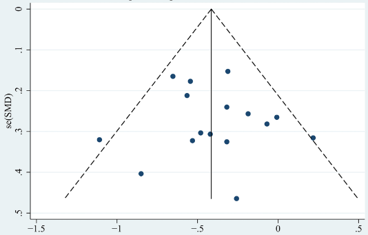
Recurrent glomerulonephritis after renal transplantation is the third most common cause of allograft loss, the most frequent of which is associated with IgA nephropathy (IgAN). This study aims to provide a systematic review of the risk factors associated with recurrent IgAN after renal transplantation. We searched English and Chinese databases, including PubMed, Embase, Web of Science, CNKI, and others, and included all case-control studies involving risk factors for recurrent IgAN after renal transplantation from the databases’ establishment to March 2022. Data were analyzed using the Stata 12.0. A total of 20 case–control studies were included in the meta-analysis, with 542 patients with recurrent IgAN and 1385 patients without recurrent IgAN. The results showed that donor age (standardized mean difference [SMD] -0.13 [95% CI -0.26, -0.001]; P = 0.048), patient age at transplantation (SMD -0.41 [95% CI -0.53, -0.29]; P < 0.001), time from diagnosis to end-stage renal disease (SMD -0.42 [95% CI -0.74, -0.10]; P = 0.010), previous transplantation (odds ratio [OR] 1.73 [95% CI 1.06, 2.81]; P = 0.027), living donor (OR 1.86 [95% CI 1.34, 2.58]; P < 0.001), related donor (OR 2.64, [95% CI 1.84, 3.79]; P < 0.001), tacrolimus use (OR 0.71 [95% CI 0.52, 0.98]; P = 0.035), basiliximab use (OR 0.39 [95% CI 0.27, 0.55]; P < 0.001), proteinuria (SMD 0.42 [95% CI 0.13, 0.71]; P = 0.005) and serum IgA level (SMD 0.48 [95% CI 0.27, 0.69]; P < 0.001) were associated with recurrent IgAN after renal transplantation. In general, tacrolimus and basiliximab use were protective factors against recurrent IgAN after renal transplantation, whereas donor age, patient age at transplantation, time from diagnosis to end-stage renal disease, previous transplantation, living donor, related donor, proteinuria, and serum IgA level were risk factors for recurrent IgAN after renal transplantation. Clinical decision making should warrant further consideration of these risk factors.

Downloads

Download data is not yet available.
Risk factors for recurrent IgA nephropathy after renal transplantation: A meta-analysis

Downloads

Published

2023-05-01

How to Cite

1.
Bai J, Wu Q, Chen J, Zheng Z, Chang J, Wang L, Zhou Y, Guo Q. Risk factors for recurrent IgA nephropathy after renal transplantation: A meta-analysis. Biomol Biomed [Internet]. 2023May1 [cited 2023Dec.5];23(3):364–375. Available from: https://www.bjbms.org/ojs/index.php/bjbms/article/view/8369

Issue

Section

Reviews

Introduction

End-stage renal disease (ESRD) is a worldwide public health concern. It imposes a significant burden on patients and often has a poor prognosis. Primary glomerulonephritis with IgA nephropathy (IgAN) is the leading cause of ESRD [1]. Currently, treatment modalities for ESRD include hemodialysis, peritoneal dialysis, and renal transplantation. Studies show that kidney transplantation is the most cost-effective approach [2–5]. More than 80% of patients with IgAN are young and middle-aged patients with fewer underlying diseases, so they can usually be ideal candidates for renal transplantation. In fact, IgAN patients account for a high proportion (approximately 13%) of candidates for renal transplantation [6]. However, recurrent glomerulonephritis after renal transplantation is the third most common cause of allograft loss, the most frequent of which is associated with IgAN [6–11]. Some studies have shown that the proportion of IgAN recurrence ranges from 9% to 53% due to different follow-up times and biopsy protocols [12].

Research suggested that recurrent IgAN after renal transplantation might be associated with younger age at transplantation, living related donor, rapidly progressive course of the original disease [12, 13], higher levels of circulating galactose-deficient IgA1 (Gd-IgA1), and other factors [14], whereas some of the above factors remained controversial. Therefore, accurate identification of risk factors associated with recurrent IgAN after kidney transplantation is a key for selecting transplant candidates and might help improve the long-term survival rate of patients.

Materials and methods

Literature search strategy

We searched English and Chinese databases, including PubMed, Embase, Medline, Web of Science, Cochrane Library, CNKI, CBMdisc, Wanfang and Weipu (VIP), and included all case-control studies on risk factors for recurrent IgAN after kidney transplantation, from the databases ’ establishment to March 2022. The risk of bias and quality was assessed according to Newcastle–Ottawa Scale. The relevant papers were identified using Medical Subject Headings (MeSH) terms: “Glomerulonephritis,” “Glomerulonephritides,” “Kidney Scarring,” “Kidney Transplantation,” “Renal Transplantation,” “Scarring, Kidney” and other free words. The idiographic search strategy retrieval is shown in Supplementary Material&#x00A0;1. Simultaneously, manual search of the included literature was performed to eliminate duplicate literature. Recurrent IgAN was defined by mesangial IgA deposition on immunofluorescence staining of allograft biopsy when clinical features of kidney transplantation recipients were hematuria, increasing proteinuria, elevated serum IgA levels and allograft dysfunction (defined by a significant increase in serum creatinine).

Table 1: Characteristics and Newcastle–Ottawa Scale quality score of included studies
Author Year NOS Country Recurrent IgAN (n) Sample (n) Period Risk factors
Okumi M et al. 2019 8 Japan 80 299 1995–2015 Donor age, living donor, related donor, previous transplantation, tacrolimus use, basiliximab use, serum IgA level
Martin-Penagos et al. 2019 8 Spain 14 35 1993—2015 Donor age, previous transplantation, tacrolimus use, basiliximab use, proteinuria
Jo et al. 2019 8 Korea 11 69 2011–2015 Previous transplantation, living donor, related donor, tacrolimus use
Di Vico et al. 2018 7 Italy 28 51 1995–2012 Donor age, age at transplantation, previous transplantation, living donor, tacrolimus use, basiliximab use, proteinuria
Garnier et al. 2018 8 France 14 67 2003–2013 Donor age, age at transplantation, previous transplantation, living donor, tacrolimus use, basiliximab use, proteinuria, serum IgA level
Temurhan et al. 2017 7 Turkey 18 41 NA Donor age, age at transplantation, living donor, tacrolimus use
Avasare et al. 2017 8 USA 14 62 2001–2012 Age at transplantation, time from diagnosis to ESRD, living donor, proteinuria
Sato et al. 2013 7 Japan 70 184 1990–2005 Donor age, age at transplantation, living donor, related donor, tacrolimus use, basiliximab use
Moroni et al. 2013 8 Italy 42 190 1981–2010 Age at transplantation, living donor, tacrolimus use, basiliximab use
Lida et al. 2020 8 Spain 23 83 1992–2016 Proteinuria
Namba et al. 2004 7 Japan 21 27 1980–2001 Age at transplantation, living donor
Coppo et al. 2007 7 Italy 30 61 NA Age at transplantation, related donor, proteinuria
Ponticelli et al. 2001 7 Italy 34 106 NA Age at transplantation, living donor, proteinuria
Wang et al. 2001 7 China 14 48 1985–1998 Age at transplantation, time from diagnosis to ESRD, related donor, serum IgA level
Ortiz et al. 2012 8 Spain 21 65 2001–2010 Donor age, age at transplantation, related donor, tacrolimus use, proteinuria
Bumgardner et al. 1998 7 USA 15 54 NA Age at transplantation, living donor, related donor
Park et al. 2021 7 Korea 13 27 2009–2016 Donor age, age at transplantation, living donor, related donor, tacrolimus use, basiliximab use, proteinuria
Moriyama et al. 2005 7 Japan 13 49 1992–1999 Donor age, age at transplantation, living donor, related donor, proteinuria, serum IgA level
Lionaki et al. 2021 8 Greece 23 96 NA Donor age, age at transplantation, time from diagnosis to ESRD, living donor, related donor, tacrolimus use
Maixnerova et al. 2021 7 Czech Republic 44 313 1991–2017 Donor age, age at transplantation, previous transplantation, living donor

NA: Not available; NOS: Newcastle–Ottawa Scale; ESRD: End-stage renal disease; IgAN: IgA nephropathy.

Literature selection and data extraction

All literature selection and necessary data extraction were performed by two independent reviewers (ZZ and JC). If there were disagreements, the reviewers discussed them, and a third researcher adjudicated them (QG, blinded to the authors and institute of studies). The following were the criteria for inclusion: 1) type of study: published case-control studies containing clinical data from the groups with recurrent IgAN and groups without recurrent IgAN; 2) the full text was available on the Internet; 3) exposure factors: risk factors for recurrent IgAN in kidney transplant recipients and outcome indicators. The following were the criteria for exclusion: 1) repeated publications; 2) insufficient full text, partial data, inconvertible data, or no control group; 3) no biopsy and patients replaced by number of grafts. During the screening process, obviously, ineligible literature was excluded first by reading titles and abstracts, and then the full text of literature that might meet the requirements was read to determine whether it met the inclusion criteria. The extracted data included literature title, publication time, first author, sample size, risk factors, number of cases in the group with recurrent IgAN and group without recurrent IgAN, etc.

Ethical statement

Ethical approval was not required for this study in accordance with local/national guidelines. Written informed consent to participate in the study was not required in accordance with local/national guidelines. The protocol was registered on PROSPERO (CRD42022315448).

Statistical analysis

We conducted data integration and analysis using Stata 12.0. We performed a meta-analysis of the risk factors that were included in more than two studies. Odds ratios (OR) and 95% confidence intervals (CI) were selected for dichotomous data on possible risk factors from included studies. Continuous data were analyzed using a standardized mean difference (SMD) and 95% CI. I2 was used to assess the heterogeneity of the included literature data. If I2 < 50%, it was considered that there was no heterogeneity, and the fixed effect model was adopted. Otherwise, the random effect model was used. To examine publication bias, the funnel plot and Egger’s test were used. Tables 2 and 3 present the results of the risk factor analysis with publication bias. In the case of statistical heterogeneity, we performed the subgroup analysis to identify the sources of heterogeneity. Moreover, the sensitivity analysis was performed to evaluate the stability of the pooled results. The graphs were created using R 3.6.3.

Figure 1.: The process of the identification and inclusion of selected studies.
Table 2: Summary of meta-analysis results of non-modifiable factors for recurrent IgA nephropathy after renal transplantation
Factors No of studies Total number of patients Heterogeneity test OR/SMD (95% CI) P value Egger’s test
P I2 t P
Donor age 11 1227 0.067 42.3 −0.13 (−0.26, −0.001) 0.048 −1.40 0.196
Patient age at transplantation 16 1441 0.229 19.6 −0.41 (−0.53, −0.29) <0.001 0.44 0.663
Time from diagnosis to ESRD 3 206 0.764 0 −0.42 (−0.74, −0.10) 0.01 −3.47 0.179
Previous transplantation 6 834 0.205 30.7 1.73 (1.06, 2.81) 0.027 0.36 0.734
Living donor 15 1635 0.641 0 1.86 (1.34, 2.58) <0.001 0.98 0.344
Related donor 10 952 0.879 0 2.64 (1.84, 3.79) <0.001 −0.99 0.350

OR: Odds ratio; SMD: Standardized mean difference; CI: Confidence interval; ESRD: End-stage renal disease.

Table 3: Summary of meta-analysis results of modifiable factors for recurrent IgA nephropathy after renal transplantation
Factors No of studies Total number of patients Heterogeneity test OR/SMD (95% CI) P value Egger’s test
P I2 t P
Tacrolimus use 11 1124 0.041 47.2 0.71 (0.52, 0.98) 0.035 −0.32 0.758
Basiliximab use 7 853 0.078 47.2 0.39 (0.27, 0.55) <0.001 0.90 0.408
Proteinuria 10 606 0.005 62.1 0.42 (0.13, 0.71) 0.005 2.41 0.043
Serum IgA level 4 463 0.582 0 0.48 (0.27, 0.69) <0.001 10.67 0.009

OR: Odds ratio; SMD: Standardized mean difference; CI: Confidence interval.

Results

Basic characteristics of the included studies and results of quality assessment

A total of 1927 patients were included in this study, including 542 patients with recurrent IgAN and 1385 patients without recurrent IgAN. Table 1 shows the basic information and characteristics of the 20 articles included and the results of quality assessment according to the Newcastle–Ottawa Scale. An overview of the results and process of literature screening is shown in Figure 1. The results of modifiable and non-modifiable risk factors are presented in Tables 2 and 3.

Non-modifiable factors

Donor age

Eleven studies [15–25] were included, with 338 patients in the recurrent IgAN group and 889 patients in the group without recurrent IgAN. The heterogeneity test showed I2 ═ 42.3%, P ═ 0.067 and was analyzed using a fixed effect model. The comparative difference between the two groups was statistically significant (SMD −0.13 [95% CI −0.26, −0.001]; P ═ 0.048]. Young donor age was associated with an increased risk for recurrent IgAN after renal transplantation (Figure 2).

Figure 2.: SMD and the corresponding 95% CIs for risk factors for recurrent IgAN. Young donor age, age at transplantation, short time from IgAN diagnosis to ESRD, high level of proteinuria, and serum IgA level were associated with an increased risk for recurrent IgAN after renal transplantation. SMD: Standardized mean difference; CI: Confidence interval; ESRD: End-stage renal disease; IgAN: IgA nephropathy.
Figure 3.: Odds ratios and the corresponding 95% CI for risk factors for recurrent IgAN. Previous transplantation, living donor, and related donor were risk factors for recurrent IgAN after renal transplantation. Tacrolimus and basiliximab use were protective factors against recurrent IgAN after renal transplantation. OR: Odds ratio; CI: Confidence interval; IgAN: IgA nephropathy.

Patient age at transplantation

Sixteen studies [17–32] were included, with 414 patients in the recurrent IgAN group and 1027 patients in the group without recurrent IgAN. The heterogeneity test showed I2 ═ 19.6%, P ═ 0.229 and was analyzed using a fixed effect model. The comparative difference between the two groups was statistically significant (SMD −0.41 [95% CI −0.53, −0.29]; P < 0.001). Young patient age at transplantation was associated with an increased risk for recurrent IgAN after renal transplantation (Figure 4).

Figure 4.: Forest plot of patient young age at transplantation as a risk factor. SMD: Standardized mean difference; CI: Confidence interval.

Time from diagnosis to ESRD

Three studies [25, 26, 31] were included, with 51 patients in the recurrent IgAN group and 155 patients in the group without recurrent IgAN. The heterogeneity test showed I2 ═ 0%, P ═ 0.764, and was analyzed using a fixed effect model. The comparative difference between the two groups was statistically significant (SMD −0.42 [95% CI −0.74, −0.1]; P ═ 0.010). Short time from diagnosis to ESRD was associated with an increased risk for recurrent IgAN after renal transplantation (Figure 2).

Previous transplantation

Six studies [15, 18, 24] were included, with 191 patients in the recurrent IgAN group and 643 patients in the group without recurrent IgAN. The heterogeneity test showed I2 ═ 30.7%, P ═ 0.205, and was analyzed using a fixed effect model. The comparative difference between the two groups was statistically significant (OR 1.73 [95% CI 1.06, 2.81]; P ═ 0.027). The previous transplantation was a risk factor for recurrent IgAN after renal transplantation (Figure 3).

Living donor

Fifteen studies [15, 17–20, 22–28, 30, 32, 33] were included, with 440 patients in the recurrent IgAN group and 1195 patients in the group without recurrent IgAN. The heterogeneity test showed I2 ═ 0%, P ═ 0.641, and was analyzed using a fixed effect model. The comparative difference between the two groups was statistically significant (OR 1.86 [95% CI 1.34, 2.58]; P < 0.001). The living donor was a risk factor for recurrent IgAN after renal transplantation (Figure 5).

Figure 5.: Forest plot of living donor as a risk factor. OR: Odds ratio; CI: Confidence interval.

Related donor

Ten studies [15, 20–23, 25, 29, 31–33] were included, with 290 patients in the recurrent IgAN group and 662 patients in the group without recurrent IgAN. The heterogeneity test showed I2 ═ 0%, P ═ 0.879, and was analyzed using a fixed effect model. The comparative difference between the two groups was statistically significant (OR 2.64 [95% CI 1.84, 3.79]; P < 0.001). The related donor was a risk factor for recurrent IgAN after renal transplantation (Figure 3).

Modifiable risk factors

Tacrolimus use

Eleven studies [15–22, 25, 27, 33] were included, with 334 patients in the recurrent IgAN group and 790 patients in the group without recurrent IgAN. The heterogeneity test showed I2 ═ 47.2%, P ═ 0.041, and was analyzed using a fixed effect model. The comparative difference between the two groups was statistically significant (OR 0.71 [95% CI 0.52, 0.98]; P ═ 0.035). The tacrolimus use was a protective factor for recurrent IgAN after renal transplantation (Figure 3).

Basiliximab use

Seven studies [15–18, 20, 22, 27] were included, with 261 patients in the recurrent IgAN group and 592 patients in the group without recurrent IgAN. The heterogeneity test showed I2 ═ 47.2%, P ═ 0.078, and was analyzed using a fixed effect model. The comparative difference between the two groups was statistically significant (OR 0.39 [95% CI 0.27, 0.55]; P < 0.001). The basiliximab use was a protective factor for recurrent IgAN after renal transplantation (Figure 3).

Proteinuria

Ten studies [16–18, 21–23, 26, 29, 30, 34] were included, with 204 patients in the recurrent IgAN group and 402 patients in the group without recurrent IgAN. The heterogeneity test showed I2 ═ 62.1%, P ═ 0.005, and was analyzed using a random effect model. The comparative difference between the two groups was statistically significant (SMD 0.42 [95% CI 0.13, 0.71]; P ═ 0.005). The high level of proteinuria was associated with an increased risk for recurrent IgAN after renal transplantation (Figure 2).

We performed a subgroup analysis to identify the sources of heterogeneity. Subgroup analysis revealed that there were no differences in the proteinuria at the time of six months (I2 ═ 68.3%, SMD 0.33 [95% CI −0.31, 0.97]; P ═ 0.312), one year (I2 ═ 21.1%, SMD 0.30 [95% CI −0.09, 0.68]; P ═ 0.130) and at biopsy (I2 ═ 0%, SMD 0.21 [95% CI −0.13, 0.55]; P ═ 0.229) after transplantation (Supplementary Material 2).

Serum IgA level

Four studies [15, 18, 31, 33] were included, with 121 patients in the recurrent IgAN group and 342 patients in the group without recurrent IgAN. The heterogeneity test showed I2 ═ 0%, P ═ 0.582, and was analyzed using a fixed effect model. The comparative difference between the two groups was statistically significant (SMD 0.48 [95% CI 0.27, 0.69]; P < 0.001). The high level of serum IgA was associated with an increased risk for recurrent IgAN after kidney transplantation (Figure 2). Subgroup analysis revealed that there was the high level of serum IgA in the recurrent IgAN group at the time of six months (SMD 0.68 [95% CI 0.08, 1.28]; P ═ 0.027), at three years (SMD 0.43 [95% CI 0.19, 0.67]; P < 0.001), and at the time of diagnosis of recurrent IgAN (SMD 0.65 [95% CI 0.02, 1.29]; P ═ 0.045) after transplantation (Supplementary Material 2).

Figure 6.: Funnel chart results for young patient age at transplantation as a risk factor. SMD: Standardized mean difference; SE: Standard error.
Figure 7.: Funnel chart results for living donor as a risk factor. OR: Odds ratio.
Figure 8.: The results for serum IgA in the trim-and-fill analysis. SE: Standard error.
Figure 9.: The results for donor age in the sensitivity analysis. CI: Confidence interval.

Results of publication bias assessment

Using the Egger’s test and funnel plots, we assessed publication bias (Figures 6 and 7, Supplementary Material 2). Furthermore, Tables 2 and 3 present the results of the risk factor analysis with publication bias. As a result, donor age, patient age at transplantation, time from diagnosis to ESRD, previous transplantation, living donor, related donor, and tacrolimus and basiliximab use did not demonstrate publication bias. The Egger’s test of proteinuria (t ═ 2.41, P ═ 0.043) and serum IgA (t ═ 10.67, P ═ 0.009) indicated the existence of publication bias. The trim-and-fill analysis was used, and no potential “missing studies” were found in proteinuria. There were two potential “missing studies” found in serum IgA in the trim-and-fill analysis. The adjustment for publication bias had no obvious impact on the pooled estimate and the results of proteinuria (adjusted pooled SMD 0.42 [95% CI 0.13–0.71]; P ═ 0.005) and serum IgA (adjusted pooled SMD 0.42 [95% CI 0.23–0.61]; P < 0.001) remained stable (Figure 8, Supplementary Material 3). Sensitivity analysis revealed that donor age, time from diagnosis to ESRD, previous transplantation, and tacrolimus use were unstable. These four factors should be interpreted with caution (Figure 9, Supplementary Material 3).

Discussion

In 1975, Berger et al. [35] first described the recurrence of IgAN in renal transplantation. In 1994, Odum et al. [36] reported up to 30% of graft loss rate secondary to recurrent IgAN in renal transplantation. Transplant engraftment after IgAN recurrence was significantly lower than in the group without recurrent IgAN. Therefore, research has attempted to identify relevant risk factors for IgAN recurrence to guide the clinical management of recurrent IgAN. Here, we provided a systematic review of the literature concerning several potential risk factors, that might help to classify the likelihood of renal transplantation patients developing recurrent IgAN. Specifically, donor age, patient age at transplantation, time from diagnosis to ESRD, previous transplantation, living donor, related donor, tacrolimus use, basiliximab use, proteinuria, and serum IgA level were included in the meta-analysis.

The present meta-analysis unraveled several organ donor-related risk factors for recurrent IgAN in renal transplant patients. Published studies showed the inconsistent results on whether donor age is a risk factor for IgAN recurrence following kidney donation. Whereas Lionaki et al. [25] reported that donor age was not associated with recurrent IgAN, other research groups demonstrated a link between donor age and the onset of recurrent IgAN [21]. Our meta-analysis results suggested that patients with recurrent IgAN were more likely to have received an organ from a young donor, than the patients without recurrent IgAN. However, the sensitivity analysis revealed that the results were unstable. The influence of donor–patient familial relationships on the incidence of recurrent IgAN is still debatable. Han et al. [37] found that donation from a living related donor was associated with a higher risk of the recurrence of IgAN (P < 0.05) compared to patients without recurrent IgAN. On the contrary, Maixnerova et al. [24] found that the living donor was not related to the recurrent IgAN. Our study showed that living donor transplantation increased the risk of recurrence of IgAN after renal transplantation, especially the living related donor (P < 0.05). Most living donor kidneys came from relatives, which led to a higher recurrence of IgAN compared to the deceased donor. On the one hand, in living related transplants, there is a higher degree of HLA matching between donors and recipients, which might be associated with the lower immunosupresion. On the other hand, IgAN has the phenomenon of familial aggregation, and genetic factors play a key role in the development of familial IgAN [38]. Therefore, the recurrence of familial IgAN was likely to occur when same-family relatives were selected as living donor kidney sources. Notably, previous studies showed that although transplantation from living related donors caused a higher recurrence of IgAN, it did not significantly increase the risk of graft loss [39].

We further identified post-transplantation recurrent IgAN risk factors associated with kidney recipients. First, consistent with previous studies, we observed that younger age at transplantation was associated with the group with recurrent IgAN compared to the group without recurrent IgAN. In a study concerning risk factors for the recurrent glomerulonephritis after renal transplantation, Allen et al. [13] found that recipient age was an independent risk factor for recurrent IgAN (P < 0.001). It could be explained that the stronger immune system of young patients, compared to the older ones, might lead to increased deposition of immune complexes. In clinical practice, immunosuppressant doses are calculated according to patients’ body weight, rarely accounting for age. Therefore, it is possible that the drug doses currently administered to younger patients might not be enough to achieve adequate immunosuppression, thus leading to a higher recurrence of IgAN. Moreover, the longer follow-up periods, which were observed in younger patients, might explain the higher diagnostic frequency of recurrent IgAN in this group, compared to the older patients [37].

In addition to previous factors, our results suggested that the time between the diagnosis of IgAN and the development of ESRD affected the recurrence of IgAN. Consistent with the previous studies, we observed a shorter time between diagnosis and ESRD onset in patients with recurrent IgAN patients than in the patients without recurrent IgAN. However, the sensitivity analysis revealed that the result was unstable; this might be due to the small number of included studies (only three). This meant that a greater risk of an IgAN recurrence was related to an initially progressing condition before transplantation. In clinical practice, the shorter the time from the diagnosis of IgAN to ESRD, the stronger the patient’s immune system, the more immune complexes will be deposited, and sufficient immunosuppression cannot be achieved, resulting in a higher recurrence rate of IgAN.

Analyses of a large sample size revealed that patients who were subjected to previous transplantation were at increased risk of IgAN recurrence [7]. Other studies, however, reported no association between previous transplantation and higher risk of recurrence [17, 24]. In the present systematic review, six studies reported previous transplantation as a risk factor for recurrent IgAN and this result was unstable. We hypothesized that the results might be biased due to limited sample size and inaccurate incidence estimates, resulting in wide confidence ranges in the studies with small samples. Previous studies showed that renal re-transplantation after the first graft failure was associated with higher survival benefit compared to dialysis [40, 41]. Additionally, no significant differences were observed in patient survival when compared to patients undergoing first renal transplantation [42–45]. Therefore, re-transplantation remains a suitable option for patients with initial graft loss due to IgAN recurrence.

When hematuria and/or proteinuria occurred, the diagnosis of recurrent IgAN might be obtained by indication biopsy [17]. However, Ortiz et al. [21] found that 52% of recurrent IgAN cases diagnosed by protocol biopsy were not accompanied by proteinuria or hematuria. Therefore, we reviewed proteinuria levels after transplantation. In the study from Coppo et al. [29], the average urinary protein excretion in patients with recurrent IgAN was significantly higher than in the control group (P ═ 0.002). Wang et al. [31] also confirmed that patients with graft dysfunction had more serious trend of proteinuria. Similarly, our meta-analysis revealed a higher proteinuria level after renal transplantation in patients with recurrent IgAN than in patients without recurrent IgAN. However, a subgroup analysis revealed that there was no difference in the proteinuria at six months, one year, and at biopsy after transplantation, which was consistent with Ortiz et al. Notably, the level of proteinuria before transplantation is also important and there is a widely held belief among clinicians that a more severe disease in patients was related to a higher risk of recurrence. This meant that the level of proteinuria pre-transplantation might be crucial in predicting recurrence in IgAN patients. In addition, whether proteinuria is a cause or a consequence of recurrent IgAN remains to be clarified. Nevertheless, strict control of proteinuria was beneficial for the prognosis of recurrent IgAN after renal transplantation [46].

The etiology of IgAN remains unclear, with the four-hit hypothesis being the most widely accepted theory on its pathophysiology. Unknown upstream mechanisms promote Gd-IgA1 production, which polymerizes and forms immunological complexes with autoantibodies. Cumulative IgA1 deposition stimulates mesangium growth and the release of several cytokines, chemokines, and extracellular matrix substances [47]. It was confirmed that Gd-IgA1 and serum IgA level in pre-transplantation served as biomarkers to predict IgAN recurrence [48]. Berthelot et al. [49] reported that lower levels of IgA–sCD89 immune complexes and higher levels of Gd-IgA1 or IgG–Gd-IgA1 complexes in pre-transplantation might indicate a higher risk of recurrence following transplantation. They discovered that sCD89 deposited in the mesangium, suggesting that sCD89–IgA complexes might play a role in the pathophysiology of IgAN recurrence. However, its detection was not widely implemented due to associated high costs and technical limitations. Instead, we advocated that serum IgA levels were potentially predictive of recurrent IgAN after renal transplantation. Our meta-analysis revealed an increase in serum IgA levels in patients with recurrent IgAN. Garnier et al. [18] found that kidney transplant recipients diagnosed with IgAN had higher levels of serum IgA, compared to patients with other nephropathies (P < 0.05). This was especially true for patients whose serum IgA levels at month 6 post-transplant were more than 222.5 mg/dL. Therefore, we proposed that high level of serum IgA was a risk factor for IgAN recurrence.

With the continuous development and use of immunosuppressive agents, the short-term engraftment rate of transplanted kidneys has been improved. However, the long-term survival rate of transplanted kidneys is still low, with the role of immunosuppressive therapy in the onset of recurrent IgAN yet to be clarified. Our study revealed that among immunosuppressant drugs, basiliximab and tacrolimus were protective against recurrent IgAN.

Basiliximab is one of the most commonly used interleukin 2 receptor antagonists (IL-2RA), widely used in immune induction therapy after renal transplantation [50]. Basiliximab targets activated T lymphocytes CD25 antigen, thus blocking the binding of IL-2. This leads to cell cycle arrest in G0 or G1 phase, thus inhibiting T cell proliferation [51]. T cells play a key role in immune response after renal transplantation, mediating cellular rejection [52]. In this context, granular complement deposition was a common pathological manifestation that might be associated with the recurrence of IgAN [53, 54]. Park et al. [22] have demonstrated that basiliximab therapy has no effect on the recurrence of IgAN after renal transplantation. This was in contrast to our findings that suggested a reduced risk of recurrent IgAN after renal transplantation with basiliximab treatment. Previous studies showed that complement activation was involved in the occurrence and development of IgAN [55, 56]. Thus, basiliximab might reduce the recurrence of IgAN by inhibiting complement activation and deposition by inhibiting T cell-mediated cellular rejection.

Tacrolimus (FK506) is a calcineurin inhibitor, often used as maintenance immunosuppression therapy after kidney transplantation [57]. Nevertheless, the protective effect of tacrolimus against recurrent IgAN after renal transplantation remained controversial. Ortiz et al. found no difference in tacrolimus use between the patients with recurrent IgAN and those without recurrent IgAN [21]. Conversely, Lionaki et al. [25] suggested that tacrolimus use might be linked to the lower rate of recurrent IgAN. While our meta-analysis revealed that tacrolimus might prevent recurrent IgAN, the result remained unstable. The specific molecular mechanism occurs via K506 binding to FK506 binding protein 12 in lymphocytes, forming a complex that binds specifically to calcineurin, inhibiting its activity. This blocks the dephosphorylation process necessary for gene expression in early lymphocytes, which in turn inhibits the activation of T cell-specific transcription factors (NF-AT) and the synthesis of interleukins. Tacrolimus further inhibits the proliferative response of T and B lymphocytes, the production of cytotoxic T cells, and the ability of T cell-dependent B cells to produce immunoglobulins [58]. Additionally, Kim et al. [59] concluded that tacrolimus use was associated with a decrease in total serum IgA1 concentration after renal transplantation. Studies confirmed that IgA deposits in the mesangial area in IgAN were mainly of the IgA1 subtype and not the IgA2 subtype [60]. These might be related to the use of tacrolimus in reducing the recurrence of IgAN.

Finally, there were some limitations in our study. As for some influencing factors, there were a small number of retrieved articles with small sample sizes. Moreover, the research results might have been affected by the quality of the original research, which showed certain bias in our analysis. Our meta-analysis could not take into account the interrelationship of factors as multivariate analysis. We further propose that studies might establish long-term follow-up periods to draw more comprehensive and objective conclusions.

Conclusion

In general, tacrolimus and basiliximab use were protective factors against recurrent IgAN after renal transplantation, whereas donor age, patient age at transplantation, time from diagnosis to ESRD, previous transplantation, living donor, related donor, proteinuria, and serum IgA level were risk factors for recurrent IgAN after renal transplantation. Donor age, time from diagnosis to ESRD, previous transplantation, and tacrolimus use should be interpreted with caution. Clinical decision making should warrant further consideration of these risk factors and further research is still needed, including studies with large patient samples, and multi-centered and high-quality randomized double-blind controlled trials.

Supplemental Data

Supplementary data are available at the following link: https://www.bjbms.org/ojs/index.php/bjbms/article/view/8369/2660.

References

  1. , (). IgA nephropathy. N Engl J Med. https://doi.org/10.1056/NEJMra020109
  2. , , (). In Denmark kidney transplantation is more cost-effective than dialysis. Dan Med J.
  3. , , , , , (). Cost-effectiveness achieved through changing the composition of renal replacement therapy in Japan. J Med Econ. https://doi.org/10.3111/13696998.2011.653512
  4. , (). An estimate of lifelong costs and QALYs in renal replacement therapy based on patients’ life expectancy. Health Policy. https://doi.org/10.1016/j.healthpol.2007.10.002
  5. , , (). Cost-effectiveness of kidney transplantation compared with chronic dialysis in end-stage renal disease. Saudi J Kidney Dis Transpl. https://doi.org/10.4103/1319-2442.160175
  6. , , , , (). Risk of renal allograft loss from recurrent glomerulonephritis. N Engl J Med. https://doi.org/10.1056/NEJMoa013036
  7. , , (). Recurrent glomerulonephritis following renal transplantation and impact on graft survival. BMC Nephrol. https://doi.org/10.1186/s12882-018-1135-7
  8. , , , , (). Changing kidney allograft histology early posttransplant: prognostic implications of 1-year protocol biopsies. Am J Transplant. https://doi.org/10.1111/ajt.13423
  9. , (). Recent advances in our understanding of recurrent primary glomerulonephritis after kidney transplantation. Kidney Int. https://doi.org/10.1016/j.kint.2016.08.030
  10. (). Recurrent disease after kidney transplantation–it is time to unite to address this problem!. Am J Transplant. https://doi.org/10.1111/j.1600-6143.2006.01571.x
  11. , , (). Factors influencing long-term outcome after kidney transplantation. Transpl Int. https://doi.org/10.1111/tri.12217
  12. , , , , (). Immunoglobulin A nephropathy. Recurrence after renal transplantation. Front Immunol. https://doi.org/10.3389/fimmu.2019.01332
  13. , , , , , (). Recurrent glomerulonephritis after kidney transplantation: risk factors and allograft outcomes. Kidney Int.. https://doi.org/10.1016/j.kint.2017.03.015
  14. , , , , (). Post-transplant immunoglobulin A deposition and nephropathy in allografts. Nephrology (Carlton). https://doi.org/10.1111/nep.13281
  15. , , , , , (). Higher immunoglobulin A nephropathy recurrence in related-donor kidney transplants: the Japan Academic Consortium of Kidney Transplantation study. Int J Urol.. https://doi.org/10.1111/iju.14066
  16. , , , , , (). A proliferation-inducing ligand increase precedes IgA nephropathy recurrence in kidney transplant recipients. Clin Transplant. https://doi.org/10.1111/ctr.13502
  17. , , , , , (). Recurrent IgA nephropathy after renal transplantation and steroid withdrawal. Clin Transplant. https://doi.org/10.1111/ctr.13207
  18. , , , , , (). Early post-transplant serum IgA level is associated with IgA nephropathy recurrence after kidney transplantation. PLoS One. https://doi.org/10.1371/journal.pone.0196101
  19. , , , , , (). A novel biomarker for post-transplant recurrent IgA nephropathy. Transplant Proc.. https://doi.org/10.1016/j.transproceed.2017.02.003
  20. , , , , (). Risk factors for recurrence of immunoglobulin A nephropathy after renal transplantation: single center study. Ther Apher Dial. https://doi.org/10.1111/j.1744-9987.2012.01139.x
  21. , , , , , (). IgA nephropathy recurs early in the graft when assessed by protocol biopsy. Nephrol Dial Transplant. https://doi.org/10.1093/ndt/gfr664
  22. , , , , (). Clinical significance of serum galactose-deficient immunoglobulin A1 for detection of recurrent immunoglobulin A nephropathy in kidney transplant recipients. Kidney Res Clin Pract. https://doi.org/10.23876/j.krcp.20.183
  23. , , , , , (). Latent IgA deposition from donor kidney is the major risk factor for recurrent IgA nephropathy in renal transplantation. Clin Transplant. https://doi.org/10.1111/j.1399-0012.2005.00403.x
  24. , , , , , (). Outcome of 313 Czech patients with IgA nephropathy after renal transplantation. Front Immunol. https://doi.org/10.3389/fimmu.2021.726215
  25. , , , , , (). Kidney transplantation outcomes in patients with IgA nephropathy and other glomerular and non-glomerular primary diseases in the new era of immunosuppression. PLoS One. https://doi.org/10.1371/journal.pone.0253337
  26. , , , , , (). Predicting post-transplant recurrence of IgA nephropathy: the importance of crescents. Am J Nephrol. https://doi.org/10.1159/000453081
  27. , , , , , (). The long-term outcome of renal transplantation of IgA nephropathy and the impact of recurrence on graft survival. Nephrol Dial Transplant. https://doi.org/10.1093/ndt/gfs472
  28. , , , , , (). Risk factors for graft loss in patients with recurrent IGA nephropathy after renal transplantation. Transplant Proc. https://doi.org/10.1016/j.transproceed.2004.05.044
  29. , , , , , (). Serological and genetic factors in early recurrence of IgA nephropathy after renal transplantation. Clin Transplant. https://doi.org/10.1111/j.1399-0012.2007.00730.x
  30. , , , , , (). Kidney transplantation in patients with IgA mesangial glomerulonephritis. Kidney Int. https://doi.org/10.1046/j.1523-1755.2001.00006.x
  31. , , , , , (). Recurrent IgA nephropathy in renal transplant allografts. Am J Kidney Dis. https://doi.org/10.1053/ajkd.2001.26885
  32. , , , (). Single-center long-term results of renal transplantation for IgA nephropathy. Transplantation. https://doi.org/10.1097/00007890-199804270-00008
  33. , , , , , (). The association of tumor necrosis factor superfamily 13 with recurrence of immunoglobulin A nephropathy in living related kidney transplantation. BMC Nephrol. https://doi.org/10.1186/s12882-019-1222-4
  34. , , , , , (). IgA nephropathy recurrence after kidney transplantation: role of recipient age and human leukocyte antigen-B mismatch. Am J Nephrol. https://doi.org/10.1159/000506853
  35. , , , (). Recurrence of mesangial deposition of IgA after renal transplantation. Kidney Int. https://doi.org/10.1038/ki.1975.35
  36. , , , , , (). Recurrent mesangial IgA nephritis following renal transplantation. Nephrol Dial Transplant.
  37. , , , , , (). Impact of recurrent disease and chronic allograft nephropathy on the long-term allograft outcome in patients with IgA nephropathy. Transpl Int. https://doi.org/10.1111/j.1432-2277.2009.00966.x
  38. , (). The genetics of IgA nephropathy: an overview from China. Kidney Dis (Basel). https://doi.org/10.1159/000381740
  39. , , (). Increased glomerulonephritis recurrence after living related donation. BMC Nephrol. https://doi.org/10.1186/s12882-016-0435-z
  40. , , , , , (). Prognosis after primary renal transplant failure and the beneficial effects of repeat transplantation: multivariate analyses from the United States renal data system. Transplantation. https://doi.org/10.1097/00007890-199812270-00014
  41. , , , (). Evaluating the survival benefit of kidney retransplantation. Transplantation. https://doi.org/10.1097/01.tp.0000235434.13327.11
  42. , , , , , (). Comparison of kidney transplantation results from the same deceased donor between patients with the first transplantation and retransplanted patients. Transplant Proc. https://doi.org/10.1016/j.transproceed.2017.12.058
  43. , , , , , (). Long-term outcome of kidney retransplantation in comparison with first kidney transplantation: a report from the Thai Transplantation Registry. Transplant Proc. https://doi.org/10.1016/j.transproceed.2012.08.029
  44. , , , , , (). Impact of renal retransplantation on graft and recipient survival. Arch Esp Urol..
  45. , , , , , (). Kidney retransplantation in comparison with first kidney transplantation. Transplant Proc. https://doi.org/10.1016/j.transproceed.2005.08.034
  46. , , , , , (). Evaluating the clinical course and prognostic factors of posttransplantation immunoglobulin A nephropathy. Transplant Proc. https://doi.org/10.1016/j.transproceed.2008.07.008
  47. , (). The role of immune modulation in pathogenesis of IgA nephropathy. Front Med. https://doi.org/10.3389/fmed.2020.00092
  48. , , , , , (). Clinical significance of serum and mesangial galactose-deficient IgA1 in patients with IgA nephropathy. PLoS One. https://doi.org/10.1371/journal.pone.0206865
  49. , , , , , (). Recurrent IgA nephropathy is predicted by altered glycosylated IgA, autoantibodies and soluble CD89 complexes. Kidney Int. https://doi.org/10.1038/ki.2015.158
  50. , , (). Immunosuppression in kidney transplantation. Prog Urol. https://doi.org/10.1016/j.purol.2010.08.048
  51. , , , , , (). A benefit-risk assessment of basiliximab in renal transplantation. Drug Saf. https://doi.org/10.2165/00002018-200427020-00002
  52. , , (). The molecular phenotypes of rejection in kidney transplant biopsies. Curr Opin Organ Transplant. https://doi.org/10.1097/MOT.0000000000000193
  53. , , , , , (). Potential impact of microarray diagnosis of T cell-mediated rejection in kidney transplants: the INTERCOM study. Am J Transplant. https://doi.org/10.1111/ajt.12387
  54. (). T-cell-mediated rejection of the kidney in the era of donor-specific antibodies: diagnostic challenges and clinical significance. Curr Opin Organ Transplant.. https://doi.org/10.1097/MOT.0000000000000189
  55. , , , , , (). Variants in complement factor H and complement factor H-related protein genes, CFHR3 and CFHR1, affect complement activation in IgA nephropathy. J Am Soc Nephrol. https://doi.org/10.1681/ASN.2014010096
  56. , (). The genetics and immunobiology of IgA nephropathy. J Clin Invest. https://doi.org/10.1172/JCI74475
  57. , , , (). Pharmacokinetics, pharmacodynamics and pharmacogenetics of tacrolimus in kidney transplantation. Curr Drug Metab. https://doi.org/10.2174/1389200219666180129151948
  58. , , , (). Tacrolimus for the prevention and treatment of rejection of solid organ transplants. Expert Rev Clin Immunol. https://doi.org/10.1586/1744666X.2016.1123093
  59. , , , , , (). Effect of immunosuppressive drugs on the changes of serum galactose-deficient IgA1 in patients with IgA nephropathy. PLoS One. https://doi.org/10.1371/journal.pone.0166830
  60. , , , , , (). IgA nephropathy. Nat Rev Dis Primers. https://doi.org/10.2215/CJN.07420716

Conflicts of interest: Authors declare no conflicts of interest.

Funding: Authors received no specific funding for this work.

Data availability: All analysis was based on previously published studies. Therefore, data sharing was not applicable to this article as no new data was created or analyzed in this study