Etiology and clinical features of non-O1/non-O139 Vibrio cholerae infection in an inland city in China

Authors

  • Yu-Han Xiang Clinical Laboratory, Affiliated Hospital of Jining Medical University, Jining, China https://orcid.org/0000-0002-0202-7042
  • Qin-Qin Hu Nursing Department, Affiliated Hospital of Jining Medical University, Jining, China https://orcid.org/0000-0001-7316-1575
  • Yan Liu Clinical Laboratory, Affiliated Hospital of Jining Medical University, Jining, China https://orcid.org/0000-0003-3001-1512
  • Rui Sheng Clinical Laboratory, Affiliated Hospital of Jining Medical University, Jining, China https://orcid.org/0000-0002-0772-1439
  • Jie Wang Clinical Laboratory, Affiliated Hospital of Jining Medical University, Jining, China
  • Wen-Jing Li School of Clinical Medicine, Jining Medical University, Jining, China https://orcid.org/0000-0002-5839-524X
  • Jian Shi School of Clinical Medicine, Jining Medical University, Jining, China
  • Xue Li School of Clinical Medicine, Jining Medical University, Jining, China
  • Shu-Hua Lu Clinical Laboratory, Affiliated Hospital of Jining Medical University, Jining, China https://orcid.org/0000-0003-3099-2461

DOI:

https://doi.org/10.17305/bb.2022.8745

Keywords:

Non-O1/non-O139 Vibrio cholerae, Vibrio cholerae, infection, bacteremia, antimicrobial susceptibility

Abstract

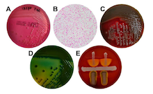
Non-O1/non-O139 Vibrio cholerae (NOVC) causes various illnesses ranging in severity from mild to life-threatening but were ignored previously. Knowledge of the NOVC infection, particularly bacteremia, is limited because of its rarity. Here we first retrospectively reported the demographic, clinical, and therapy characteristics of patients with NOVC infection. Isolated NOVC stains were identified by a series of biochemical, mass spectrometry (MS), and serum agglutination tests. The results of 11 patients with NOVC infection (including 8 with bacteremia) with a median age of 68 years were included in this report. Most isolated NOVC strains had antibiotic susceptibility. Patients with NOVC-positive were distributed in various departments, most occurring in gastroenterology (6 cases). Hepatic disease was the most common comorbid disease, followed by diabetes (3 cases) and biliary tract disease (3 cases). Two cases were previously healthy. The most common symptom at presentation was fever. All patients presented with abnormal changes in hematology and inflammatory parameters. Cephalosporins were the most frequently used antibiotics. Ten patients had a favorable outcome after treatment; one died from complicated underlying diseases. In summary, we recommend the timely identification of NOVC strains using MALDI-TOF-MS. The suspicion of NOVC bacteremia cannot be ruled out regardless of the host’s immune status. An alternative therapeutic regimen for this infection may be β-lactam antibiotics or combined with β-lactamase inhibitors. Regardless, the specific therapeutic regimen should be based on the antibiogram data.

Downloads

Download data is not yet available.

Author Biographies

Wen-Jing Li, School of Clinical Medicine, Jining Medical University, Jining, China


Jian Shi, School of Clinical Medicine, Jining Medical University, Jining, China


Xue Li, School of Clinical Medicine, Jining Medical University, Jining, China


Etiology and clinical features of non-O1/non-O139 Vibrio cholerae infection in an inland city in China

Downloads

Additional Files

Published

2023-07-03

How to Cite

1.
Xiang Y-H, Hu Q-Q, Liu Y, Sheng R, Wang J, Li W-J, Shi J, Li X, Lu S-H. Etiology and clinical features of non-O1/non-O139 Vibrio cholerae infection in an inland city in China. Biomol Biomed [Internet]. 2023Jul.3 [cited 2023Dec.1];23(4):698–704. Available from: https://www.bjbms.org/ojs/index.php/bjbms/article/view/8745

Issue

Section

Translational and Clinical Research

Introduction

Vibrio cholerae (V. cholerae) is a pathogenic gram-negative bacillus autochthonous to the aquatic environment that infects humans through contaminated water or food [1]. Based on the lipopolysaccharide surface O-antigen, over 200 serogroups of V. cholerae exist [2]. V. cholera O1 and O139 are the two major serogroups responsible for epidemic cholera outbreaks worldwide [3]. In contrast, other V. cholerae serogroups, designated as non-O1/non-O139 V. cholerae (NOVC), are not associated with epidemic cholera [4]. NOVC are genetically diverse strains that are generally non-pathogenic in healthy hosts [5]. Therefore, the clinical significance of NOVC was ignored previously; until recently, when NOVC infections emerged in public discussions [6].

As emerging evidence shows, NOVC has several virulence genes (e.g., toxR, hlyA, and ompU) encoding the cholera toxin that synergistically contributes to the infection process [7, 8]. Therefore, NOVC could cause various illnesses ranging in severity from mild in immunocompetent patients to life-threatening in immunocompromised hosts [9]. Based on previous reports, NOVC infection frequently presents as hypothermia, hyperthermia, diarrhea, abdominal pain, and other presentations (e.g., abscesses, necrotizing fasciitis, cellulitis, and others) [10, 11]. In contrast, it sporadically presents as a rare or atypical clinical manifestation (e.g., bacteremia and epigastric pain) [12]. Although bacteremia caused by NOVC is rare among these infections, it almost invariably occurs in immunodeficient patients and is potentially fatal [13, 14]. NOVC bacteremia rarely occurs in healthy patients [12]. Because of its rare occurrence, the epidemiology, clinical manifestations, and pathogenesis of NOVC infection, particularly bacteremia, remain unclear [15]. Currently, existing knowledge on NOVC bacteremia is mainly from limited cases [16, 17]. Meanwhile, no large-scale trials have yet to be conducted; thus, recommendations regarding the choice of NOVC infection therapy are inconclusive [10]. Therefore, a comprehensive review of patients with NOVC infection will be valuable for developing early diagnosis and treatment.

NOVC infection is associated with exposure to seawater and seafood consumption [9]. In recent years, several emerging reports have also shown that NOVCs can grow in inland waters [18]. Mainland China, especially inland areas, rarely report NOVC infection cases [6]. Until 2020, only 35 NOVC infection cases were linked to inland water sources worldwide, as reviewed by Vezzulli et al. [19]. Here, we first report the demographic, clinical, epidemiological, and therapeutic characteristics of a patient cohort with NOVC infection in the inland city of Shandong, China, to contribute to the understanding and timely diagnoses of NOVC infection.

Materials and methods

Patients

We reviewed the clinical records of all patients diagnosed with NOVC infection from January 1, 2019, to December 31, 2021. A total of 11 cases (7 men, 4 women; median age, 68 [range, 37–73] years) with laboratory-confirmed NOVC infection were identified from three tertiary hospitals in Shandong, China. The demographic and clinical characteristics, including age, gender, disease history, clinical features, laboratory results, therapeutic regimen, and prognosis, were obtained from the medical records and entered into standardized data collection forms. If data were missing from the records or clarification was needed, we gathered data by direct communication with attending doctors. Two investigators (Yu-Han Xiang and Qin-Qin Hu) independently extracted and cross-checked the information for each case to ensure the accuracy of the data.

Bacterial isolates and identification

NOVC strains were isolated from individuals with NOVC infection as per the following procedures. Blood, bile, pus, and stool samples were collected and processed in a microbiology laboratory within 30 min to isolate V. cholerae. Samples were ground under aseptic conditions and then inoculated on China blue agar plates (a selective medium similar to MacConkey agar [20]). China blue agar (Merck Millipore, Germany) is composed of meat extract (3 g/L), peptone from casein (5 g/L), sodium chloride (5 g/L), lactose (10 g/L), China-blue (0.375 g/L), and agar (12.0 g/L). An initial presumptive phenotype identification was performed according to colony morphology. The morphological characteristics of the isolated colonies were determined based on the color, shape, texture, size, and gram-staining of the colony. The hemolysis test was also performed to evaluate pathogenicity using a blood agar culture plate containing 4% Blood Agar Base (Merck Millipore, Germany) and 6% defibrinated goat blood. Thiosulfate-citrate bile salts-sucrose agar (TCBS) was used as the selective medium to isolate V. cholerae. Christie–Atkins–Munch–Peterson (CAMP) test was used to identify V. cholerae isolates by phenotype, using Escherichia coli (E. coli, ATCC25922) and Staphylococcus aureus (ATCC25923) as the quality control. CAMP-positive strains were assumed to belong to NOVC serogroups.

All isolates were subsequently examined by an automated Vitek-2 Compact system (Biomeriuex, France) and confirmed by matrix-assisted laser desorption ionization-time-of-flight mass spectrometry (MALDI-TOF-MS, BioMérieux, France). E. coli (ATCC8739) was used as the quality control for MALDI-TOF-MS. Meanwhile, the serotype of all isolates was confirmed by a serum agglutination test. V. cholerae strains that did not agglutinate with either O1 or O139 antisera were assumed to belong to NOVC serogroups.

Antimicrobial susceptibility tests

Antimicrobial susceptibility of NOVC strains was determined using the AST-GN13 panel in the automated Vitek-2 Compact system following the recommendations of the Clinical and Laboratory Standards Institute (CLSI, 31st edition). Antibiotics were selected according to the first-line and second-line drugs used for NOVC infection, including amikacin, ampicillin, ampicillin/sulbactam, aztreonam, cefazolin, cefepime, cefotetan, ceftazidime, cefuroxime, cefoperazone/sulbactam, ciprofloxacin, gentamicin, imipenem, levofloxacin, meropenem, piperacillin, piperacillin/tazobactam, tobramycin, and sulfamethoxazole. The antibiotic susceptibility was judged as susceptible, intermediate, or resistant based on the breakpoints recommended in CLSI. E. coli (ATCC25922) and S. aureus (ATCC25923) were used as controls.

Ethical statement

The study has been performed in accordance with the Declaration of Helsinki. The study protocol has been approved by the Institutional Review Board of the Affiliated Hospital of Jining Medical University (No. 2022C213). Informed consent was waived by the Institutional Review Board of the Affiliated Hospital of Jining Medical University due to the retrospective nature of the study.

Results

Phenotype and molecular identification and antimicrobial susceptibility of NOVC

The phenotype identification of isolated colonies was determined by various media cultivations. The sample collected upon admission yielded small, round, tabular, and faint yellow colonies on the China blue agar plates (Figure 1A). Meanwhile, the blood culture grew gram-negative curved or straight bacilli (Figure 1B), which were non-lactose fermenting and showed β-hemolytic, oxidase-positive colonies on blood agar (Figure 1C). However, the culture on TCBS agar presented as celadon (sucrose-fermenting) colonies (Figure 1D). The CAMP test indicated a weakly positive result and appeared as a crescent-shaped zone of complete synergistic hemolysis (Figure 1E); thus, NOVC was suspected. The strains were then identified using an automated Vitek-2 Compact system. All strains had been routinely typed as V. cholerae using the Vitek-2 Compact system, with identification rates of 94%–98% (Figure 2A).

Figure 1.: Phenotype identification of V. cholerae strains. (A) China blue agar showed small, round, tabular, and faint yellow colonies. (B) Gram stain showing Gram-negative curved bacilli (magnification, ×1000). (C) Blood agar showing β-hemolytic colonies. (D) TCBS agar presented as celadon (sucrose-fermenting) colonies. (E) CAMP test showed a crescent-shaped zone of complete synergistic hemolysis. CAMP: Christie–Atkins–Munch–Peterson test; TCBS: Thiosulfate-citrate bile salts-sucrose agar.
Figure 2.: Molecular identification of V. cholerae strains. (A) Representative result of Vitek-2 Compact system analysis (case 5). (B) Representative result of MALDI-TOF-MS analysis. MALDI-TOF-MS: Matrix-assisted laser desorption ionization-time-of-flight mass spectrometry.

Further species confirmation was obtained by the MALDI-TOF-MS analysis, with confidence levels of 99.9% for all strains (Figure 2B). A serum agglutination test was performed to classify the serotype of the pathogen. The pathogen indicated self-coagulation in normal saline, gradually weakening after several passages. The strain displayed no agglutination with the O1 or O139 antisera. Thus, the strains were classified as NOVC. Antimicrobial susceptibility tests indicated that two strains were intermediate to ciprofloxacin, two were resistant to ampicillin and sulfamethoxazole, and the others were all sensitive to the tested antibiotics. Multidrug-resistant isolates were not detected.

Demographic and clinical characteristics

Table 1 shows the demographic, clinical, and outcomes of all identified patients. Identified cases of NOVC infection included seven males and four females, with a median age of 68 years (range, 37–73 years). Patients with NOVC-positive were distributed in various departments, including gastroenterology (six cases), intensive care unit (two cases), infection (one case), intervention (one case), and gastrointestinal surgery (one case). Nine (81.8%) of 11 patients had at least one underlying comorbid disease. Hepatic disease (seven cases) was the most frequent comorbid disorder, followed by diabetes (three cases), biliary tract disease (three cases), hypertension (two cases), and one case each of pancreatic cancer, severe pneumonia, Behcet’s disease, bronchial asthma, and cerebral infarction. Only two cases were previously healthy. The most common symptoms at presentation were fever, fever with chills or rigors, edema of both lower limbs, and gastrointestinal symptoms, such as abdominal pain and diarrhea. According to the laboratory results, all patients presented with abnormal changes in hematology and inflammatory parameters (Table 2). Specifically, 7 (63.6%) of 11 patients indicated an increase in white blood cell counts, 10 (90.9%) an increase in neutrophil ratios, and 7 (63.6%) a decrease in platelet counts. In addition, platelet counts were elevated in two (18.2%) patients. Of nine patients who were evaluable for C-reactive protein levels (CRP), eight (88.9%) patients were CRP-positive (CRP ≥8 mg/L). The procalcitonin (PCT) levels were available in seven patients, and a positive PCT (PCT ≥0.5 ng/mL) was found in all seven (100%) patients. Only one patient had an increased level of hepatic function parameters.

Table 1: The demographic, clinical, and outcomes of all identified patients
Cases Age (years) Sex Underlying disease Clinical features Treatment regimens Treatment course (days) Outcomes Samples
Case 1 41 Male Pancreatic cancer with liver metastasis Fever Ceftriaxone 6 Cured Blood
Case 2 56 Male Liver cirrhosis, hepatic encephalopathy, diabetes Fever, dark stools, hematemesis Ceftazidime 8 Cured Blood
Case 3 70 Female Autoimmune liver disease, biliary cirrhosis, hypertension Fever, edema of both lower limbs Imipenem/Cilastatin 5 Cured Blood
Case 4 39 Male Acute liver failure, severe pneumonia, hepatitis B, Behcet’s disease Fever, edema of both lower limbs, skin icterus Piperacillin/Tazobactam 6 Early withdrawal Blood
Case 5 72 Male Liver cancer, liver cirrhosis, hypertension, diabetes Fever with chills or rigors, burning sensation in urination Piperacillin/Tazobactam 14 Cured Blood
Case 6 73 Female Liver cirrhosis, ascites, diabetes Fever, abdominal distension, diarrhea Ceftriaxone 7 Cured Blood
Case 7 69 Female None Abdominal pain, dizzy, headache, nausea Sulbactam/Cefoperazone 3 Improved Blood
Case 8 70 Female Cholangiocarcinoma, bronchial asthma Fever with chills or rigors Sulbactam/Cefoperazone 9 Cured Blood
Case 9 39 Male Cholecystitis, common bile duct stones with cholangitis Abdominal pain Cefoxitin 4 Cured Bile
Case 10 37 Male None Abdominal pain Meloxicillin/Ornidazole 5 Cured Pus
Case 11 68 Male Liver cirrhosis, cerebral infarction, splenectomy and cholecystectomy Fever, diarrhea Ceftriaxone 5 Cured Stool
Table 2: The laboratory results of all identified patients
Cases WBC (RefR: 3.5–9.5 ×10⁹/L) Neutrophils (RefR: 40%–75%) PLT (RefR: 125–350 ×109/L) CRP (RefR: <8 mg/L) PCT (RefR: <0.05 ng/nl) AST (RefR: 15–40 U/L) ALT (RefR: 9–50 U/L) GGT (RefR: 10–60 U/L)
Case 1 9.96 79.70 84.00 NA NA 40 33 57
Case 2 6.09 88.80 36.00 31.00 0.84 40 32 43
Case 3 10.78 86.50 44.00 50.00 0.52 38 34.6 19
Case 4 11.42 82.50 105.0 12.13 3.96 88 125.5 121
Case 5 8.73 91.70 64.00 67.36 3.04 31 32 70
Case 6 3.96 76.41 26 13.43 NA 32 33 52
Case 7 22.00 93.63 59 20.60 6.08 28 24 50
Case 8 12.00 81.67 415 109.40 0.56 29 25 24
Case 9 10.90 88.40 263 13.1 0.881 24 25.9 12
Case 10 16.15 86.94 352 NA NA 20 18 19
Case 11 3.62 36.74 219 0.71 NA 18 24 20

WBC: White blood cell; RefR: Reference range; PLT: Platelet; CRP: C-reactive protein; PCT: Procalcitonin; AST: Aspartate aminotransferase; ALT: Alanine aminotransferase; GGT: Gamma-glutamyl transpeptidase; NA: Not available.

Treatment and outcome

All 11 patients were treated with broad-spectrum antibiotics (Table 1). The most commonly used antibiotics were cephalosporins, such as ceftriaxone (cases 1, 6, and 11), ceftazidime (case 2), and cefoxitin (case 9). Five patients were empirically treated with a compound preparation of antibiotic and enzyme inhibitors, including imipenem/cilastatin (case 3), piperacillin/tazobactam (cases 4 and 5), and sulbactam/cefoperazone (cases 7 and 8). In addition, case 10 was synchronously given mezlocillin and ornidazole. Because of complicated underlying diseases, case 4 experienced multiple organ dysfunction syndromes and had to undergo mechanical ventilation but progressed to a critical condition. According to the decision of the patient’s family, the resuscitation attempt was refused. Another patient (case 7) showed improvement with a shorter 3-day treatment and was discharged according to the patient’s choice. The clinical symptoms of the remaining nine patients recovered after symptomatic treatment.

Discussion

This is the first published study to provide a systematic description of the demographic, clinical, and therapy characteristics of 11 hospitalized patients with confirmed NOVC infection in an inland city in China, contributing to the understanding of NOVC infection.

The accurate and timely identification of NOVC is crucial for the early diagnosis of NOVC infection [10]. However, traditional biochemical methods are time-consuming, leading to a delayed diagnosis or underreporting NOVC. In this study, MALDI-TOF MS was used for the rapid identification of human-pathogenic V. cholerae to obtain consistent results with biochemical identification by the Vitek-2 Compact system. Similar to a previous report [21], MALDI-TOF MS is a robust tool for rapidly identifying Vibrio species. The minimal awareness among clinicians and the non-reporting of clinical cases might be another explanation for the underreporting of NOVC [12]. Although NOVC infections are sporadic, we still remind clinicians to be aware of the possibility of infections due to this pathogen.

Evidence shows that cirrhosis is the leading risk factor for NOVC bacteremia [9, 22]. Similarly, in our cohort, we observed that over half of the patients had hepatic disease. NOVCs are non-pathogenic in healthy hosts and rarely lead to fatal bacteremia [5]. Two healthy hosts were diagnosed with NOVC infection but recovered soon after short antimicrobial treatments. However, these cases were not alone, as previously reported by Deshayes et al. or Namdari et al. [10, 23]. Thus, it is recommended that the suspicion of bacteremia cannot be ruled out in immunocompetent hosts when NOVC infection.

NOVC is usually susceptible to most antimicrobial agents [9]. In the present study, we observed a similar phenomenon: over half of the strains were sensitive to the tested antibiotics. Several NOVC strains with antibiotic resistance have recently been described in environmental and clinical settings, involving trimethoprim, SXT [13], ampicillin [24], and even multidrug-resistant (MDR) strains [25]. Although the MDR strain was not detected in our cases, two strains were identified as ampicillin- and sulfamethoxazole-resistant, which was consistent with its known antimicrobial susceptibility pattern [26]. Thus, it is recommended that antimicrobial therapy in patients with NOVC infection must be based on antimicrobial susceptibility patterns, considering the heterogeneity of antibiotic resistance.

Based on the published cases of NOVC bacteremia, antimicrobial therapy demonstrated extreme heterogeneity in antibiotic type, dosage, and treatment duration [10]. In the present cohort, cephalosporin appeared to be the first-choice antibiotic and was associated with a favorable outcome, consistent with the previous experience [27, 28]. In addition, many of our cases received dual-agent therapy with antibiotic and enzyme inhibitors, especially in immunocompromised patients, as recommended by Couzigou et al. [29]. We noticed that treatment duration varies according to the patient’s background, clinical presentation, and severity, ranging from 3 to 14 days. Thus, the duration of treatment remains a matter of debate. Except for our one patient with complicated underlying diseases, all patients had a favorable outcome, regardless of the type, dosage, and treatment duration of antimicrobial agents. Based on the experience from our center, 1–2 weeks of β-lactam antibiotics or combined with β-lactamase inhibitors might be an alternative therapeutic regimen for this infection. Although recommendations regarding the choice of therapy are not conclusive, patients with NOVC infection, even bacteremia, usually have a favorable outcome in the current treatment pattern.

Several limitations of this study should be recognized. Firstly, a small number of patients were included due to the rarity of this disease. Secondly, the molecular characterization of NOVC strains might provide more support for NOVC identification, however, the lack of virulence gene profiles of NOVC was another limitation for this study since self-paying patients refused the gene testing. The virulence gene profiles of NOVC strains would be concerned in further studies. Thirdly, radiology results were not available due to the retrospective design, limiting the comprehensive description of this infection. Additionally, the lack of patient exposure history might limit the elaboration of infection transmission of NOVC in inland cities, so this needs further investigation.

Conclusion

In this single-center study, we report for the first time on the largest cohort of patients with confirmed NOVC infection in an inland city in China. Although NOVC infections were sporadic, we still remind clinicians to be aware of the possibility of bacteremia, regardless of immunocompromised or immunocompetent individuals. MALDI-TOF MS is a robust tool for the rapid identification of this pathogen. In addition, β-lactam antibiotics combined with β-lactamase inhibitors may be an alternative therapeutic regimen for this infection. Regardless, the specific therapeutic regimen should be based on the antibiogram data.

References

  1. , , , , (). Antibiotic resistance in Vibrio cholerae: understanding the ecology of resistance genes and mechanisms. Vaccine. https://doi.org/10.1016/j.vaccine.2019.06.031
  2. , , , , (). Cholera. Lancet (London, England). https://doi.org/10.1016/S0140-6736(17)30559-7
  3. , , , , , (). Corrigendum: revisiting the global epidemiology of cholera in conjunction with the genomics of Vibrio cholerae. Front Public Health. https://doi.org/10.3389/fpubh.2019.00237
  4. , , , , (). Global emergence of environmental non-O1/O139 Vibrio cholerae infections linked with climate change: a neglected research field?. Environ Microbiol. https://doi.org/10.1111/1462-2920.15040
  5. , , , , , (). Non-O1, non-O139 Vibrio cholerae bacteremia in an urban academic medical center in the United States. IDCases. https://doi.org/10.1016/j.idcr.2019.e00527
  6. , , , , , (). Non-O1/non-O139 Vibrio cholerae bacteraemia in mainland China from 2005 to 2019: clinical, epidemiological and genetic characteristics. Epidemiol Infect. https://doi.org/10.1017/S0950268820001545
  7. , , , (). Environmental and clinical strains of Vibrio cholerae non-O1, non-O139 from Germany possess similar virulence gene profiles. Front Microbiol. https://doi.org/10.3389/fmicb.2019.00733
  8. , , (). Virulence gene profiles, biofilm formation, and antimicrobial resistance of Vibrio cholerae non-O1/non-O139 bacteria isolated from West Bengal, India. Heliyon. https://doi.org/10.1016/j.heliyon.2018.e01040
  9. , , , , , (). Non-O1, non-O139 Vibrio cholerae bacteraemia in a cirrhotic patient. J Med Microbiol. https://doi.org/10.1099/jmm.0.021014-0
  10. , , , , , (). Non-O1, non-O139 Vibrio cholerae bacteraemia: case report and literature review. SpringerPlus. https://doi.org/10.1186/s40064-015-1346-3
  11. , , , (). Non-O1, non-O139 Vibrio cholerae septicemia at a tertiary care center in Beirut, Lebanon; a case report and review. J Infect Public Health. https://doi.org/10.1016/j.jiph.2018.01.001
  12. , , , , (). Atypical manifestation of Vibrio cholerae: fear the water!. Acta Clin Belg. https://doi.org/10.1080/17843286.2018.1483563
  13. , , , , , (). The first case of bacteraemia due to non-O1/non-O139 Vibrio cholerae in a type 2 diabetes mellitus patient in mainland China. Int J Infect Dis. https://doi.org/10.1016/j.ijid.2014.04.015
  14. , , , , (). Non O1, non O139 Vibrio cholerae bacteraemia in an infant; case report and literature review. J Pakistan Med Assoc.
  15. , , , , , (). Non-O1, non-O139 Vibrio cholerae (NOVC) bacteremia: case report and literature review, 2015–2019. Infect Drug Resist. https://doi.org/10.2147/IDR.S245806
  16. , , , (). Nontoxigenic Vibrio cholerae septicemia in an immunocompromised patient. Case Rep Infect Dis. https://doi.org/10.1155/2012/698746
  17. , , , (). Non-O1, non-O139 Vibrio cholerae bacteremia in a chronic hemodialysis patient. Rev Argent microbiol. https://doi.org/10.1590/S0325-75412011000200002
  18. , , , , , (). Inland cholera in freshwater environs of North India. Vaccine. https://doi.org/10.1016/j.vaccine.2019.06.038
  19. , , , , (). Global emergence of environmental non-O1/O139 Vibrio cholerae infections linked with climate change: a neglected research field?. https://doi.org/10.1111/1462-2920.15040
  20. , , , , , (). Clinical performance of BD Kiestra InoqulA automated system in a Chinese tertiary hospital. Infect Drug Resist. https://doi.org/10.2147/IDR.S245173
  21. , , , , , (). Evaluation of the Bruker biotyper matrix-assisted laser desorption ionization-time of flight mass spectrometry system for identification of blood isolates of Vibrio species. J Clin Microbiol. https://doi.org/10.1128/JCM.00105-15
  22. , , , , (). Non-O1 Vibrio cholerae bacteremia in patients with cirrhosis: 5-yr experience from a single medical center. Am J Gastroenterol.
  23. , , (). A cytotoxin-producing strain of Vibrio cholerae non-o1, non-o139 as a cause of cholera and bacteremia after consumption of raw clams. J Clin Microbiol. https://doi.org/10.1128/JCM.38.9.3518-3519.2000
  24. , , , , , (). Molecular analysis of non-O1/non-O139 Vibrio cholerae isolated from hospitalised patients in China. BMC Microbiol. https://doi.org/10.1186/1471-2180-13-52
  25. , , , (). Multiresistant Vibrio cholerae non-O1/non-O139 from waters in South India: resistance patterns and virulence-associated gene profiles. J Basic Microbiol. https://doi.org/10.1002/jobm.200900085
  26. , , , , , (). Non-O1/non-O139 Vibrio cholerae carrying multiple virulence factors and V. cholerae O1 in the Chesapeake Bay, Maryland. Appl Environ Microbiol. https://doi.org/10.1128/AEM.03540-14
  27. , , , , , (). Vibrio cholerae non-O1, non-O139 bacteraemia associated with pneumonia, Italy 2016. Infection. https://doi.org/10.1007/s15010-016-0961-4
  28. , , , , , (). Two cases of bacteriemia caused by nontoxigenic, non-O1, non-O139 Vibrio cholerae isolates in Ho Chi Minh City, Vietnam. J Clin Microbiol. https://doi.org/10.1128/JCM.01915-14
  29. , , , , (). Non-O:1 and non-O:139 Vibrio cholerae septicemia and pyomyositis in an immunodeficient traveler returning from Tunisia. Travel Med Infect Dis. https://doi.org/10.1016/j.tmaid.2006.06.002

Conflicts of interest: Authors declare no conflicts of interest.

Funding: This study was supported by the Jining Key Research and Development Program (2021YXNS027), the Shandong Health Science and Technology Development Program (202211000699), and the Jining Key Research and Development Program (2022YXNS023).

Data availability: The data sets used and/or analyzed during the current study are available from the corresponding author upon reasonable request