Myocarditis and coronavirus disease 2019 vaccination: A systematic review and meta-summary of cases

Authors

  • Pandit Bagus Tri Saputra Department of Cardiology and Vascular Medicine, Faculty of Medicine, Universitas Airlangga–Dr Soetomo General Academic Hospital, Surabaya, Indonesia https://orcid.org/0000-0002-5815-0592
  • Roy Bagus Kurniawan Faculty of Medicine, Universitas Airlangga, Surabaya, Indonesia https://orcid.org/0000-0002-4220-8286
  • Desy Trilistyoati Faculty of Medicine, Universitas Airlangga, Surabaya, Indonesia
  • Makhyan Jibril Al Farabi Department of Cardiology and Vascular Medicine, Faculty of Medicine, Universitas Airlangga–Dr Soetomo General Academic Hospital, Surabaya, Indonesia
  • Hendri Susilo Department of Cardiology and Vascular Medicine, Universitas Airlangga Hospital, Surabaya, Indonesia https://orcid.org/0000-0002-5603-9487
  • Mochamad Yusuf Alsagaff Department of Cardiology and Vascular Medicine, Faculty of Medicine, Universitas Airlangga–Dr Soetomo General Academic Hospital, Surabaya, Indonesia; Department of Cardiology and Vascular Medicine, Universitas Airlangga Hospital, Surabaya, Indonesia
  • Yudi Her Oktaviono Department of Cardiology and Vascular Medicine, Faculty of Medicine, Universitas Airlangga–Dr Soetomo General Academic Hospital, Surabaya, Indonesia
  • Henry Sutanto Department of Cardiology, CARIM School for Cardiovascular Diseases, Maastricht University, Maastricht, ‎Netherlands
  • Arief Gusnanto School of Mathematics, University of Leeds, Leeds, UK https://orcid.org/0000-0001-5748-784X
  • Citrawati Dyah Kencono Wungu Department of Physiology and Medical Biochemistry, Faculty of Medicine, Universitas Airlangga, Surabaya, Indonesia; Institute of Tropical Disease, Universitas Airlangga, Surabaya, Indonesia https://orcid.org/0000-0001-5180-957X

DOI:

https://doi.org/10.17305/bb.2022.8779

Keywords:

myocarditis, COVID-19 vaccination, vaccine, SARS-CoV2, side effect

Abstract

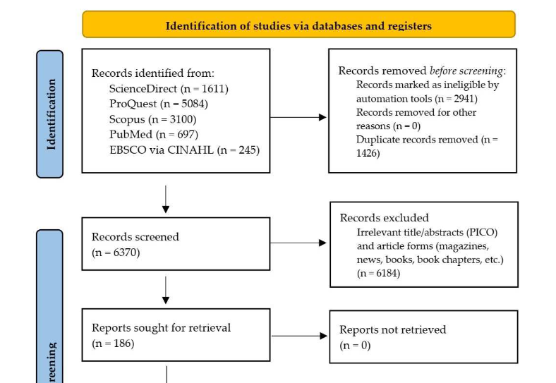
Vaccination is significant to control, mitigate, and recover from the destructive effects of coronavirus disease 2019 (COVID-19). The incidence of myocarditis following COVID-19 vaccination has been increasing and growing public concern; however, little is known about it. This study aimed to systematically review myocarditis following COVID-19 vaccination. We included studies containing individual patient data of myocarditis following COVID-19 vaccination published between January 1, 2020 and September 7, 2022 and excluded review articles. Joanna Briggs Institute critical appraisals were used for risk of bias assessment. Descriptive and analytic statistics were performed. A total of 121 reports and 43 case series from five databases were included. We identified 396 published cases of myocarditis and observed that the majority of cases was male patients, happened following the second dose of mRNA vaccine administration, and experienced chest pain as a symptom. Previous COVID-19 infection was significantly associated (p < 0.01; OR, 5.74; 95% CI, 2.42–13.64) with the risk of myocarditis following the administration of the first dose, indicating that its primary mechanism is immune-mediated. Moreover, 63 histopathology examinations were dominated by non-infective subtypes. Electrocardiography and cardiac marker combination is a sensitive screening modality. However, cardiac magnetic resonance is a significant noninvasive examination to confirm myocarditis. Endomyocardial biopsy may be considered in confusing and severe cases. Myocarditis following COVID-19 vaccination is relatively benign, with a median length of hospitalization of 5 days, intensive care unit admission of <12%, and mortality of <2%. The majority was treated with nonsteroidal anti-inflammatory drugs, colchicine, and steroids. Surprisingly, deceased cases had characteristics of being female, older age, non-chest pain symptoms, first-dose vaccination, left ventricular ejection fraction of <30%, fulminant myocarditis, and eosinophil infiltrate histopathology.

Downloads

Download data is not yet available.
Myocarditis and coronavirus disease 2019 vaccination: A systematic review and meta-summary of cases

Downloads

Additional Files

Published

2023-07-03

How to Cite

1.
Saputra PBT, Kurniawan RB, Trilistyoati D, Farabi MJA, Susilo H, Alsagaff MY, Oktaviono YH, Sutanto H, Gusnanto A, Dyah Kencono Wungu C. Myocarditis and coronavirus disease 2019 vaccination: A systematic review and meta-summary of cases. Biomol Biomed [Internet]. 2023Jul.3 [cited 2023Nov.30];23(4):546–567. Available from: https://www.bjbms.org/ojs/index.php/bjbms/article/view/8779

Issue

Section

Reviews

Introduction

Since first identified and reported in China in late 2019, coronavirus disease 2019 (COVID-19) has rapidly and extensively spread among countries, escalating into a pandemic. The COVID-19 pandemic has severely affected economic, social, political, and cultural sectors [1] and has persisted in attracting worldwide attention.

To control, mitigate, and recover from the destructive effects of COVID-19, vaccination is critical. Recent evidence regarding the ineffectiveness of COVID-19 vaccines for some strains. However, COVID-19 vaccination may pose beneficial effects, particularly, for high-risk populations, including older adults and individuals with multiple comorbidities [2]. To support international vaccination coverage, the World Health Organization (WHO) issued authorization for COVID-19 vaccine emergency use [3] in combination with government regulation to accelerate [4] COVID-19 vaccination up to 12 billion doses, which continues to increase globally [5]. With the rapid increase in vaccination shots comes several reported adverse events complained by patients, ranging from minor to major events [6].

COVID-19 vaccination induced several adverse symptoms including pain at the injection site, fever, myalgia, arthralgia, headache, and abdominal discomfort [6, 7]. Several vaccine-related diseases have also been reported in different organs, including the respiratory tract (e.g., asthma attack, diffuse alveolar hemorrhage, and eosinophilic pneumonia), gastrointestinal tract (e.g., appendicitis, autoimmune hepatitis, intestinal ulcer, and perforation), skin (e.g., alopecia areata, bullous pemphigoid, psoriasis, and eosinophilic cellulitis), and endocrine organs (e.g., Graves’ disease, hypophysitis, hypothyroidism, and diabetes mellitus) [8]. Emerging myocarditis cases have received media attention. Various literature reported myocarditis following COVID-19 vaccination, which is considered one of the serious adverse events [9]. The number of these reports has been increasing as more individuals get vaccinated, thereby growing public concern regarding COVID-19 vaccination [10].

Therefore, to provide appropriate information and a standing position for the public and clinicians, including myocarditis following COVID-19 vaccination, any vaccination-related issues should be thoroughly investigated. However, most reports on myocarditis following COVID-19 vaccination were case reports and case series, and little is known regarding the nature and characteristics of this phenomenon. Therefore, this systematic review aimed to describe the characteristics of myocarditis following COVID-19 vaccination by collecting detailed individual patient data (IPD) from those available reports and literature to create a systematic review and meta-summary of cases of myocarditis following COVID-19 vaccination.

Materials and methods

This review followed the Preferred Reporting Items for Systematic Reviews and Meta-Analyses (PRISMA) 2020 [11] and was registered and can be accessed in the PROSPERO database (CRD42021271806). No ethical approval was required as we only analyzed secondary data from published literature.

Eligibility criteria

The following study types were included in this review: case reports, case series, and observational studies (cross-sectional, case-control, and cohort studies). Screening of eligible studies was based on the following inclusion criteria: (1) patients of all ages with myocarditis following any COVID-19 vaccination type as participants according to the definition from European Society of Cardiology (ESC) guidelines for myocarditis [12] or Centers for Disease Control and Prevention (CDC) updates on myocarditis following COVID-19 vaccination [13]; (2) individual-specific patient data were available; (3) all COVID-19 vaccine types (mRNA and non-mRNA vaccines); and (4) reported in English. Diagnosis of myocarditis was based on the combination of clinical signs, electrocardiography (ECG), cardiac markers, echocardiography/angiocardiography, cardiac magnetic resonance (CMR), and, if available, histopathologic examination (autopsy or endomyocardial biopsy) according to the ESC [12] and CDC updates on myocarditis [13].

We excluded nonscientific articles (popular magazines), randomized-controlled trials, reviews, unavailable text, duplication, and studies that report unclear populations (e.g., pericarditis without myocardial involvement or heart failure and myocardial injury instead of myocarditis). Myocarditis that was not due to COVID-19 was excluded. If needed, we contacted the authors to ask for additional patient information.

Search strategy

On September 7, 2022, a literature search was systematically performed on ScienceDirect, Scopus, PubMed, CINAHL via EBSCO, and ProQuest using keywords and medical subject headings with English language restrictions. Published articles from January 1, 2020, the month of COVID-19 discovery, to September 7, 2022 were included. The search terms were (“COVID19” OR “COVID-19” OR “coronavirus disease 2019” OR “SARS-CoV-2”) AND (“vaccine” OR “vaccination”) AND (“myocarditis” OR “myocardial injury” OR “myopericarditis”).

Study selection

Titles and abstracts were independently screened for eligibility. Subsequently, studies with potentially eligible abstracts were reviewed for full-text articles according to previously determined inclusion and exclusion criteria. Any disagreements were resolved by consensus of those authors.

Data extraction

The following data were extracted from included studies: the number of cases; sex; age; region; comorbidities; previous COVID-19 infection; current COVID-19 infection status; vaccine types; vaccine doses; from the day(s) of vaccination to specific myocarditis symptoms; from the day(s) of vaccination to the emergency department (ED) admission; from the day(s) of specific symptom onset to ED from specific myocarditis symptoms; preceding symptoms; presenting symptoms; cardiac markers, including troponin I, high-sensitive troponin I (hsTnI), troponin T, high-sensitive troponin T (hsTnT), creatinine phosphokinase (CPK), creatinine kinase myocardial band (CKMB), brain natriuretic peptide (BNP), and Pro-BNP; D-dimer; white blood cells (WBCs); erythrocyte sedimentation rate (ESR); C-reactive protein (CRP); ECG/Holter stress test; echocardiography/angiography; CMR; cardiac histopathologic examination; length of hospitalization (LoH); management; and outcomes (deceased or recovery).

In the context of day(s) from the event (e.g., vaccination day) to the event (e.g., specific symptoms), the first event was determined as day one. The specific symptoms included chest pain, dyspnea, palpitation, syncope, or cardiac arrest [13], which led patients to the ED. Preceding symptoms were determined as any symptoms or signs that occurred before specific symptoms. CMR data were collected and mainly classified as edema, hyperemia, fibrosis/scar, and pericardial involvement [12], [14]. Patients were classified into probable and confirmed cases according to the CDC criteria [13], and their myocarditis score was also evaluated based on the ESC criteria [12]. All collected data were independently compared as pre-piloted forms, and any discrepancy was resolved through consensus.

Quality assessment

We independently analyzed and judged the risk of bias of included studies using the Joanna Briggs Institute (JBI) for case reports and case series critical appraisal instruments [15]. The JBI critical appraisal for case reports consists of eight questions, whereas JBI critical appraisal for case series consists of ten questions. However, we combined questions number 4 and 5 in the JBI appraisal as they represent the same domain (bias selection of participants into the study), and we did not include question number 10, considering statistical analysis is not an appropriate characteristic in this context.

Figure 1.: Preferred reporting items for systematic reviews and meta-analyses flow diagram of the study selection process.

This critical appraisal consists of yes/no/unclear answers for each question [16]. The studies were classified as low, medium, and high risks of bias according to the total “yes” answers of ≥74%, 50%–74%, and ≤49% of total questions, respectively.

Statistical analysis

All collected IPD from the included study were combined in a meta-summary. Patients with confirmed cases were compared with probable cases. A p-value threshold of 0.05 was considered statistically significant. The Shapiro–Wilk test was used to determine the normality distribution of the data [17]. Categorical variables were presented as frequencies and percentages. Continuous variables were presented as means and standard deviations; otherwise, median and range (minimum–maximum) were considered. As indicated, categorical data were compared using Chi-square or Fisher’s exact test [18]. Moreover, normally distributed data were compared by employing the independent T-test. Otherwise, the Mann–Whitney test would be used when the data were abnormally distributed by the Shapiro–Wilk test. We identified demographic and comorbid factors that may contribute to the occurrence of myocarditis following the first COVID-19 vaccination by multivariate analysis (logistic regression). Only variables with a p-value of <0.25 were included in the multivariate analysis [19]. The Spearman or Pearson correlation test was used to evaluate the correlation between clinical parameters and outcomes by considering data distribution. We used a statistical software, IBM SPSS Statistics for Windows, version 22 (IBM Corp., Armonk, NY, USA), during statistical analysis processes.

Results

Study selection

In the primary search, 6370 studies were identified after the removal of 2941 irrelevant research articles by the automation feature of each search engine and 1426 duplications. A total of 6184 articles were excluded during the title and abstract screening, leaving 186 studies for full-text reviews. Subsequently, 24 studies were excluded, while 164 studies consisted of 121 case reports [20–130] and 43 case series [131–178], with a total of 396 cases included (Figure 1) in this meta-summary. Some studies, such as those by Diaz et al. [179] and Montgomery et al. [180] fulfilled the inclusion criteria and consisted of several participants. However, we excluded them because specific individual data could not be extracted. We only included patients who fulfilled the myocarditis criteria, such as in the case series reported by Patel et al. [178].

Meta-summary of cases

IPD from 396 cases was included in this systematic review (Table S2). The basic data and comparison of demographic, clinical characteristics, and management between probable and confirmed cases are presented in Table 1.

Table 1: Characteristics and comparisons between published probable and confirmed cases of myocarditis following vaccination
CDC VAERS Myocarditis Status# [13]
Variables Reported cases* Percentage or median (%)** Probable (%) Confirmed (%) p-valuea
Age 370 22 (12–88)b 20 (12–88)b 23 (12–80)b 0.04c
Male 384 361 (87.6) 73 (90.1) 263 (86.8) 0.57h
**WHO regional area 383
Africa (AFR) 1 (0.3) 1 (1.2) 0 N/A
America (AMR) 150 (39.2) 33 (40.2) 117 (38.9) N/A
South-East Asia (SEAR) 2 (0.5) 1 (1.2) 1 (0.3) N/A
Europe (EUR) 155 (40.5) 27 (32.9) 128 (42.5) N/A
Eastern Mediterranean (EMR) 16 (4.2) 10 (12.2) 6 (2.0) N/A
Western Pacific (WPR) 59 (15.4) 10 (12.2) 49 (16.3) N/A
**Vaccine type 394
BNT16 2b2 270 (68.5) 61 (75.3) 209 (66.8) N/A
mRNA-1273 97 (24.6) 16 (19.8) 81 (25.9) N/A
Ad26.COV2. S 8 (2.0) 2 (2.5) 6 (1.9) N/A
AZD1222 14 (3.6) 2 (2.5) 12 (3.8) N/A
BBV152 1 (0.3) 0 (0) 1 (0.3) N/A
rAd26 and rAd5 1 (0.3) 0 (0) 1 (0.3) N/A
NVX-C oV2373 1 (0.3) 0 (0) 1 (0.3) N/A
Inactivated SARS-CoV-2 2 (0.5) 0 (0) 2 (0.6) N/A
**Previous SARS-CoV-2 infection 167 33 (19.8) 4 (11.1) 29 (22.1) 0.22h
COVID-19 vaccination history 384
First dose 89 (23.2) 16 (19.8) 73 (24.1) 0.50h
Second/Third doses 295 (76.8) 65 (80.2) 230 (75.2)
Comorbidities 231
None 131 (56.7) 24 (42.9) 107 (61.1) 0.03g,h
Psychiatric/functional disorders 14 (6.1) 4 (7.1) 10 (5.7) 0.75
Immune system/inflammatory disorders 14 (6.1) 6 (10.7) 8 (4.6) 0.11
Hypertension 13 (5.6) 5 (8.9) 8 (4.6) 0.31
Bronchial asthma 13 (5.6) 6 (10.7) 7 (4.0) 0.09
Myocarditis 11 (4.8) 5 (8.9) 6 (3.4) 0.14
Dyslipidemia 10 (4.3) 3 (5.4) 7 (4.0) 0.71
Miscellaneous atopic disease 6 (2.6) 1 (1.8) 5 (2.9) 1.00
GERD/gastritis 5 (2.2) 1 (1.8) 4 (2.3) 1.00
Dysrhytmia 5 (2.2) 1 (1.8) 4 (2.3) 1.00
Hypothyroidism 5 (2.2) 1 (1.8) 4 (2.3) 1.00
Genetic/chromosomal disease 5 (2.2) 2 (3.6) 3 (1.7) 0.60
Obesity 4 (1.7) 3 (5.4) 1 (0.6) 0.05g
DMT2/insulin resistance 4 (1.7) 3 (5.4) 1 (0.6) 0.05g
Coronary arterial disease 4 (1.7) 3 (5.4) 1 (0.6) 0.05g
Malignancy 4 (1.7) 0 4 (2.3) 0.58
Infections 3 (1.3) 0 3 (1.7) 1.00
Congestive heart failure 2 (0.9) 2 (3.6) 0 0.06
Multiple sclerosis 2 (0.9) 2 (3.6) 0 0.06
Obstructive sleep apnea 1 (0.4) 0 1 (0.6) 1.00
Aortic root dilatation 1 (0.4) 0 1 (0.6) 1.00
Vitiligo 1 (0.4) 1 (1.8) 0 0.24
COPD 1 (0.4) 1 (1.8) 0 0.24
Recurrent pneumothorax 1 (0.4) 0 1 (0.6) 1.00
Encephalopathy 1 (0.4) 1 (1.8) 0 0.24
Preceding symptoms 215
None 71 (33.0) 20 (43.5) 51 (30.2) 0.13h
Fever 96 (44.7) 15 (32.6) 81 (47.9) 0.09h
Myalgia 47 (21.9) 8 (17.4) 39 (23.1) 0.53h
Malaise 31 (14.4) 5 (10.9) 26 (15.4) 0.60
Headache 24 (11.2) 5 (10.9) 19 (11.2) 1.00h
Chills 21 (9.8) 5 (10.9) 16 (9.5) 0.78
Nausea 10 (4.7) 2 (4.3) 8 (4.7) 1.00
Diarrhea 10 (4.7) 2 (4.3) 8 (4.7) 1.00
Vomiting 9 (4.2) 4 (8.7) 5 (3.0) 0.10
Dry cough 9 (4.2) 1 (2.2) 8 (4.7) 0.69
Palpitation 9 (4.2) 0 9 (5.3) 0.21
Shortness of breath 9 (4.2) 3 (6.5) 6 (3.6) 0.41
Limb pain 7 (3.3) 1 (2.2) 6 (3.6) 1.00
Diaphoresis 5 (2.3) 1 (2.2) 4 (2.4) 1.00
Injection site pain 4 (1.9) 0 4 (2.4) 0.58
Arthralgia 4 (1.9) 0 4 (2.4) 0.58
Sore throat 4 (1.9) 0 4 (2.4) 0.58
Back pain 3 (1.4) 1 (2.2) 2 (1.2) 0.52
Abdominal pain 2 (0.9) 1 (2.2) 1 (0.6) 1.00
Hematuria 1 (0.5) 0 1 (0.6) 1.00
Epistaxis 1 (0.5) 0 1 (0.6) 1.00
Presenting symptoms 387
Chest pain 323 (83.5) 68 (84.0) 255 (83.3) 1.00h
Fever 57 (14.7) 9 (11.1) 48 (15.7) 0.39h
Dyspnea or SoB 56 (14.5) 12 (14.8) 44 (14.4) 1.00h
Malaise/fatigue 35 (9.0) 11 (13.6) 24 (7.8) 0.17h
Headache 25 (6.5) 5 (6.2) 20 (6.5) 1.00h
Tachycardia/palpitation 23 (5.9) 6 (7.4) 17 (5.6) 0.60
Gastrointestinal symptoms 18 (4.7) 4 (4.9) 14 (4.6) 1.00
Myalgia 15 (3.9) 2 (2.5) 13 (4.2) 0.75
Chills 10 (2.6) 3 (3.7) 7 (2.3) 0.44
Diaphoresis 7 (1.8) 1 (1.2) 6 (2.0) 1.00
Syncope 7 (1.8) 1 (1.2) 6 (2.0) 1.00
Hypotension 6 (1.6) 1 (1.2) 5 (1.6) 1.00
Limb pain 4 (1.0) 1 (1.2) 3 (1.0) 1.00
Shock 4 (1.0) 1 (1.2) 3 (1.0) 1.00
Arrest 3 (0.8) 0 3 (1.0) 1.00
Athralgia 1 (0.3) 0 1 (0.3) 1.00
Paresthesia 1 (0.3) 1 (1.2) 0 0.21
Heart failure 1 (0.3) 0 1 (0.3) 1.00
Cough 1 (0.3) 1 (1.2) 0 0.21
Back pain 1 (0.3) 0 1 (0.3) 1.00
From vaccination to specific symptoms (days) 344 3 (1–90)b 3 (1–60)b 3 (1–90)b 0.00c,g
From vaccination to the ED (days) 211 4 (1–90)b 3 (2–60)b 4 (1–90)b 0.28c
Specific symptoms to the ED (days) 202 0 (0–21)b 0 (0–18)b 0 (0–21)b 0.03c,g
Clinical laboratoryf
CRP 262 205 (78.2) 36 (81.8) 169 (77.5) 0.68h
White blood cells 110 44 (40.0) 10 (45.5) 34 (38.6) 0.73h
ESR 67 35 (52.2) 10 (66.7) 25 (48.1) 0.33h
D-dimer 44 24 (54.5) 11 (68.8) 13 (46.4) 0.26h
**Cardiac-specific examinationf
hsTroponin I 48 43 3 (100.0) 40 (88.9) 1.00
Troponin I 177 165 (93.2) 47 (97.9) 118 (91.5) 0.18
hsTroponin T 43 42 (97.7) 3 (75.0) 39 (100.0) 0.09
Troponin T 74 67 (90.5) 9 (81.8) 58 (92.1) 0.28
CPK 68 58 (85.3) 10 (90.9) 48 (84.2) 1.00
CKMB 57 48 (84.2) 9 (81.8) 39 (84.8) 1.00
BNP 82 42 (51.2) 5 (38.5) 37 (53.6) 0.48h
NT-proBNP 65 47 (72.3) 4 (50.0) 43 (75.4) 0.20
Left ventricular rejection fraction (LVEF) (%) 265 55 (5.0–72.0) 57 (10–70) 54 (5.0–72.0) 0.11c
LVEF < 50.00 % 265 86 (32.7) 12 (26.7) 74 (33.9) 0.44h
ESC score 396 3 (1–5) 2 (1–4)b 3 (1–5)b <0.01c,g
Length of hospitalization (day) 248 5 (0–79) 3 (1–16)b 5 (0–79)b <0.01c,g
Management 322
None/observation 33 (10.2) 9 (11.8) 24 (9.8) 0.76h
NSAIDs 177 (55.0) 50 (65.8) 127 (51.6) 0.04g,h
Colchicine 81 (25.2) 19 (25.0) 62 (25.2) 1.00h
Corticostesroids 64 (19.9) 11 (14.5) 53 (21.5) 0.24 h
Beta-blockers 54 (16.8) 8 (10.5) 46 (18.7) 0.14h
ACEIs/ARBs 50 (15.5) 6 (7.9) 44 (17.9) 0.55h
Intensive care unit 39 (12.1) 11 (14.5) 28 (11.4) 0.60h
IVIGs 34 (10.6) 5 (6.6) 29 (11.8) 0.28h
Diuretics 19 (5.9) 2 (2.6) 17 (6.9) 0.26
Inotropic agents 15 (4.7) 3 (3.9) 12 (4.9) 1.00
Gastric acid suppressors 14 (4.3) 6 (7.9) 8 (3.3) 0.11
Antibiotics 11 (3.4) 3 (3.9) 8 (3.3) 0.73
Anticoagulants 10 (3.1) 0 10 (4.1) 0.13
ECMO 10 (3.1) 1 (1.3) 9 (3.7) 0.46
MHS 10 (3.1) 2 (2.6) 8 (3.3) 1.00
Paracetamol 9 (2.8) 4 (5.3) 5 (2.0) 0.22
Antiplatelets 9 (2.8) 1 (1.3) 8 (3.3) 0.69
Miscellaneous analgesia 8 (2.5) 2 (2.6) 6 (2.4) 1.00
Anti-arrhythmias 7 (2.2) 1 (1.3) 6 (2.4) 1.00
Vasodilators 6 (1.9) 2 (2.6) 4 (1.6) 0.62
Intubation/ventilator 5 (1.6) 2 (2.6) 3 (1.2) 0.34
Hemodialysis 4 (1.2) 2 (2.6) 2 (0.8) 0.24
CPR 4 (1.2) 1 (1.3) 3 (1.2) 1.00
Heart failure 2 (0.6) 0 2 (0.8) 1.00
Statin 2 (0.6) 1 (1.3) 1 (0.4) 0.42
Immunosuppressant 2 (0.6) 0 2 (0.8) 1.00
Bronchodilators 1 (0.3) 1 (1.3) 0 0.24
Interleukin antagonist 1 (0.3) 0 1 (0.4) 1.00
MgSO4 1 (0.3) 1 (1.3) 0 0.24
Vitamin D 1 (0.3) 0 1 (0.4) 1.00

#The definition of probable and confirmed cases is based on the Centers for Disease Control and Prevention classification of myocarditis [13]. *A case is defined as individual patient data (IPD). Reported cases refer to the number of cases that reported a specific variable. Notably, not all cases describe all variables and that made each variable have a different number of reported cases. For example, reported data on age, sex, and previous COVID-19 infection are available in 370, 384, and 167 cases, respectively. The numbers of reported cases (which act as the denominator for each group) between the probable and confirmed groups are also different (not shown). **This refers to the percentage of variable (with the reported cases of variable as the denominator). Furthermore, this refers to the median in variables with continuous data (e.g., age, from vaccination to the specific symptom, from vaccination to the ED, from specific symptoms to the ED, ESC score, and length of hospitalization). aThe p-values (rightmost column) of the result of testing the hypothesis between the probable and confirmed groups a Fisher’s exact test; bmedian (minimum–maximum); cMann–Whitney test; dmean ± standard deviation; eindependent T-test; fan increase of more than their reference ranges; gsignificant at p ≤ 0.05; hChi-square test. N/A: Not applicable; ED: Emergency department; DMT2: Diabetes mellitus type 2; GERD: Gastroesophageal reflux disease; COPD: Chronic obstructive pulmonary disease; WBC: White blood cell; ESR: Erythrocyte sedimentation rate; CRP: C-reactive protein; CKMB: Creatinine kinase myocardial band; BNP: B-type natriuretic peptide; NT-proBNP: N-Terminal pro-BNP; ESC: European Society of Cardiology; NSAIDs: Nonsteroidal anti-inflammatory drugs; IVIG: Intravenous immunoglobulin; ACE: Angiotensin-converting enzyme; ECMO: Extracorporeal membrane oxygenation; MHS: Mechanical hemodynamic support; CPR: Cardiopulmonary resuscitation.

Notably, not all cases describe all variables and that made each variable have a different number of reported cases. For example, reported data on age, sex, and previous COVID-19 infection are available in 370, 384, and 167 cases, respectively. That also applies to other variables, which are subsequently named as reported cases in Table 1. Furthermore, due to different reported cases in each variable, the numbers of probable and confirmed cases (which act as the denominator for each group) are different in each variable. The number of probable and confirmed cases for each variable is not shown.

Demographic characteristics

The majority of reported myocarditis was mRNA-type vaccines, including BNT162b2 (68.5%) and mRNA-1273 (23.9%). Moreover, we observed myocarditis following the administration of viral vector-based vaccine (6%, consisting of 8, 14, and 1 cases of Ad26.COV2.S, AZD1222, and rAd26-rAd5, respectively), inactivated virus vaccine (3 cases), and recombinant protein vaccine (1 case of NVX-CoV2373). The median age was 22 (range 12–88) years, and the majority (87.6%) were male patients. There were 121 cases of children under 18 years old, with eight cases of children under 13 years old. According to WHO regional area, the majority of reported articles were from Europe (40.5), America (39.2%), and Western Pacific (15.2%). Less than 10% of reports were from Asian and African regions.

Clinical characteristics

Of 231 reported cases, 43.3% had comorbidities, including psychiatric disorders (6.1%), immune/inflammatory disorders (6.1%), hypertension (5.6%), bronchial asthma (5.6%), history of myocarditis (4.8%), dyslipidemia (4.3%), and others (Table 1). There were 33 of 167 (19.8%) reported cases with previous COVID-19 infection. The majority (76.8%) of the cases were associated with the second dose of the COVID-19 mRNA vaccine.

Preceding symptoms accompanied 33% of cases; the most reported preceding symptoms were fever (44.7%), myalgia (21.9%), malaise (14.4%), headache (11.2%), and chills (9.8%). Other preceding symptoms are listed in Table 1.

Chest pain was the most common presenting (83.5%) (Table 1) and myocarditis-specific (97%) symptom (Table S3). Other specific symptoms included dyspnea (14.5%), palpitation (5.9%), hypotension/syncope/heart failure (4.4%), and diaphoresis (1.8%). Three patients (0.8%) had cardiac arrest events in the ED. The three most common constitutional symptoms were fever (14.7%), malaise (9%), and headache (6.5%).

The median of vaccination day to the presence of specific symptoms and from the vaccination day to the ED admission was 3 (1–90) and 4 (1–90) days, respectively. Meanwhile, most cases reported that the patients came to the ED on the day of specific symptom onset. The confirmed group had a significantly shorter interval of the day(s) of vaccination to specific symptoms and day(s) of specific symptoms to the ED (p < 0.01 and p < 0.03, respectively) than the probable group (Table 1).

Laboratory examination

In this meta-summary, all inflammatory markers, except for WBC (40%), were increased in more than half of the cases: CRP (78.2%), D-Dimer (54.5%), and ESR (52.2%). No differences in inflammatory marker abnormality between the probable and confirmed groups were noted (Table 1).

Electrocardiography

Of 352 ECG results, typical abnormalities were noted in 290 (82.3%) cases (Table 2). ST-segment abnormalities were observed as 83% of abnormalities and 68.5% of overall ECG results. The most common ST-segment abnormality was ST-segment elevation (81.7%), whereas 36% was diffuse ST-segment elevation. T-wave abnormalities were noted in 10% of the ECG results. Intraventricular conduction abnormalities (LBBB, RBBB, and nonspecific wide QRS complex) were more common than atrioventricular block (grades I–III) (6.8% vs 1.4%, respectively). Additionally, ventricular-origin arrhythmia was more common than atrial-origin arrhythmia (6.3% vs 1.7%, respectively).

Table 2: Cardiac-specific examination findings
Examination Number of cases (%)
ECG
Available ECGa 352 (96)
PR-segment depression 26 (7.4)
ST-segment abnormality 241 (68.5)
ST depression 11 (3.1)
ST elevation* 197 (56)
Nonspecific ST abnormality 29 (8.2)
T abnormality 35 (9.9)
Peak T wave 5 (1.4)
T inversion 24 (6.8)
Unspecified abnormality 16 (4.5)
Intraventricular conduction 24 (6.8)
Left-BBB 3 (0.9)
Right-BBB 14 (4)
Unpacified wide QRS complex (≥0.12 s) 7 (2)
Atrioventricular block (grades I–III) 5 (1.4)
Premature atrial contraction 2 (0.6)
Atrial fibrillation 2 (0.6)
Supraventricular tachycardia 2 (0.6)
Premature ventricular contraction 6 (1.7)
Ventricular tachycardia 14 (4)
Ventricular fibrillation 2 (0.6)
Other arrhythmias (e.g., long QT interval, W wave inversion, early repolarization, poor R regression, AVD, JER, and poor QRS) 9 (2.6)
Abnormal ECG 290 (82.3)
Cardiac markers
Available cardiac markerb 379 (97.2)
Abnormal cardiac marker 372 (98.4)
Echocardiography/angiography
Available echocardiography/angiographyc 326 (82.1)
Hypokinesia 116 (35.6)
Pericardial effusion 33 (10)
LVEF < 55% 113 (34.7)
Abnormal echocardiography/angiography 113 (34.7)
CMR
Available CMRd 286 (75.9)
LGE/fibrosis/scar 242 (84.6)
Myocardial edema 126 (44.1)
EGE/hyperemia 12 (4.2)
Pericardial involvement 21 (7.3)
Abnormal CMR 259 (90)
Histopathology examination
Available histopathology examinatione 63 (16)
Lymphocytic 17 (27)
Lymphohistyocytic 12 (19)
Giant cell 4 (6.4)
Eosinophilic 3 (4.8)
Mixed 3 (3.8)
Neutrophil predominance: histiocyte/lymphocyte 2 (3.2)
Histiocyte predominance 1 (1.6)
Not mentioned 15 (23.8)
Abnormal histopathology examination 56 (89)

*Diffuse ST-segment elevation is the most common type and is observed in 71 reported cases (36% of the total ST-segment elevation). aAvailable ECG refers to the number of cases that reported ECG examination. Its percentage is derived from the number of cases divided by the total cases (396 cases). The number of cases serves as the denominator for ECG finding/abnormality; bAvailable cardiac marker refers to the number of cases that reported cardiac marker examination. Its percentage is derived from the number of cases divided by the total cases (396 cases). The number of cases serves as the denominator for cardiac marker abnormality; cAvailable echocardiography/angiography refers to the number of cases that reported echocardiography/angiography examination. Its percentage is derived from the number of cases divided by the total cases (396 cases). The number of cases serves as the denominator for echocardiography/angiography findings/abnormality; dAvailable CMR refers to the number of cases that reported CMR examination. Its percentage is derived from the number of cases divided by the total cases (396 cases). The number of cases serves as the denominator for CMR findings/abnormality; eAvailable CMR refers to the number of cases that reported CMR examination. Its percentage is derived from the number of cases divided by the total cases (396 cases). The number of cases serves as the denominator for CMR findings/abnormality; fAvailable histopathology examination refers to the number of cases that reported histopathology examination. Its percentage is derived from the number of cases divided by the total cases (396 cases). The number of cases serves as the denominator for histopathology examination findings/abnormality. BBB: Bundle branch block; AVD: Atrioventricular dissociation; JER: Junctional escape rhythm; CMR: Cardiac magnetic resonance; LGE: Late gadolinium enhancement; EGE: Early gadolinium enhancement.

Cardiac markers

Eight cardiac markers were reported in this meta-summary (e.g., troponin I, hsTnI, troponin T, hsTnT, CPK, CKMB, BNP, and pro-BNP) (Table 1). Among abnormal cardiac markers, the three most commonly reported markers were troponin I (165 cases), troponin T (67 cases), and CPK (58 cases). All troponins increased in more than 90% of reported cases, whereas CPK and CKMB increased in 85.3% and 84.2% of reported cases, respectively. Moreover, hsTnT was only reported in 43 cases; however, all of those cases increased. Of 379 reported cardiac markers, 372 (98.4%) showed abnormal results.

Radiology examination

Echocardiography or angiography information was available in 326 of 396 cases, of which 113 (34.7%) revealed abnormal results (Table 2). The three most common abnormalities were hypokinesia (35.6%), reduced left ventricular ejection fraction (LVEF) (34.7%), and pericardial effusion (10%). The median LVEF was 55%.

Of 286 CMR examinations, 259 (90%) revealed abnormal results. Based on ESC and CDC recommendations for CMR in myocarditis, the prevalence of fibrosis/scar (late gadolinium enhancement [LGE]), myocardial edema (increased global or regional myocardial signal intensity), and hyperemia (early gadolinium enhancement) in this meta-summary were 84.6%, 44.1%, and 4.2%, respectively. LGE was the most common typical myocarditis sign on CMR examination. Pericardial involvement was observed in 7.3% of CMR examinations.

Histopathology examination

Histopathology examinations were reported in 63 (16%) cases, of which 88% revealed abnormal results. Lymphocytic and lymphohistiocytic myocarditis were the two most common histopathology subtypes, accounting for 17 (27%) and 12 (19%) examinations, respectively (Table S3). Fulminant myocarditis with extensive cardiomyolysis or necrosis was observed in 13 (20%) cases, whereas the rest of fulminant myocarditis had relatively minimal inflammation. One case had normal EMB; however, it became fulminant myocarditis during autopsy [70].

Table 3: Bivariate and multivariate analyses of patients characteristics and myocarditis following the first-dose vaccination
Bivariate Analysis
Variables Total First dose Second/third doses p-value
Age (years)a 369 28 (12–80) 21 (12–88) 0.00
Sexb
Male 335 (87.5%) 68 (77.3%) 267 (90.5%) 0.00
Female 48 (12.5%) 20 (22.7%) 28 (9.5%)
Comorbid factorsb
Presence 91 (40.8%) 19 (31.1%) 72 (44.4%) 0.10
Absence 132 (59.2%) 42 (68.9%) 90 (55.6%)
Previous COVID-19 infectionb
Yes 33 (20.4%) 18 (43.9%) 15 (12.4%) 0.00
No 129 (79.6%) 23 (56.1%) 106 (87.6%)
Multivariate Analysis
Variables p-value Odds ratio Lower 95% CI Upper 95% CI
Age 0.95 1.0 0.97 1.03
Sex
Male 0.03 0.18 0.04 0.82
Female Reference
Previous COVID-19 infection
Yes 0.00 5.74 2.42 13.64
No Reference

aMann–Whitney test; bFisher’s exact test. COVID-19: Coronavirus disease 2019; CI: Confidence interval.

Diagnosis and management

According to the CDC classification of myocarditis, 312 and 84 cases were classified as confirmed and probable groups, respectively (Table S2). Based on the ESC score for myocarditis, an ESC score of 3 (39.7%) was the most common, followed by 2 (29.5 %), 4 (18.4%), 1 (8.6%), and 5 (3.8%). The confirmed group had a significantly higher ESC score than the probable group (p ═ 0.00).

No specific intervention and/or observation were reported in 33 (11.8%) cases. The majority of patients were treated with nonsteroidal anti-inflammatory drugs (NSAIDs) (55%), colchicine (25%), and steroids (20%). Intravenous immunoglobulins (IVIGs) were administered in 34 (10.6%) cases. Common cardiovascular drugs were beta-blockers (16.8%), ACEis/ARBs (15.5%), and diuretics (6%). Thirty-nine (12%) cases were admitted to the intensive care unit (ICU), whereas extracorporeal membrane oxygenation (ECMO), mechanical hemodynamic support, and cardiopulmonary resuscitation (CPR) were provided to 10, 10, and 4 cases, respectively. Antiarrhythmic drugs, antiplatelets, and anticoagulants were also used in a small number of cases (Table 1). The probable group used NSAIDs more than the confirmed group (p ═ 0.04).

Outcomes

Seven cases (<2%) were deceased (4 females and 3 males, respectively), whereas the other 98% recovered. BNT162b2 (4 cases) [42, 53, 125, 145] was the most common vaccine type in deceased cases, followed by Ad26.COV2.S (2 cases) [106, 113] and mRNA-1273 (1 case) [70]. Four of the seven deceased cases were older than the age of 55. All deaths occurred following the first dose of vaccination except for one case [145]. The LVEF was <30% in all deceased cases except for one case [125]. All cases had no significant comorbidity except for one case [106]. All the available data showed that the patients went to the ED on the same day of symptom onset (chest pain, 2 cases [42, 53]; hypotension, 1 case [145]; shortness of breath, 1 case; fever, 1 case [125]; palpitation, 1 case [113]; and cardiac arrest, 1 case [70]). Five of the seven cases that reported histopathology examination revealed abnormal findings (including one case that revealed normal findings in endomyocardial biopsy although revealed fulminant myocarditis in autopsy) [70]. Three of the five cases had fulminant appearance myocarditis, whereas extensive inflammation or necrotic eosinophil was noted in all cases [42, 53, 70, 106, 113, 125, 145].

Bivariate and multivariate analyses of factors affecting myocarditis following the first-dose vaccination compared with second/third vaccinations

We analyzed demographic and comorbidity associated with myocarditis following the first dose of COVID-19 vaccination compared with the second and third doses of vaccination. Bivariate analysis showed that older age (p < 0.01), female sex (p < 0.01), and previous COVID-19 infection (p < 0.001) were significantly associated with the risk of myocarditis following the first dose of vaccination (Table 3). After adjusting with other variables in multivariate analysis, compared with the second and third doses of vaccination, previous COVID-19 infection was positively associated (p < 0.01; odds ratio [OR], 5.74; 95% confidence interval [CI], 2.42–13.64) whereas being male was negatively associated (p ═ 0.03; OR, 0.16; 95% CI, 0.04–0.82) with the risk of myocarditis following the first dose of vaccination (Table 3).

Correlation analysis

The median LoH was 5 (0–79) days. The confirmed group had a significantly longer LoH than the probable group (p < 0.01; median, minimum–maximum: 5, 0–79 vs 3, 1–16 days). Spearman correlation showed that the LoH was positively correlated with age (p < 0.001; r ═ 0.277) and cumulative ESC score (p < 0.001; r ═ 0.251) (Table 4), whereas it was negatively correlated with LVEF (p < 0.001; r ═ −0.351).

Discussion

This is a meta-summary of myocarditis following COVID-19 vaccination from current literature. Of note, this is not the first systematic review of the relevant topic. However, most published articles mainly investigated demographic characteristics or were limited to abstract conferences. This pooled 396 IPD provides more data for analysis (Table 1).

Hyperimmunity or immune-mediated process is suspected as one of the principal mechanisms of myocarditis following vaccination [181] through the activation of aberrant immune responses to produce severe acute respiratory syndrome coronavirus 2 (SARS-CoV-2) spike antibodies that cross-react with structurally similar cardiac-self antigens, including α-myosin heavy chain, actin [181], or even myocardial cell-expressed spike protein [151]. The nature of the higher antigenicity of mRNA vaccine in producing spike antibodies than that of other vaccine types [182] may explain our finding with higher myocarditis incidence following COVID-19 mRNA vaccination than that of other observed vaccine forms (Table 1).

Table 4: Spearman correlation of the length of hospitalization with LVEF, ESR, CRP, WBC, age, and ESC score
Age LVEF ESC
r ═ 0.28 r ═ −0.351 r ═ 0.251
LoH p < 0.01 p < 0.001 p < 0.001
n ═ 234 n ═ 146 n ═ 234

The ESC score refers to the total typical finding according to the ESC guidelines. LoH: Length of hospitalization; LVEF: Left ventricular ejection fraction; ESR: Erythrocyte sedimentation rate; CRP: C-reactive protein; WBC: White blood cell; ESC: European Society of Cardiology.

The studies included in this systematic review report cases from different populations worldwide, suggesting that the risk of myocarditis following COVID-19 vaccination is not population specific. Since we noted a higher incidence of this myocarditis in male than that in female patients (Table 1), there may be influences of hormonal differences between sex groups as the previous study explains the roles of testosterone that promotes a more aggressive immune response by inducing CD4+ cells and inhibiting anti-inflammatory immune cells, whereas estrogen suppresses pro-inflammatory lymphocytes [183]. The slightly higher proportion of female patients who developed myocarditis following the first vaccine shot than that of male patients may be because of a marginal effect (Table 3). Moreover, we reported that the median age was 22 years (Table 1). The fact that myocarditis following COVID-19 vaccination had a high incidence in the young population may also indicate the theory of hyperimmune mechanism; however, further research is needed to confirm this. Younger populations were observed to have more potent immune responses than older populations [184].

Moreover, young age was a valuable finding for diagnosing myocarditis following COVID-19 vaccination as cardiovascular disease is commonly associated with older age [12], whereas more than 60% of cases were below the age of 30. Previous COVID-19 infection may sensitize the immune system, producing a stronger immune response, even on the first vaccination dose. Our study observed that previous COVID-19 infection possessed a six-fold odd of having myocarditis following the first COVID-19 vaccine compared with the group without a previous history of COVID-19 infection (Table 3). A previous meta-analysis reported that Takotsubo cardiomyopathy, which may have similar symptoms to myocarditis, is more prevalent in female patients following the first dose of vaccination than that in young male patients following the second dose of COVID-19 vaccination [185].

The results in Table 1 indicate that comorbidities are present in less than half the number of cases. The most common comorbidity is autoimmune or inflammatory disorders, including asthma, hypothyroidism, and vitiligo. They constitute one-third of cases with comorbidities. One of the possible explanations is the aberrant immune response of patients with autoimmune-related diseases contributes to myocarditis [186]. Surprisingly, psychiatric or functional disorders, particularly attention deficit hyperactivity disorder (ADHD) and depression, constitute the second (14%) most common comorbidities, which may be related to current evidence that depression [187, 188] and ADHD [189, 190] are associated with autoimmune disorders.

Moreover, patients with a history of myocarditis constitute 11% of comorbid cases. Although those numbers were slightly smaller than the number of hypertensive cases, they could be considered high as hypertension prevalence was higher than that of myocarditis. Immune-response sensitization may contribute to recurrent myocarditis [186]. None of the studies reported myocarditis following COVID-19 vaccination in pregnancy. Pregnancy seems neither a risk factor for developing myocarditis nor it is a protective factor. Regarding immune mediation as the basic mechanism, the immune system suppression state in pregnancy [191, 192] may diminish the aberrant immune response to COVID-19 vaccination.

The preceding symptoms of myocarditis following COVID-19 vaccination were not specific (e.g., fever, myalgia, and chill) and only occurred in approximately half of the cases (Table 1). Chest pain should be the first specific clue of myocarditis following COVID-19 vaccination diagnosis and the main reason nearly all patients (80.0%) came to the ED (Table 1). Considering that cardiovascular comorbidities are relatively high, it is mandatory to distinguish chest pain between myocarditis and other cardiovascular diseases [12]. Chest pain was commonly felt less than four days following the vaccine shot, and most patients came to the ED on the same day of specific symptom onset (Table 1). Interestingly, confirmed patients tended to seek help in the ED faster than probable patients (Table 1).

As previously stated, myocardial inflammation is the primary underlying process and is expected to be depicted by laboratory examination results. We observed higher CRP levels in 78.2% of patients, whereas WBC and ESR increments were reported in approximately 40% of patients (Table 1). Indeed, inflammatory parameters may be less sensitive and specific. Cardiac-specific markers are considered far more sensitive and increased in most cases, particularly troponins (Tables 1 and 2). Natriuretic peptides were highly sensitive to rule out heart failure [193], whereas only a few cases posed symptoms. In addition to cardiac markers, admission ECG is an inexpensive and fairly sensitive examination. ST-segment abnormality could be observed in 70% of cases, mainly consisting of diffuse ST-segment elevation (Table 3). Diffuse ST-segment elevation is a specific sign of myocarditis. However, it only presented in half of the cases. The combination of cardiac markers and ECG was a sensitive early screening modality for ruling out myocarditis following COVID-19 vaccination. However, coronary angiography may be needed to rule out acute coronary syndrome since both would result in similar findings.

Histopathology examination revealed abnormal findings in 90% of cases (Table 2). Myocardial inflammation in myocarditis has a patchy nature [194], and a negative result may be because of inadequate sample collection or sampling bias during sample fixation, processing, or preparation [140, 195]. Nonetheless, histopathology examination has an imperative standing position as the gold standard diagnosis modality, drawing possible etiology of myocarditis, and considering appropriate management [183]. Acute lymphocytic and lymphohistiocytic myocarditis are the commonest histopathology subtypes in this review (Table 2), and both of them seem to be associated with immune-mediated etiologies [195, 196]. The immune-mediated mechanism is often associated with minimal inflammation or myocytolysis [195], which was also observed in this meta-summary [54, 57, 68, 88, 118, 120, 121, 145, 148, 151]. In 9 of 15 patients, the cardiomyocytes expressed SARS-CoV-2 spike proteins [151] that may provoke cardiac tissue inflammation following COVID-19 mRNA vaccination. Although human data are limited, surprisingly, cardiomyocytes in mice also expressed spike proteins intracellularly following a vaccine shot [197]. In most cases, a shift from CD8+ T cells to CD4+ T cells was noted [151], wherein CD4+ T cell is one of the major drivers in immune-mediated myocarditis [198]. Human leukocyte antigen-DR (HLA-DR), an HLA class II region commonly observed in several immune-mediated diseases, was increasingly expressed in 11 of 14 patients following COVID-19 vaccination [151]. Additionally, perforin-mediated myocardial lysis associated with acute idiopathic [199] and viral myocarditis [200, 201] was not observed in 15 patients who received a vaccine shot [151]. As the temporal association between vaccination is strong and the absence of other alternative causes (e.g., negative viral panel), the COVID-19 vaccine is probably the primary inducer of those complex immune-mediated processes.

Furthermore, infiltrating eosinophils were noted in 24% of all histopathology reports (Table 3) and 100% of all deceased cases. Eosinophils are known as one of the effectors in hypersensitivity, and their activation promotes the secretion of their large granules to provoke inflammatory and cytotoxic effects [202], which may explain why the presence of eosinophils may be associated with higher mortality.

According to the latest ESC and CDC criteria, CMR is a significant alternative modality to EMB in myocarditis diagnosis. In this review, CMR sensitivity is similar to EMB (Table 3). CMR provides noninvasive tissue characterization, including inflammatory stages and patterns, which may be used to rule out the ischemic cause, a common etiology that mimics myocarditis [12]. In the present cohort, myocardial fibrosis or scar represented by LGE, followed by myocardial edema were the two most common typical findings to confirm the diagnosis of myocarditis following COVID-19 vaccination. Diagnosis should not rely on pleural involvement as it is not a specific sign of myocarditis and is only positive in 10% of cases.

In a recent study, the vast majority of patients were treated with NSAIDs, thereby accelerating the resolution of myocardial inflammation [203] (Table 1). However, its position remains inconclusive in myocarditis [12]. IVIGs were commonly used for ICU patients to modulate the immune system and inflammatory response [204], which is proposed as the basic pathophysiology of myocarditis following COVID-19 vaccination [183]. Colchicine seems promising in treating pericarditis [205] and decreasing inflammation in myocarditis [206]. Notwithstanding, more clinical trials are needed regarding the finding of its toxicity in a pre-clinical study [207]. The American Heart Association considers steroids in severe cases although steroid position was unclear as most cases of myocarditis following COVID-19 vaccination were mild to moderate. In summary, the chief management of myocarditis remains supportive, and specific guideline-based therapy was directed for common complications, such as heart failure and arrhythmia [12]. Nevertheless, to conclude the optimal management for myocarditis following mRNA vaccination, further research is needed.

The confirmed group had a longer LoH than the probable group (Table 1). One of the possible explanations was that patients with CMR and EMB findings may be treated more carefully and appropriately or they tended to have severe clinical manifestations; therefore, they have longer LoH. Interestingly, LoH is correlated with cumulative ESC scores (Table 4). Patients with longer LoH tended to have older age and lower LVEF (Table 4). Generally, older age and lower LVEF are associated with more severe outcomes [208, 209], including myocarditis following COVID-19 vaccination.

Histopathology examination revealed that myocarditis following COVID-19 vaccination is associated with a relatively good prognosis. Dominant lymphocyte infiltration cells as the most common subtype observed in this meta-summary is associated with better outcomes than other subtypes [194], especially since the majority of cases showed minimal inflammation and cardiomyolysis. Moreover, negative perforin cells observed in myocarditis following COVID-19 vaccination were associated with better LVEF improvement [210] as in this review. These may explain why most myocarditis following COVID-19 vaccination had relatively benign characteristics.

Deceased cases are minor (<2%) and seem to have distinct characteristics (Table S2). Compared with overall cases, deceased cases seem likely to occur in female patients (67% vs 12%), those without previous COVID-19 infection who present following the first dose of vaccine shot (86% vs 23%), and in older age (70% death in patients aged >55 years). The geriatric population seems to have a higher mortality rate due to myocarditis since this may be because of the senescence physiologic state in the geriatric population during compensated hemodynamic collapses and other pathological conditions. Notably, all geriatric deaths had an LVEF of <30% (Table&#x00A0;S2). Owing to unspecific symptoms, underdiagnosis may also occur and increase mortality [42]. Therefore, early warning and identification are needed.

We observed that the deceased cases have sudden and severe clinical manifestations, which may be because of the following reasons: (1) all of them went to the ED on the day of specific onset, (2) severely reduced LVEF (<30%) in 4 of 5 cases, (3) extensive myocardial damage in the majority of the cases, (4) progressive histopathology examination from normal to fulminant myocarditis, and (5) rapidly progressed disease to death in less than 2 days after admission in more than half of the cases. Several cases have been treated with heart failure drugs (Table 1). As the majority of EMB following COVID-19 vaccination showed non-infectious myocarditis and the presence of eosinophil infiltrates in all deceased cases, early immunosuppressant drugs may be required in addition to heart failure and intensive therapy [12], particularly in fulminant myocarditis. However, EMB remains the gold standard for diagnosing and determining etiology and appropriate therapy in myocarditis [12, 195].

The limitation of this meta-summary was that it consisted of case reports and case series, which are the lowest levels in the hierarchy of evidence and may lack reported data. However, Sampayo-Cordero et al. [211] reported that aggregating case reports or case series and quantitatively analyzing them showed similar results to the meta-analysis of clinical study. A meta-summary of case reports was helpful in the setting of scarce data [212, 213]. We anticipated the bias by assessing the included articles. In the present meta-summary, no confirmed cases were diagnosed by histopathology examination; however, CMR was a recommended [13] and valuable noninvasive modality to confirm myocarditis following COVID-19 vaccination.

This is not the first review of the relevant topic. Overall, the results of this systematic review were concordant with those of published reviews. For example, myocarditis following vaccination has a preponderance of young adolescents and males; occurs following the second dose of mRNA vaccine shot; and is characteristically manifested by chest pain, elevated cardiac markers, and benign outcomes [214–218]. However, almost all of the included articles did not gather the specific IPD and focused on the pooled prevalence of each variable. In this systematic review, we collected the IPD of each case. Therefore, this meta-summary of cases allowed for statistical analysis to evaluate the comparison (e.g., comparison between probable and definitive cases), correlation (e.g., the correlation between LVEF and LoH to potential prognostic variables), and association (e.g., multivariate analysis of cases with previous COVID-19 infection had a six-fold odds of myocarditis following the first dose of vaccine shot than those without COVID-19 history) that were scarcely observed in previous reviews. Furthermore, this systematic review quantitatively analyzed more variables, including from the day of specific pain to the ED admission and prodromal symptoms, which were not reported in other reviews. Lastly, the detailed variable description (Table&#x00A0;S2 and S3), including 63 histopathology examinations, was one of the strengths of this systematic review.

Conclusion

Patients complaining of chest pain, particularly young males, following the second dose of mRNA COVID-19 vaccination should be suspected of myocarditis. Cases of myocarditis following the first vaccination dose are associated with previous COVID-19 infection and being female. Comorbidities are present in half of the cases, and half of them are dysregulated immune-associated diseases. More attention should be provided to older females who come to the ED following the first dose of COVID-19 vaccination, even when their comorbidities are not significant and they present non-chest pain symptoms, as those characteristics dominate most deceased cases. The combination of ECG and cardiac markers, particularly troponins, is a highly sensitive screening modality, and their normal values may be used to rule out myocarditis, whereas CMR is preferred as a non-invasive examination to confirm myocarditis. Histopathology examination has an imperative value as the gold standard modality to diagnose, determine possible etiology, and guide appropriate management, particularly in severe and confusing cases. Non-infective subtypes dominate histopathology examinations with eosinophil involvement. Histopathology and epidemiology evidence indicate that immune-mediated process is the underlying mechanism of myocarditis following COVID-19 vaccination. The management is mainly supportive.

Supplemental Data

Table S1: Critical appraisal of included studies. Critical appraisal table of included case reports according to the Joanna Briggs Institute Critical Appraisal Tools 2017
No Study Q1 Q2 Q3 Q4 Q5 Q6 Q7 Total (Yes) Total (%) Category
1 Adzaki et al. 2021 Unclear Yes Yes No Yes Yes Yes 5 71.43 Medium
2 Agdamag et al. 2022 Yes Yes Yes Yes Yes Yes Yes 7 100.00 Low
3 Albert et al. 2021 Unclear Unclear Yes Yes Yes Yes Yes 5 71.43 Medium
4 Alizadeh et al. 2022 Unclear Yes Yes Yes Yes Yes Yes 6 85.71 Low
5 Ameratunga et al. 2022 Unclear Yes Yes Yes Yes Yes Yes 6 85.71 Low
6 Ammirati et al. 2021 Unclear Yes Yes Yes No No Yes 4 57.14 Medium
7 Ansari et al. 2022 Unclear Yes Yes Yes Yes Yes Yes 6 85.71 Low
8 Asaduzzaman et al. 2022 Unclear Yes Yes Yes No No Yes 4 57.14 Medium
9 Azir et al. 2021 Unclear Yes Yes Yes Unclear No Yes 4 57.14 Medium
10 Bae et al. 2022 Unclear Yes Yes Yes Yes Yes Yes 6 85.71 Low
11 Banala et al. 2022 Unclear Yes Yes Yes Yes Yes Yes 6 85.71 Low
12 Bartlett et al. 2022 Unclear Yes Yes Yes Yes Yes Yes 6 85.71 Low
13 Bitar et al. 2022 Unclear Yes Unclear Yes Yes Yes Yes 5 71.43 Medium
14 Brage et al. 2022 Yes Yes Yes Yes Yes Yes Yes 7 100.00 Low
15 Bucur et al. 2022 Yes Yes Yes Yes No Yes Yes 6 85.71 Low
16 Cereda 2021 Unclear Yes No Yes Yes Yes Yes 5 71.43 Medium
17 Chacar et al. 2021 Yes Yes No Yes Yes Yes Yes 6 85.71 Low
18 Chellapandian et al. 2021 Yes Yes Yes Yes Yes Yes Yes 7 100.00 Low
19 Choi et al. 2021 Yes Yes Yes Yes Yes Yes Yes 7 100.00 Low
20 Chow et al. 2022 Unclear Yes Yes Yes Yes Yes Yes 6 85.71 Low
21 Cimaglia 2021 Unclear Yes Yes Yes No Yes Yes 5 71.43 Medium
22 D’angelo et al. 2021 Unclear Yes No Yes Yes Yes Yes 5 71.43 Medium
23 Das et al. 2022 Yes Yes Yes Yes Yes Yes Yes 7 100.00 Low
24 Deb et al. 2021 Unclear Yes Yes No Yes Yes Yes 5 71.43 Medium
25 Dlewati et al. 2022 Unclear Yes Yes No Yes Yes Yes 5 71.43 Medium
26 Ehrlich et al. 2021 Unclear Yes Yes Yes Unclear Unclear Yes 4 57.14 Medium
27 Elhouderi 2022 Yes No Yes No Yes Yes Yes 5 71.43 Medium
28 Etuk et al. 2022 Unclear Yes Yes Yes Yes Yes Yes 6 85.71 Low
29 Fadah et al. 2022 Unclear Yes Yes Yes Yes Yes Yes 6 85.71 Low
30 Farooq et al. 2022 Unclear No Yes Yes Yes Yes Yes 5 71.43 Medium
31 Fritz et al. 2022 Unclear Yes Yes Yes Yes Yes Yes 6 85.71 Low
32 Garcia et al. 2021 Unclear Yes Yes Yes Unclear Yes Yes 5 71.43 Medium
33 Gautam et al. 2021 Yes Yes Yes Yes No No Yes 5 71.43 Medium
34 Generette et al. 2022 Unclear Yes Yes Yes Yes Yes Yes 6 85.71 Low
35 Gill Jashan 2022 Unclear Yes Yes Yes Yes Yes Yes 6 85.71 Low
36 Habedank et al. 2021 Unclear Yes Yes Yes No Yes Yes 5 71.43 Medium
37 Habib 2021 Yes Yes Yes Yes Yes Yes Yes 7 100.00 Low
38 Hasnie et al. 2021 Unclear Yes Yes Yes Yes Yes Yes 6 85.71 Low
39 Hassanzadeh et al. 2022 Yes Yes Yes No Yes Yes Yes 6 85.71 Low
40 Hirsch et al. 2022 Yes Yes Yes Yes Yes Yes Yes 7 100.00 Low
41 Horiuchi et al. 2022 Unclear Yes No Yes No Yes Yes 4 57.14 Medium
42 Hoshino et al. 2022 Unclear Yes Yes Yes Yes Yes Yes 6 85.71 Low
43 Hung et al. 2021 Unclear Yes Yes Yes Yes Yes Yes 6 85.71 Low
44 Isaak et al. 2021 Unclear Yes No Yes No Yes Yes 4 57.14 Medium
45 Iwamuro et al. 2022 Unclear Yes No Yes Yes Yes Yes 5 71.43 Medium
46 Jamal et al. 2022 Unclear Yes Yes Yes Yes Yes Yes 6 85.71 Low
47 Kadwalwala et al. 2021 Unclear Yes Yes Yes Yes Yes Yes 6 85.71 Low
48 Kang et al. 2022 Unclear Yes No Yes Yes Yes Yes 5 71.43 Medium
49 Kawakami et al. 2022 Unclear Yes Unclear Yes Yes Yes Yes 5 71.43 Medium
50 Kawano et al. 2022 Yes Yes Yes Yes Yes Yes Yes 7 100.00 Low
51 Kawauchi et al. 2022 Unclear Yes Yes Yes Unclear Yes Yes 5 71.43 Medium
52 Kelle et al. 2022 Yes Unclear Yes Yes Unclear Unclear Yes 4 57.14 Medium
53 Kim Cheol et al. 2021 Unclear Yes Yes Yes Unclear Yes Yes 5 71.43 Medium
54 Kim Dongwon et al. 2021 Yes Yes Unclear Yes Yes Yes Yes 6 85.71 Low
55 Kim Hyun et al. 2022 Unclear Yes Yes Yes Yes Yes Yes 6 85.71 Low
56 Kim Hyung et al. 2022 Unclear Yes Yes Yes Yes Yes Yes 6 85.71 Low
57 Kimball et al. 2022 Unclear Yes Yes Yes Yes Yes Yes 6 85.71 Low
58 Kojima et al. 2022 Yes Yes Unclear Yes Yes Yes Yes 6 85.71 Low
59 Korsoglou et al. 2022 Unclear Yes Yes Yes Yes Yes Yes 6 85.71 Low
60 Kyaw et al. 2022 Unclear Yes Yes No Yes Yes Yes 5 71.43 Medium
61 Lim et al. 2021 Unclear Yes No Yes No Yes Yes 4 57.14 Medium
62 Lin et al. 2022 Yes Yes Yes No Yes Yes Yes 6 85.71 Low
63 Loch et al. 2022 Unclear Yes Yes Yes Yes Yes Yes 6 85.71 Low
64 Mangesha et al. 2022 Yes Yes Yes Yes Yes Yes Yes 7 100.00 Low
65 Marsukjai et al. 20222 Yes Yes Yes Yes Unclear Yes Yes 6 85.71 Low
66 McCullough et al. 2021 Yes Yes Yes Unclear Yes Yes Yes 6 85.71 Low
67 McLean 2021 Unclear Yes Yes Yes Yes Yes Yes 6 85.71 Low
68 Mimouni et al. 200 Unclear Yes Yes Yes Yes Yes Yes 6 85.71 Low
69 Minocha et al. 2021 Unclear Yes Yes Yes Unclear Yes Yes 5 71.43 Medium
70 Miqdad et al. 2022 Unclear Unclear Yes Unclear Yes Yes Yes 4 57.14 Medium
71 Mohammed et al. 201 Yes Yes Yes Yes Yes Yes Yes 7 100.00 Low
72 Morton et al. 2022 Yes Unclear Yes Yes Yes Yes Yes 6 85.71 Low
73 Murakami et al. 2022 Yes Unclear Yes Yes Yes Yes Yes 6 85.71 Low
74 Murase et al. 2022 Yes Unclear Yes Yes Yes Unclear Yes 5 71.43 Medium
75 Muthukumar et al. 2021 Unclear Yes Yes Yes Yes Yes Yes 6 85.71 Low
76 Nagasaka et al. 2022 Yes Yes Yes Yes Yes Yes Yes 7 100.00 Low
77 Naghashzadeh et al. 2022 Yes Yes Yes Yes Yes Yes Yes 7 100.00 Low
78 Nassar et al. 2021 Yes Yes Unclear No Yes Yes Yes 5 71.43 Medium
79 Nguyen et al. 2021 Yes Yes Yes Unclear Unclear Unclear Yes 4 57.14 Medium
80 Nitish et al. 2022 Yes Yes Yes Yes Yes Yes Yes 7 100.00 Low
81 Nunn et al. 2022 Yes Yes Yes Yes Unclear Unclear Yes 5 71.43 Medium
82 Oh etal al. 2022 Yes Yes Yes Yes Yes Yes Yes 7 100.00 Low
83 Ohnishi et al. 2022 Unclear Yes Yes Yes Yes Yes Yes 6 85.71 Low
84 Ohtani et al. 2022 Yes Yes Yes Yes Yes Yes Yes 7 100.00 Low
85 Oka et al. 2022 Yes Yes Yes Yes Yes Yes Yes 7 100.00 Low
86 Olmos et al. 2022 Yes Yes Yes Yes Yes Yes Yes 7 100.00
87 Olagunju et al. 2022 Yes Yes Yes Unclear Yes Unclear Yes 5 71.43 Medium
88 Onderko et al. 2021 Yes Yes Yes Yes Unclear Yes Yes 6 85.71 Low
89 Pantsios et al. 2022 Yes Yes Yes Yes Unclear Yes Yes 6 85.71 Low
90 Park and You 2022 Yes Yes Yes Unclear Yes Unclear Yes 5 71.43 Medium
91 Pasha et al. 2022 Yes Yes Yes Yes Yes Unclear Yes 6 85.71 Low
92 Patel et al. 2022 Unclear Unclear Yes Yes Yes Yes Yes 5 71.43 Medium
93 Patrignani et al. 2021 Unclear Yes Yes Yes Unclear Yes Yes 5 71.43 Medium
94 Rasbi et al. 2022 Unclear Yes Yes Unclear Yes Yes Yes 5 71.43 Medium
95 Riddell et al. 2022 Unclear Yes Yes Yes Yes Yes Yes 6 85.71 Low
96 Sano et al. 2022 Yes Yes Yes Yes Unclear Yes Yes 6 85.71 Low
97 Satomi et al. 2022 Yes Yes Yes Yes Yes Yes Yes 7 100.00 Low
98 Schmitt et al. 2021 Yes Yes Yes Yes Yes Yes Yes 7 100.00 Low
99 Sciaccaluga et al. 2022 Yes Unclear Yes Yes Yes Yes Yes 6 85.71 Low
100 Sharbatdaran et al. 2022 Yes Yes Yes Yes Yes Yes Yes 7 100.00 Low
101 Shumkova et al. 2021 Unclear Yes Yes Yes Yes Yes Yes 6 85.71 Low
102 Singh et al. 2021 Unclear Yes Yes Yes No Yes Yes 5 71.43 Medium
103 Sokolska et al. 2021 Unclear Yes Yes Yes No No Yes 4 57.14 Medium
104 Takase et al. 2021 Yes Unclear Yes Yes Yes Yes Yes 6 85.71 Low
105 Tinoco et al. 2021 Unclear Yes Yes Yes Yes Yes Yes 6 85.71 Low
106 Tomohiko et al. 2022 Yes Yes Yes Yes Yes Yes Yes 7 100.00 Low
107 Torres et al. 2021 Unclear Yes Yes No Yes Yes Yes 5 71.43 Medium
108 Ture et al. 2022 Unclear Yes Yes No Yes Yes Yes 5 71.43 Medium
109 Uesako et al. 2022 Yes Yes Yes Yes Yes Yes Yes 7 100.00 Low
110 Ujueta et al. 2021 Yes Yes Yes Yes Yes Yes Yes 7 100.00 Low
111 Watanabe et al. 2022 Yes Yes Yes Yes Yes Yes Yes 7 100.00 Low
112 Watkins et al. 2021 Unclear Yes Yes Unclear Yes Yes Yes 5 71.43 Medium
113 Williams et al. 2021 Unclear Unclear Yes Yes Yes Yes Yes 5 71.43 Medium
114 Wong et al. 2022 Yes Unclear Yes Yes Yes Yes Yes 6 85.71 Low
115 Wu Bryan et al. 2022 Unclear Yes Yes Yes Yes Yes Yes 6 85.71 Low
116 Wu Chia et al. 2022 Yes Yes Yes Yes Yes Yes Yes 7 100.00 Low
117 Yamamoto et al. 2022 Yes Unclear Yes Yes Unclear Unclear Yes 4 57.14 Medium
118 Yamamoto et al. 2022 Yes Yes Yes Yes Yes Yes Yes 7 100.00 Low
119 Yen et al. 2022 Yes Yes Yes Yes Unclear Unclear Yes 5 71.43 Medium
120 Zaveri et al. 2021 Yes Yes Yes Yes Unclear Yes Yes 6 85.71 Low
Total (%) 46.7 88.3 89.2 85.8 76.7 86.7 100.8

Question 1 Were patient’s demographic characteristics clearly described?

Question 2 Was the patient’s history clearly described and presented as a timeline?

Question 3 Was the current clinical condition of the patient on presentation clearly described?

Question 4 Were diagnostic tests or assessment methods and the results clearly described?

Question 5 Was the intervention(s) or treatment procedure(s) clearly described?

Question 6 Was the post-intervention clinical condition clearly described?

Question 7 Does the case report provide takeaway lessons?

Result analysis

Low risk of bias 74

Medium risk of bias 45

High risk of bias 2

Tables S2 and S3. Owing to the numerous reported data, we provided our supplementary files 2 and 3 in the form of.xls document that could be accessed at https://drive.google.com/drive/folders/1BNINLw22ul4-cFWL2yerImPUDoQ2x77I?usp=shari.

Acknowledgements

The authors would like to thank the Department Cardiology of Airlangga University for supporting this research.

References

  1. , , , , , (). How the COVID-19 pandemic effected economic, social, political, and cultural factors: a lesson from Iran. Int J Soc Psychiatry. https://doi.org/10.1177/0020764020939984
  2. (). Bivalent Covid-19 vaccines—A cautionary tale. N Engl J Med. https://doi.org/10.1056/NEJMp2215780
  3. (). . WHO lists additional COVID-19 vaccine for emergency use and issues interim policy recommendations. 2021. Available from: https://www.who.int/news/item/07-05-2021-who-lists-additional-covid-19-vaccine-for-emergency-use-and-issues-interim-policy-recommendations
  4. , , , , , (). . Panduan Penerapan Protokol Kesehatan Di Masjid. 1st ed. In: I’tishom R, Rosyid AN, editors. Surabaya: Airlangga University Press; 2022. https://play.google.com/books/reader?id=LhNvEAAAQBAJ&pg=GBS.PA9&printsec=frontcover.
  5. (). . More than 12.7 billion shots given: Covid-19 tracker. Bloomberg. 2022. https://www.bloomberg.com/graphics/covid-vaccine-tracker-global-distribution/
  6. , , , (). Review of COVID-19 vaccine subtypes, efficacy and geographical distributions. Postgrad Med J.. https://doi.org/10.1136/postgradmedj-2021-140654
  7. , , , (). . Antihoaks pada vaksinasi COVID-19. In: Harianto A, Meinarsari G, editors. Surabaya: Airlangga University Press. https://play.google.com/books/reader?id=XxBsEAAAQBAJ&pg=GBS.PR6&printsec=frontcover.
  8. , , , , (). What should we do after the COVID-19 vaccination? Vaccine-associated diseases and precautionary measures against adverse reactions. Vaccines. https://doi.org/10.3390/vaccines10060866
  9. (). News from the centers for disease control and prevention. J Am Med Assoc. https://doi.org/10.1001/jama.2022.0936
  10. , , , , , (). Myocarditis following COVID-19 vaccination: a systematic review (October 2020–October 2021). Hear Lung Circ. https://doi.org/10.1016/j.hlc.2022.02.002
  11. , , (). The PRISMA 2020 statement: an updated guideline for reporting systematic reviews. BMJ. https://doi.org/10.1136/bmj.n71
  12. , , , , , (). Current state of knowledge on aetiology, diagnosis, management, and therapy of myocarditis: a position statement of the European Society of Cardiology working group on myocardial and pericardial diseases. Eur Heart J. https://doi.org/10.1093/eurheartj/eht210
  13. (). . COVID-19 vaccine safety updates advisory committee on immunization practices (ACIP). 2021. Available from: cdc.gov/coronavirus
  14. , , , , , (). Cardiovascular magnetic resonance in nonischemic myocardial inflammation: expert recommendations. J Am Coll Cardiol. https://doi.org/10.1016/j.jacc.2018.09.072
  15. , , , , , (). . Chapter 7: systematic reviews of etiology and risk. In: Munn Z, editor. Joanna Briggs Institute Reviewer’s Manual. Adelaide (SA): The Joanna Briggs Institute. https://jbi-global-wiki.refined.site/space/MANUAL
  16. , , , , , (). Methodological quality of case series studies: an introduction to the JBI critical appraisal tool. JBI Evid Synth. https://doi.org/10.11124/JBISRIR-D-19-00099
  17. (). Mathematical statistics and data analysis. New Delhi: Cengage Learning;. https://doi.org/10.1080/00401706.1989.10488573
  18. , , (). Principles of biostatistics. Boca Raton (FL): CRC Press;.
  19. , , , (). Purposeful selection of variables in logistic regression. Source Code Biol Med. https://doi.org/10.1186/1751-0473-3-17
  20. , , , (). Acute myocardial injury following COVID-19 vaccination: a case report and review of current evidence from vaccine adverse events reporting system database. J Prim Care Community Health. https://doi.org/10.1177/21501327211029230
  21. , , , , , (). Perimyocarditis following first dose of the mRNA-1273 SARS-CoV-2 (moderna) vaccine in a healthy young male: a case report. BMC Cardiovasc Disord. https://doi.org/10.1186/s12872-021-02183-3
  22. , , , , , (). Temporal relation between second dose BNT162b2 mRNA Covid-19 vaccine and cardiac involvement in a patient with previous SARS-COV-2 infection. IJC Hear Vasc. https://doi.org/10.1016/j.ijcha.2021.100774
  23. , , , , , (). Acute myocarditis after administration of the BNT162b2 vaccine against COVID-19. Rev Esp Cardiol. https://doi.org/10.1016/j.rec.2021.04.005
  24. , , (). Acute myocarditis after SARS-CoV-2 vaccination in a 24-year-old man. Port J Cardiol. https://doi.org/10.1016/j.repc.2021.07.005
  25. , , , , , (). Acute myocarditis after the second dose of SARS-CoV-2 vaccine: serendipity or atypical causal relationship?. Anatol J Cardiol. https://doi.org/10.5152/AnatolJCardiol.2021.99
  26. , , , , (). Acute myocarditis following administration of BNT162b2 vaccine. IDCases. https://doi.org/10.1016/j.idcr.2021.e01197
  27. , , , , , (). Acute myocarditis following Comirnaty vaccination in a healthy man with previous SARS-CoV-2 infection. Radiol Case Rep. https://doi.org/10.1016/j.radcr.2021.07.082
  28. , , , , , (). Acute myocarditis following mRNA-1273 SARS-CoV-2 vaccination. CJC Open. https://doi.org/10.1016/j.cjco.2021.07.008
  29. , , , , , (). COVID-19 mRNA vaccine and myocarditis. Eur J Case Rep Intern Med. https://doi.org/10.12890/2021
  30. , , , , , (). COVID-19 vaccine-induced myocarditis: case report with literature review. Diabetes Metab Syndr Clin Res Rev. https://doi.org/10.1016/j.dsx.2021.102205
  31. , , (). Every rose has its thorns—acute myocarditis following COVID-19 vaccination. Kardiol Pol. https://doi.org/10.33963/KP.a2021.0075
  32. , , , , , (). In-depth evaluation of a case of presumed myocarditis after the second dose of COVID-19 mRNA vaccine. Circulation. https://doi.org/10.1161/CIRCULATIONAHA.121.056038
  33. , , , (). Myocarditis after BNT162b2 vaccination in a healthy male. Am J Emerg Med. https://doi.org/10.1016/j.ajem.2021.06.051
  34. , , , , , (). Myocarditis after SARS-CoV-2 vaccination: a vaccine-induced reaction?. Can J Cardiol. https://doi.org/10.1016/j.cjca.2021.05.010
  35. , , (). Myocarditis following COVID-19 vaccination. IJC Hear Vasc. https://doi.org/10.1016/j.ijcha.2021.100872
  36. , , , (). Myocarditis following COVID-19 vaccination. Radiol Case Rep. https://doi.org/10.1016/j.radcr.2021.05.033
  37. , (). Myopericarditis in a previously healthy adolescent male following COVID-19 vaccination: a case report. Acad Emerg Med. https://doi.org/10.1111/acem.14322
  38. , , , (). Recurrence of acute myocarditis temporally associated with receipt of the mRNA coronavirus disease 2019 (COVID-19) vaccine in a male adolescent. J Pediatr. https://doi.org/10.1016/j.jpeds.2021.06.035
  39. , (). Long QT interval and syncope after a single dose of COVID-19 vaccination: a case report. Pan Afr Med J. https://doi.org/10.11604/pamj.2021.40.67.31546
  40. , , , , , (). Fulminant myocarditis following coronavirus disease 2019 vaccination: a case report. Eur Hear J Case Rep. https://doi.org/10.1093/ehjcr/ytac007
  41. , , , , , (). A case of myocarditis after COVID-19 vaccination: incidental or consequential?. Heliyon. https://doi.org/10.1016/j.heliyon.2022.e09537
  42. , , , , , (). First identified case of fatal fulminant necrotizing eosinophilic myocarditis following the initial dose of the Pfizer-BioNTech mRNA COVID-19 vaccine (BNT162b2, comirnaty): an extremely rare idiosyncratic hypersensitivity reaction. J Clin Immunol. https://doi.org/10.1007/s10875-021-01187-0
  43. , , , , (). Case report: transient increase of CMR T1 mapping indices in a patient with COVID-19 mRNA vaccine induced acute myocarditis. Front Cardiovasc Med. https://doi.org/10.3389/fcvm.2022.880717
  44. , , , , , (). COVID-19 mRNA vaccine-associated encephalopathy, myocarditis, and thrombocytopenia with excellent response to methylprednisolone: a case report. J Neuroimmunol. https://doi.org/10.1016/j.jneuroim.2022.577883
  45. , , , (). STEMI mimic: focal myocarditis in an adolescent patient after mRNA COVID-19 vaccine. J Emerg Med. https://doi.org/10.1016/j.jemermed.2021.09.017
  46. , , , , , (). Simultaneous occurrence of immune-mediated thrombocytopenia and myocarditis after mRNA-1273 COVID-19 vaccination: a case report. J Korean Med Sci. https://doi.org/10.3346/jkms.2022.37.e169
  47. , , , (). Outcome of post-Covid vaccination myocarditis in an adolescent male. Clin Pediatr (Phila).. https://doi.org/10.1177/00099228221116207
  48. , , , (). Myopericarditis with significant left ventricular dysfunction following Covid-19 vaccination: a case report. Am J Case Rep. https://doi.org/10.12659/AJCR.934066
  49. , , , , , (). Serial changes in troponin I in COVID-19 vaccine-associated myocarditis. Cardiol Res. https://doi.org/10.14740/cr1412
  50. , , , (). Recurrent MRI-documented myocarditis following Pfizer-BioNTech SARS-CoV-2 vaccination. Am J Med Sci. https://doi.org/10.1016/j.amjms.2022.06.024
  51. , , , , (). First report of acute myocarditis post-Pfizer-BioNTech COVID-19 vaccination in the Kingdom of Bahrain. Cureus. https://doi.org/10.7759/cureus.20313
  52. , , , , (). Myocarditis following COVID-19 mRNA (mRNA-1273) vaccination. Clin Case Rep. https://doi.org/10.1002/ccr3.5741
  53. , , , , , (). Myocarditis-induced sudden death after BNT162b2 mRNA COVID-19 vaccination in Korea: case report focusing on histopathological findings. J Korean Med Sci. https://doi.org/10.3346/jkms.2021.36.e286
  54. , (). Lymphohistiocytic myocarditis possibly due to moderna mRNA-1273 vaccine. Am J Clin Pathol. https://doi.org/10.1093/ajcp/aqac029
  55. , , , , , (). Post-RNA (mRNA) vaccination myocarditis: CMR features. Diagnostics. https://doi.org/10.3390/diagnostics12051034
  56. , , , , (). COVID-19 mRNA vaccine-associated myocarditis presenting as STEMI in a 48-year-old male. Case Rep Cardiol. https://doi.org/10.1155/2022/2284530
  57. , , , , , (). Biopsy-proven lymphocytic myocarditis following first mRNA COVID-19 vaccination in a 40-year-old male: case report. Clin Res Cardiol. https://doi.org/10.1007/s00392-021-01936-6
  58. , , (). Myopericarditis after mRNA COVID-19 vaccine in a patient with recent history of COVID-19. Cureus. https://doi.org/10.7759/cureus.25264
  59. , , (). A rare case of myocarditis after the first dose of moderna vaccine in a patient with two previous COVID-19 infections. Cureus. https://doi.org/10.7759/cureus.24802
  60. , , , (). Acute myopericarditis after first dose of mRNA-1273 SARS-CoV-2 vaccine in a young adult. Cureus. https://doi.org/10.7759/cureus.22111
  61. , , , , , (). COVID-19 vaccine-induced pneumonitis, myositis and myopericarditis. Cureus. https://doi.org/10.7759/cureus.20979
  62. , , , , , (). Severe myopericarditis following the third dose of an mRNA COVID-19 vaccine: utility of a multimodal treatment approach. BMJ Case Rep. https://doi.org/10.1136/bcr-2022-249533
  63. , , , , , (). A late presentation of COVID-19 vaccine-induced myocarditis. Cureus. https://doi.org/10.7759/cureus.17890
  64. , , , (). Myopericarditis with hemorrhagic pericardial effusion following BNT162b2 mRNA COVID-19 vaccine. IDCases. https://doi.org/10.1016/j.idcr.2022.e01511
  65. , , (). Transient myopericarditis following vaccination for COVID-19. J Med Cases. https://doi.org/10.14740/jmc3876
  66. , , , (). A case of myocarditis in a 60-year-old man 48 h after mRNA vaccination against SARS-CoV2. Clin Res Cardiol. https://doi.org/10.1007/s00392-021-01946-4
  67. , , , (). Myocarditis following AstraZeneca (an adenovirus vector vaccine) COVID-19 vaccination: a case report. Clin Case Rep. https://doi.org/10.1002/ccr3.5744
  68. , , , , , (). Giant cell myocarditis after first dose of BNT162b2—a case report. Eur J Heart Fail. https://doi.org/10.1002/ejhf.2590
  69. , , , (). Fulminant myocarditis after the first dose of mRNA-1273 vaccination in a patient with previous COVID-19: a case report. Eur Hear J - Case Rep. https://doi.org/10.1093/ehjcr/ytac290
  70. , , , , , (). An autopsy case report of fulminant myocarditis: following mRNA COVID-19 vaccination. J Cardiol Cases. https://doi.org/10.1016/j.jccase.2022.06.006
  71. , (). A case of myopericarditis with pleuritis following AstraZeneca Covid-19 vaccination. QJM An Int J Med. https://doi.org/10.1093/qjmed/hcab278
  72. , , , , , (). A 17-year-old male with acute myocarditis following mRNA-1273 vaccination in Japan. J Cardiol Cases. https://doi.org/10.1016/j.jccase.2022.03.012
  73. , , (). The rise of mRNA COVID-19 vaccine-associated myocarditis and its implications on the future use of this new vaccine platform. Cureus. https://doi.org/10.7759/cureus.25631
  74. , , , (). Multimodality imaging and histopathology in a young man presenting with fulminant lymphocytic myocarditis and cardiogenic shock after mRNA-1273 vaccination. BMJ Case Rep. https://doi.org/10.1136/bcr-2021-246059
  75. , , , , , (). Fulminant giant cell myocarditis following heterologous vaccination of ChAdOx1 nCoV-19 and Pfizer-BioNTech COVID-19. Medicina (Kaunas). https://doi.org/10.3390/medicina58030449
  76. , , , , , (). Acute myocarditis in a patient following mRNA-1273 SARS-CoV-2 vaccination. Intern Med. https://doi.org/10.2169/internalmedicine.9000-21
  77. , , , , , (). Fulminant myocarditis 24 days after coronavirus disease messenger ribonucleic acid vaccination. Intern Med. https://doi.org/10.2169/internalmedicine.9800-22
  78. , , , , , (). Course of cardiac magnetic resonance imaging findings in acute myocarditis after COVID-19 mRNA vaccination. Intern Med. https://doi.org/10.2169/internalmedicine.9797-22
  79. , , , , , (). Biopsy-proven fulminant myocarditis requiring mechanical circulatory support following COVID-19 mRNA vaccination. CJC Open. https://doi.org/10.1016/j.cjco.2022.02.004
  80. , , , , , (). A case report for myopericarditis after BNT162b2 COVID-19 mRNA vaccination in a Korean young male. J Korean Med Sci. https://doi.org/10.3346/jkms.2021.36.e277
  81. , , , , (). A case of heart transplantation for fulminant myocarditis after ChAdOx1 nCoV-19 vaccination. J Korean Med Sci. https://doi.org/10.3346/jkms.2022.37.e104
  82. , , , , , (). A case report for acute myopericarditis after NVX-CoV2373 (novavax® ) COVID-19 vaccination. J Korean Med Sci. https://doi.org/10.3346/jkms.2022.37.e265
  83. , , , (). Intermittent complete heart block with ventricular standstill after Pfizer COVID-19 booster vaccination: a case report. J Am Coll Emerg Physicians Open. https://doi.org/10.1002/emp2.12723
  84. , , , , , (). Case report: myocarditis associated with COVID-19 mRNA vaccination following myocarditis associated with campylobacter jejuni. Front Cardiovasc Med. https://doi.org/10.3389/fcvm.2022.837759
  85. , , (). Case report: disappearance of late gadolinium enhancement and full functional recovery in a young patient with SARS-CoV-2 vaccine-related myocarditis. Front Cardiovasc Med. https://doi.org/10.3389/fcvm.2022.852931
  86. , , , , , (). COVID-19 mRNA vaccine-associated myocarditis. Cureus. https://doi.org/10.7759/cureus.21009
  87. , , , , , (). Case report: acute fulminant myocarditis and cardiogenic shock after messenger RNA coronavirus disease 2019 vaccination requiring extracorporeal cardiopulmonary resuscitation. Front Cardiovasc Med. https://doi.org/10.3389/fcvm.2021.758996
  88. , , , , , (). Ventricular tachycardia from myocarditis following COVID-19 vaccination with tozinameran (BNT162b2, Pfizer-BioNTech). Pacing Clin Electrophysiol. https://doi.org/10.1111/pace.14486
  89. , , , (). Transient severe myocarditis and intraventricular thrombus associated with SARS-CoV-2 vaccination. Singapore Med J. https://doi.org/10.11622/smedj.202204210-0033-z
  90. , , , , , (). Covaxin-induced lymphocytic myocarditis. Cureus. https://doi.org/10.7759/cureus.26759
  91. , , , (). Severe myocarditis after SARS-CoV-2 vaccination in a 49-year-old woman. CMAJ. https://doi.org/10.1503/cmaj.211687
  92. , , , , , (). Myocarditis, pulmonary hemorrhage, and extensive myositis with rhabdomyolysis 12 days after first dose of Pfizer-BioNTech BNT162b2 mRNA COVID-19 vaccine: a case report. Am J Case Rep. https://doi.org/10.12659/AJCR.934399
  93. , , , , , (). Case report: probable myocarditis after Covid-19 mRNA vaccine in a patient with arrhythmogenic left ventricular cardiomyopathy. Front Cardiovasc Med. https://doi.org/10.3389/fcvm.2021.759119
  94. , , , (). A paediatric case of myopericarditis post-COVID-19 mRNA vaccine. Cardiol Young. https://doi.org/10.1017/S1047951122000312
  95. , , , , , (). Perimyocarditis following COVID-19 vaccination. Clin Med Insights Cardiol. https://doi.org/10.1177/11795468211056634
  96. , , , , , (). Acute myocarditis after COVID-19 vaccination: a case report. La Rev Médecine Interne. https://doi.org/10.1016/j.revmed.2021.10.003
  97. , , , , , (). Acute myocarditis associated with the Pfizer/BioNTech vaccine. Kardiol Pol. https://doi.org/10.33963/KP.A2021.0095
  98. , , , (). Acute myocarditis after receiving first dose of BNT162b2 mRNA vaccine. J Cardiol Cases. https://doi.org/10.1016/j.jccase.2021.12.009
  99. , , (). Acute fulminant myocarditis after ChAdOx1 nCoV-19 vaccine: a case report and literature review. Front Cardiovasc Med. https://doi.org/10.3389/fcvm.2022.856991
  100. , , , (). Case report of acute myocarditis after administration of coronavirus disease 2019 vaccine in Japan. Eur Hear J - Case Rep. https://doi.org/10.1093/ehjcr/ytab534
  101. , , (). Case report: mRNA COVID-19 vaccine-related acute pericarditis with evolution to myopericarditis. Eur Hear J - Case Rep. https://doi.org/10.1093/ehjcr/ytac298
  102. , , , (). Prominent J waves and ventricular fibrillation caused by myocarditis and pericarditis after BNT162b2 mRNA COVID-19 vaccination. Can J Cardiol. https://doi.org/10.1016/j.cjca.2022.02.005
  103. , , , , , (). Recurrence of myopericarditis following mRNA COVID-19 vaccination in a male adolescent. CJC Open. https://doi.org/10.1016/j.cjco.2021.12.002
  104. , , , (). A rare case of COVID-19 vaccine-induced myopericarditis in a young adult. Radiol Case Rep. https://doi.org/10.1016/j.radcr.2022.03.039
  105. , , , (). Cardiac magnetic resonance findings in acute myocarditis after mRNA COVID-19 vaccination. J Cardiol Cases. https://doi.org/10.1016/j.jccase.2022.01.015
  106. , , , , (). COVID-19 vaccine-induced myocarditis: case report with literature review. Diabetes Metab Syndr Clin Res Rev. https://doi.org/10.1016/j.dsx.2021.102205
  107. , , , (). Myocarditis after BNT162b2 vaccination in a healthy male. Am J Emerg Med. https://doi.org/10.1016/j.ajem.2021.06.051
  108. , , (). Myocarditis following the second dose of COVID-19 vaccination in a Japanese adolescent. Cureus. https://doi.org/10.7759/cureus.23474
  109. , , , , , (). Myocarditis following rAd26 and rAd5 vector-based COVID-19 vaccine: case report. ESC Hear Fail. https://doi.org/10.1002/ehf2.13821
  110. , , (). A case of myopericarditis following administration of the Pfizer COVID-19 vaccine. Arch Clin Cases. https://doi.org/10.22551/2022.34.0901.10195
  111. , , , (). Acute myocarditis following the administration of the second BNT162b2 COVID-19 vaccine dose. Cureus. https://doi.org/10.7759/cureus.18880
  112. , , , , , (). Fulminant myocarditis in a patient with a lung adenocarcinoma after the third dose of modern COVID-19 vaccine. a case report and literature review. Curr Probl Cancer Case Rep. https://doi.org/10.1016/j.cpccr.2022.100153
  113. , , , , (). Lymphohistocytic myocarditis after Ad26.COV2.S viral vector COVID-19 vaccination. IJC Hear Vasc. https://doi.org/10.1016/j.ijcha.2021.100869
  114. , , , , , (). Self-limited myocarditis after the first dose of coronavirus disease 2019 messenger RNA-1273 vaccine in a healthy male. Acta Cardiol Sin. https://doi.org/10.6515/ACS.202203/_38(2).20210912A
  115. , , , , , (). Acute myocarditis associated with COVID-19 vaccination: a case report. J Cardiol Cases. https://doi.org/10.1016/j.jccase.2021.11.006
  116. , , , (). Acute necrotizing eosinophilic myocarditis after COVID-19 vaccination. Eur Heart J. https://doi.org/10.1093/eurheartj/ehac239
  117. , , , , , (). Fulminant myocarditis after the second dose of COVID-19 mRNA vaccination. Clin Case Rep. https://doi.org/10.1002/ccr3.5378
  118. , , , , (). Acute near-fatal multisystem inflammatory syndrome and fulminant myocarditis post ChAdOx1 nCoV-19 vaccination in a SARS-CoV-2 Naïve individual. Intern Med J. https://doi.org/10.1111/imj.15883
  119. , , , , , (). Severe acute myocarditis after the third (booster) dose of mRNA COVID-19 vaccination. Vaccines. https://doi.org/10.3390/vaccines10040575
  120. , , , , , (). Acute myocarditis associated with COVID-19 vaccination: a case report. J Cardiol Cases. https://doi.org/10.1016/j.jccase.2021.11.006
  121. , , , , , (). Acute myocarditis after COVID-19 vaccination with mRNA-1273 in a patient with former SARS-CoV-2 infection. ESC Hear Fail. https://doi.org/10.1002/ehf2.13613
  122. , , , , , (). Acute myocarditis following vaccination with the first dose of the mRNA-1273 vaccine. J Investig Med High Impact Case Rep. https://doi.org/10.1177/23247096221092291
  123. , , , , , (). Acute myopericarditis after the second dose of mRNA COVID-19 vaccine mimicking acute coronary syndrome. Case Rep Cardiol. https://doi.org/10.1155/2022/1600734
  124. , (). A case of myocarditis presenting with a hyperechoic nodule after the first dose of COVID-19 mRNA vaccine. J Korean Med Sci. https://doi.org/10.3346/jkms.2022.37.e131
  125. , , , , , (). An autopsy case of fulminant myocarditis after severe acute respiratory syndrome coronavirus 2 vaccine inoculation. Pathol Int. https://doi.org/10.1111/pin.13267
  126. , , (). Recurrent myocarditis following COVID-19 infection and the mRNA vaccine. Cureus. https://doi.org/10.7759/cureus.26650
  127. , , , , (). COVID-19 mRNA vaccine (comirnaty)-induced myocarditis. Med J Aust. https://doi.org/10.5694/mja2.51394
  128. , , , (). Myocarditis post moderna vaccination: review of criteria for diagnosis. Cureus. https://doi.org/10.7759/cureus.19633
  129. , , , , , (). Concomitant myocarditis and painless thyroiditis after AstraZeneca coronavirus disease 2019 vaccination: a case report. J Med Case Rep. https://doi.org/10.1186/s13256-022-03438-z
  130. , , , , , (). Cardiogenic shock revealing myocarditis after mRNA vaccination against covid-19: case report and brief review for the first case in Morocco. Ann Med Surg. https://doi.org/10.1016/j.amsu.2021.103210
  131. , , , , , (). Acute myocarditis after a second dose of the mRNA COVID-19 vaccine: a report of two cases. Clin Imag. https://doi.org/10.1016/j.clinimag.2021.06.019
  132. , , , , , (). Myocarditis following COVID-19 mRNA vaccination Saif. Vaccine. https://doi.org/10.1016/j.vaccine.2021.05.087
  133. , , , , (). Perimyocarditis in adolescents after pfizer-BioNTech COVID-19 vaccine. J Pediatric Infect Dis Soc. https://doi.org/10.1093/jpids/piab060
  134. , , , , , (). Symptomatic acute myocarditis in 7 adolescents after pfizer-biontech covid-19 vaccination. Pediatrics. https://doi.org/10.1542/peds.2021-052478
  135. (). Acute myocarditis associated with anti-covid-19 vaccination. Clin Exp Vaccine Res. https://doi.org/10.7774/CEVR.2021.10.2.196
  136. , , , , , (). A series of patients with myocarditis following SARS-CoV-2 vaccination with mRNA-1279 and BNT162b2. JACC Cardiovasc Imag. https://doi.org/10.1016/j.jcmg.2021.06.003
  137. , , , , , (). Myocarditis associated with mRNA COVID-19 vaccination. Radiology. https://doi.org/10.1148/radiol.2021211430
  138. , , , , , (). Myocarditis and other cardiovascular complications of the mRNA-based COVID-19 vaccines. Cureus. https://doi.org/10.7759/cureus.15576
  139. , , (). Myocarditis and pericarditis after vaccination for COVID-19. J Am Coll Emerg Physicians Open. https://doi.org/10.1002/emp2.12498
  140. , , , , , (). Myocarditis temporally associated with COVID-19 vaccination. J Am Heart Assoc. https://doi.org/10.1161/JAHA.120.019506
  141. , , , , (). Myopericarditis after the pfizer messenger ribonucleic acid coronavirus disease vaccine in adolescents. J Pediatr. https://doi.org/10.1016/j.jpeds.2021.06.083317
  142. , , , , , (). Patients with acute myocarditis following mRNA COVID-19 vaccination. JAMA Cardiol. https://doi.org/10.1001/jamacardio.2021.2828
  143. , , , , , (). Occurrence of acute infarct-like myocarditis following COVID-19 vaccination: just an accidental co-incidence or rather vaccination-associated autoimmune myocarditis?. Clin Res Cardiol. https://doi.org/10.1007/s00392-021-01916-w
  144. , , , , , (). Myopericarditis in young adults presenting to the emergency department after receiving a second COVID-19 mRNA vaccine. Acad Emerg Med. https://doi.org/10.1111/acem.14307
  145. , , , , , (). Fulminant myocarditis and systemic hyperinflammation temporally associated with BNT162b2 mRNA COVID-19 vaccination in two patients. Int J Cardiol. https://doi.org/10.1016/j.ijcard.2021.08.018
  146. (). Myocarditis after BNT162b2 and mRNA-1273 COVID-19 vaccination: a report of 7 cases. Ann Med Surg. https://doi.org/10.1016/j.amsu.2022.103657
  147. , , , , (). Acute myopericarditis after COVID-19 vaccine in teenagers. Case Rep Cardiol. https://doi.org/10.1155/2021/8268755
  148. , , , , , (). Myocarditis after COVID-19 mRNA vaccination in three young adult males: significance of biopsy in vaccine-associated myocarditis. Pathol Int. https://doi.org/10.1111/pin.13234
  149. , , , , , (). CMR imaging 6 months after myocarditis associated with the BNT162b2 mRNA COVID-19 vaccine. Pediatr Cardiol. https://doi.org/10.1007/s00246-022-02878-0
  150. , , , , , (). Case series of myocarditis following mRNA COVID vaccine compared to pediatric multisystem inflammatory syndrome: multicenter retrospective study. Vaccines. https://doi.org/10.3390/vaccines10081207
  151. , , , , , (). Intramyocardial inflammation after COVID-19 vaccination: an endomyocardial biopsy-proven case series. Int J Mol Sci. https://doi.org/10.3390/ijms23136940
  152. , , , (). Case report: COVID-19 vaccination associated fulminant myocarditis. Front Cardiovasc Med. https://doi.org/10.3389/fcvm.2021.769616
  153. , , , , , (). Association of myocarditis with BNT162b2 messenger RNA COVID-19 vaccine in a case series of children. JAMA Cardiol. https://doi.org/10.1001/jamacardio.2021.3471
  154. , , , , (). COVID-19 vaccination-induced myopericarditis: an imager’s perspective. CJC Open. https://doi.org/10.1016/j.cjco.2022.01.007
  155. , , , , , (). Acute cardiac side effects after COVID-19 mRNA vaccination: a case series. Eur J Med Res. https://doi.org/10.1186/s40001-022-00695-y
  156. , , , , , (). Myocarditis and pericarditis in association with COVID-19 mRNA-vaccination: cases from a regional pharmacovigilance centre. Glob Cardiol Sci Pract. https://doi.org/10.21542/gcsp.2021.18
  157. , , , , , (). Case series of potential cardiac inflammation associated with various SARS-CoV-2 vaccinations assessed by cardiac MRI. Front Cardiovasc Med. https://doi.org/10.3389/fcvm.2022.829392
  158. , , , , , (). Myocarditis following mRNA COVID-19 vaccination: call for endomyocardial biopsy. ESC Hear Fail. https://doi.org/10.1002/ehf2.13791
  159. , , , , , (). Myocarditis after BNT162b2 and mRNA-1273 vaccination. Circulation. https://doi.org/10.1161/CIRCULATIONAHA.121.055913
  160. , , , , , (). Myocarditis following COVID-19 vaccination—a case series. Vaccine. https://doi.org/10.1016/j.vaccine.2021.09.004
  161. , , , , , (). A report on short-term follow-up cardiac imaging and clinical outcomes of myocarditis after coronavirus disease 2019 vaccination. J Cardiovasc Med. https://doi.org/10.2459/jcm.0000000000001341
  162. , , , , (). A case series of myocarditis following third (booster) dose of COVID-19 vaccination: magnetic resonance imaging study. Front Cardiovasc Med. https://doi.org/10.3389/fcvm.2022.839090
  163. , , , , , (). Myopericarditis after the Pfizer messenger ribonucleic acid coronavirus disease vaccine in adolescents. J Pediatr. https://doi.org/10.1016/j.jpeds.2021.06.083
  164. , , , , , (). Myocarditis following COVID-19 mRNA vaccination. Baylor Univ Med Cent Proc. https://doi.org/10.1080/08998280.2021.1990743
  165. , , , , , (). Patients with myocarditis associated with COVID-19 vaccination. J Am Coll Cardiol. https://doi.org/10.1016/j.jacc.2022.02.004
  166. , , , , (). Cardiovascular magnetic resonance findings in young adult patients with acute myocarditis following mRNA COVID-19 vaccination: a case series. J Cardiovasc Magn Reson. https://doi.org/10.1186/s12968-021-00795-4
  167. , , , , , (). Myocarditis in the setting of recent COVID-19 vaccination. Case Rep Cardiol. https://doi.org/10.1155/2021/6806500
  168. , , , , (). Cardiac magnetic resonance characteristics of acute myocarditis occurring after mRNA-based COVID-19 vaccines immunization. Cardiol J. https://doi.org/10.5603/CJ.a2021.0152
  169. , , , , , (). Clinical presentation and cardiac imaging findings in patient cases of perimyocarditis and pericarditis with a temporal association to SARS-CoV-2 vaccination. Front Cardiovasc Med. https://doi.org/10.3389/fcvm.2022.964412
  170. , , , , , (). Case report: five patients with myocarditis after mRNA COVID-19 vaccination. Front Pediatr. https://doi.org/10.3389/fped.2022.977476
  171. , , , , , (). Case report: two case reports of acute myopericarditis after mRNA COVID-19 vaccine. Front Cardiovasc Med. https://doi.org/10.3389/fcvm.2022.827237
  172. , , , , , (). Case report: importance of MRI examination in the diagnosis and evaluation of COVID-19 mRNA vaccination induced myocarditis: our experience and literature review. Front Cardiovasc Med. https://doi.org/10.3389/fcvm.2022.844626
  173. , , , , (). COVID-19-Vaccination-induced myocarditis in teenagers: case series with further follow-up. Int J Environ Res Public Health. https://doi.org/10.3390/ijerph19063456
  174. , , , , , (). Case report: myocarditis after COVID-19 vaccination—case series and literature review. Front Med. https://doi.org/10.3389/fmed.2022.836620
  175. , , , , (). COVID-19 vaccine cardiac complications: a case series on implications of marijuana in adolescents with myopericarditis. Cureus. https://doi.org/10.7759/cureus.24665
  176. , , , , , (). Clinical profiles and CMR findings of young adults and pediatrics with acute myocarditis following mRNA COVID-19 vaccination: a case series. Vaccines. https://doi.org/10.3390/vaccines10020169
  177. , , , , , (). Symptomatic myocarditis post COVID-19 vaccination. Cureus. https://doi.org/10.7759/cureus.24052
  178. , , , (). Myocarditis and pericarditis following mRNA vaccination in autoimmune inflammatory rheumatic disease patients: a single-center experience. Rheumatol Autoimmun. https://doi.org/10.1002/rai2.12042
  179. , , , , , (). Myocarditis and pericarditis after vaccination for COVID-19. J Am Med Assoc. https://doi.org/10.1001/jama.2021.13443
  180. , , , , , (). Myocarditis following immunization with mRNA COVID-19 vaccines in members of the us military. JAMA Cardiol. https://doi.org/10.1001/jamacardio.2021.2833
  181. , (). Potential antigenic cross-reactivity between SARS-CoV-2 and human tissue with a possible link to an increase in autoimmune diseases. Clin Immunol. https://doi.org/10.1016/j.clim.2020.108480
  182. , (). Myocarditis after COVID-19 mRNA vaccination: clinical observations and potential mechanisms. Nat Rev Cardiol. https://doi.org/10.1038/s41569-021-00662-w
  183. , , , (). The quest for new approaches in myocarditis and inflammatory cardiomyopathy. J Am Coll Cardiol. https://doi.org/10.1016/j.jacc.2016.09.937
  184. (). The effect of aging of the immune system on vaccination responses. Hum Vaccines Immunother. https://doi.org/10.4161/hv.24696
  185. , , , , , (). Global reports of takotsubo (stress) cardiomyopathy following COVID-19 vaccination: a systematic review and meta-analysis. IJC Hear Vasc. https://doi.org/10.1016/j.ijcha.2022.101108
  186. , , , (). Autoimmunity in acute myocarditis: how immunopathogenesis steers new directions for diagnosis and treatment. Curr Cardiol Rep. https://doi.org/10.1007/s11886-020-01278-1
  187. , (). Depression in autoimmune diseases. Curr Top Behav Neurosci. https://doi.org/10.1007/7854/_2016/_7
  188. , , , (). Major depressive disorders accompanying autoimmune diseases—Response to treatment. Prog Neuro-Psychopharmacol Biol Psychiatry. https://doi.org/10.1016/j.pnpbp.2019.109678
  189. , , , , (). Associations between allergic and autoimmune diseases with autism spectrum disorder and attention-deficit/hyperactivity disorder within families: a population-based cohort study. Int J Environ Res Public Health. https://doi.org/10.3390/ijerph19084503
  190. , , , , , (). Familial co-aggregation of attention-deficit/hyperactivity disorder and autoimmune diseases: a cohort study based on Swedish population-wide registers. Int J Epidemiol. https://doi.org/10.1093/ije/dyab151
  191. , , , , , (). Susceptibility to COVID-19 in pregnancy, labor, and postpartum period: immune system, vertical transmission, and breastfeeding. Front Glob Women’s Heal. https://doi.org/10.3389/fgwh.2021.602572
  192. (). Immune suppression in pregnancy and cancer: parallels and insights. Transl Oncol. https://doi.org/10.1016/j.tranon.2020.100759
  193. , , , (). Natriuretic peptides (BNP and NT-proBNP): measurement and relevance in heart failure. Vasc Health Risk Manag. https://doi.org/10.2147/vhrm.s5789
  194. , , , , , (). Myocarditis and inflammatory cardiomyopathy: current evidence and future directions. Nat Rev Cardiol. https://doi.org/10.1038/s41569-020-00435-x
  195. , , , (). The spectrum of myocarditis: from pathology to the clinics. Virchows Arch. https://doi.org/10.1007/s00428-019-02615-8
  196. , , (). Acute lymphocytic myocarditis. J Geriatr Cardiol. https://doi.org/10.11909/j.issn.1671-5411.2018.07.009
  197. , , , , , (). Intravenous injection of coronavirus disease 2019 (COVID-19) mRNA vaccine can induce acute myopericarditis in mouse model. Clin Infect Dis. https://doi.org/10.1093/cid/ciab707
  198. , (). Regulatory role of CD4+ T cells in myocarditis. J Immunol Res. https://doi.org/10.1155/2018/4396351
  199. , , , , (). Perforin-mediated myocardial damage in acute myocarditis. Lancet. https://doi.org/10.1016/0140-6736(90)92486-2
  200. , , , , , (). Coxsackievirus B3-induced myocarditis: perforin exacerbates disease, but plays no detectable role in virus clearance. Am J Pathol. https://doi.org/10.1016/S0002-9440(10)65585-X
  201. , , , , (). Expression of cytokine mRNAs in murine hearts with acute myocarditis caused by Coxsackievirus B3. J Pathol. https://doi.org/10.1002/(SICI)1096-9896(199709)183:1&#x2329;105::AID--PATH1094&#x232A;3.0.CO;2-E
  202. , , (). Analysing the eosinophil cationic protein—a clue to the function of the eosinophil granulocyte. Respir Res. https://doi.org/10.1186/1465-9921-12-10
  203. , , , , , (). Non-steroidal anti-inflammatory drug use in acute myopericarditis: 12-month clinical follow-up. Open Hear. https://doi.org/10.1136/openhrt-2018-000990
  204. , , , , , (). Use of intravenous immunoglobulin in human disease: a review of evidence by members of the primary immunodeficiency committee of the American academy of allergy, asthma and immunology. J Allergy Clin Immunol. https://doi.org/10.1016/j.jaci.2006.01.015
  205. (). Colchicine for pericarditis. Trends Cardiovasc Med. https://doi.org/10.1016/j.tcm.2014.09.011
  206. , , , , (). Myocarditis and colchicine: a new perspective from cardiac MRI. J Cardiovasc Magn Reson. https://doi.org/10.1186/1532-429X-18-S1-O100
  207. , , , , , , (). Colchicine aggravates coxsackievirus B3 infection in mice. Int J Cardiol. https://doi.org/10.1016/j.ijcard.2016.04.144
  208. , , , , , (). Association between serum inflammatory parameters and the disease severity in COVID-19 patients. J Clin Lab Anal. https://doi.org/10.1002/jcla.24162
  209. , , (). Hospitalisation in patients with heart failure with preserved ejection fraction. Clin Med Insights Cardiol. https://doi.org/10.1177/1179546817751609
  210. , , , , , (). High perforin-positive cardiac cell infiltration and male sex predict adverse long-term mortality in patients with inflammatory cardiomyopathy. J Am Heart Assoc. https://doi.org/10.1161/JAHA.116.005352
  211. , , , , , (). Agreement between results of meta-analyses from case reports and clinical studies, regarding efficacy and safety of idursulfase therapy in patients with mucopolysaccharidosis type II (MPS-II). a new tool for evidence-based medicine in rare diseases. Orphanet J Rare Dis. https://doi.org/10.1186/s13023-019-1202-6
  212. (). Metasummary is a useful way of synthesising quantitative and qualitative data. BJOG An Int J Obstet Gynaecol. https://doi.org/10.1111/1471-0528.15461
  213. , , , (). From qualitative meta-summary to qualitative meta-synthesis: introducing a new situation-specific theory of barriers and facilitators for self-care in patients with heart failure. Qual Health Res. https://doi.org/10.1177/1049732318800290
  214. , , , , , (). A systematic review and meta-analysis of the association between SARS-CoV-2 vaccination and myocarditis or pericarditis. Am J Prev Med. https://doi.org/10.1016/j.amepre.2022.09.002
  215. , (). COVID-19 vaccine induced myocarditis in young males: a systematic review. Eur J Clin Invest. https://doi.org/10.1111/eci.13947
  216. , , (). Myocarditis in SARS-CoV-2 infection vs. COVID-19 vaccination: a systematic review and meta-analysis. Front Cardiovasc Med. https://doi.org/10.3389/fcvm.2022.951314
  217. , , , , , (). Global reports of myocarditis following COVID-19 vaccination: a systematic review and meta-analysis. Diabetes Metab Syndr Clin Res Rev. https://doi.org/10.1016/j.dsx.2022.102513
  218. , , , , , (). Myopericarditis after COVID-19 mRNA vaccination among adolescents and young adults: a systematic review and meta-analysis. JAMA Pediatr. https://doi.org/10.1001/jamapediatrics.2022.4768

Conflicts of interest: Authors declare no conflicts of interest.

Funding: Authors received no specific funding for this work.

Data availability: All data are available in this manuscript and supplementary files