Refining PD-1/PD-L1 assessment for biomarker-guided immunotherapy: A review
DOI:
https://doi.org/10.17305/bb.2023.9265Keywords:
PD-1, PD-L1, biomarkers, diagnosis, treatment response, digital pathology.Abstract
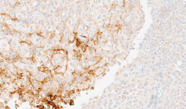
Anti-programmed cell death ligand 1 (anti-PD-L1) immunotherapy is an increasingly crucial in cancer treatment. To date, the Federal Drug Administration (FDA) has approved four PD-L1 immunohistochemistry (IHC) staining protocols, commercially available in the form of "kits", facilitating testing for PD-L1 expression. These kits comprise four PD-L1 antibodies on two separate IHC platforms, each utilizing distinct, non-interchangeable scoring systems. Several factors, including tumor heterogeneity and the size of the tissue specimens assessed, can lead to PD-L1 status misclassification, potentially hindering the initiation of therapy. Therefore, the development of more accurate predictive biomarkers to distinguish between responders and non-responders prior to anti-PD-1/PD-L1 therapy warrants further research. Achieving this goal necessitates refining sampling criteria, enhancing current methods of PD-L1 detection, and deepening our understanding of the impact of additional biomarkers. In this article, we review potential solutions to improve the predictive accuracy of PD-L1 assessment in order to more precisely anticipate patients' responses to anti-PD-1/PD-L1 therapy, monitor disease progression and predict clinical outcomes.
Citations
Downloads
Downloads
Additional Files
Published
Issue
Section
Categories
License
Copyright (c) 2023 Marek Zdrenka, Adam Kowalewski, Navid Ahmadi, Rizwanullah Sidiqi, Łukasz Chmura, Jędrzej Borowczak, Mateusz Maniewski, Łukasz Szylberg

This work is licensed under a Creative Commons Attribution 4.0 International License.
How to Cite
Accepted 2023-06-23
Published 2024-01-03


