Diagnostics of common microdeletion syndromes using fluorescence in situ hybridization: single center experience in a developing country

Authors

  • Amina Kurtovic-Kozaric Department of Pathology, Clinical Center of the University of Sarajevo
  • Lejla Mehinovic Clinical Center of the University of Sarajevo
  • Meliha Stomornjak-Vukadin Clinical Center of the University of Sarajevo
  • Ilvana Kurtovic-Basic Clinical Center of the University of Sarajevo
  • Feriha Catibusic Clinical Center of the University of Sarajevo
  • Mirza Kozaric Clinical Center of the University of Sarajevo
  • Senka Mesihovic-Dinarevic Clinical Center of the University of Sarajevo
  • Mensuda Hasanhodzic University Clinical Center Tuzla
  • Darinka Glamuzina University Clinical Hospital Mostar

DOI:

https://doi.org/10.17305/bjbms.2016.994

Keywords:

Microdeletion syndrome, DiGeorge, Williams, Prader-Willi, Angelman, Wolf-Hirschhorn, Fluorescent in situ hybridization

Abstract

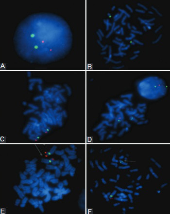
Microdeletion syndromes are caused by chromosomal deletions of less than 5 megabases which can be detected by fluorescence in situ hybridization (FISH). We evaluated the most commonly detected microdeletions for the period from June 01, 2008 to June 01, 2015 in the Federation of Bosnia and Herzegovina, including DiGeorge, Prader-Willi/Angelman, Wolf-Hirschhorn, and Williams syndromes. We report 4 patients with DiGeorge syndromes, 4 patients with Prader-Willi/Angelman, 4 patients with Wolf-Hirschhorn syndrome, and 3 patients with Williams syndrome in the analyzed 7 year period. Based on the positive FISH results for each syndrome, the incidence was calculated for the Federation of Bosnia and Herzegovina. These are the first reported frequencies of the microdeletion syndromes in the Federation of Bosnia and Herzegovina.

Downloads

Download data is not yet available.

References

Dallapiccola B, Mingarelli R, Novelli G. The link between cytogenetics and mendelism. Biomed &Pharmacother 1995; 49:83-93.

http://dx.doi.org/10.1016/0753-3322(96)82592-3

Hunter AG. Outcome of the routine assessment of patients with mental retardation in a genetics clinic. Am J Med Genet 2000; 90:60–8.

http://dx.doi.org/10.1002/(SICI)1096-8628(20000103)90:1<60::AID-AJMG11>3.0.CO;2-P

Kirchhoff M, Bisgaard AM, Bryndorf T, Gerdes T. MLPA analysis for a panel of syndromes with mental retardation reveals imbalances in 5.8% of patients with mental retardation and dysmorphic features, including duplications of the Sotos syndrome and Williams-Beuren syndrome regions. Eur J Med Genet 2007; 50:33–42.

http://dx.doi.org/10.1016/j.ejmg.2006.10.002

Flint J, Wilkie AO, Buckle VJ, Winter RM, Holland AJ, McDermid HE. The detection of subtelomeric chromosomal rearrangements in idiopathic mental retardation. Nat Genet 1995; 9:132–40. http://dx.doi.org/10.1038/ng0295-132

Vissers LE, De Vries BB, Osoegawa K, Janssen IM, Feuth T, Choy CO, et al. Array-based comparative genomic hybridization for the genomewide detection of submicroscopic chromosomal abnormalities. Am J Hum Genet 2003; 73:1261–70.

http://dx.doi.org/10.1086/379977

Tézenas Du, Montcel S, Mendizabai H, Aymé S, Lévy A, Philip N. Prevalence of 22q11 microdeletion. J Med Genet 1996; 33:719. http://dx.doi.org/10.1136/jmg.33.8.719

Goodship J, Cross I, LiLing J, Wren C. A population study of chromosome 22q11 deletions in infancy. Arch Dis Child 1998; 79:348-51. http://dx.doi.org/10.1136/adc.79.4.348

Oskarsdóttir S, Vujic M, Fasth A. Incidence and prevalence of the 22q11 deletion syndrome: a population-based study in western Sweden. Arch Dis Child 2004; 89:148–151.

http://dx.doi.org/10.1136/adc.2003.026880

Devriendt K, Fryns JP, Mortier G, van Thienen MN, Keymolen K. The annual incidence of DiGeorge/velocardiofacial syndrome. J Med Genet 1998; 35:789–790.

http://dx.doi.org/10.1136/jmg.35.9.789-a

Schinzel A. Catalogue of unbalanced chromosome aberrations in man. 2nd ed. New York: W de Gruyter; 2001.

Jones KL. Smith's recognizable patterns of human malformation. 6th ed. Philadelphia: Elsevier Saunders; 2006.

Ryan AK, Goodship JA, Wilson DI, Philip N, Levy A, Seidel H, et al. Spectrum of clinical features associated with interstitial chromosome 22q11 deletion: a European collaborative study. J Med Genet 1997; 34:798-4.

http://dx.doi.org/10.1136/jmg.34.10.798

Chen KS, Manian P, Koeuth T, Potocki L, Zhao Q, Chinault AC,et al. Homologous recombination of a flaking repeat gene cluster is a mechanism for a common continuous gene deletion syndrome. Nat Genet 1997; 17:154-63.

http://dx.doi.org/10.1038/ng1097-154

Shaw CJ, Bi W, Lupski RJ. Genetic proof of unequal meiotic crossover in reciprocal deletion and duplication of 17p11.2. Am J Hum Genet 2002; 71:1072-81.

http://dx.doi.org/10.1086/344346

Cassidy SB, Driscoll DJ. Prader-Willi syndrome. Eur J Hum Genet 2009; 1:3-13.

http://dx.doi.org/10.1038/ejhg.2008.165

Vogels A, Fryns PJ. The Prader-Willi syndrome and the Angelman Syndrome. Genetic Counsel 2002; 13:385-96.

Battaglia A, Carey JC, Wright TJ. Wolf-Hirschhorn (4p-) syndrome. Adv Pediatr 2001; 48: 75–113.

Battaglia A, Filippi T, Carey JC. Update on the clinical features and natural history of Wolf-Hirschhorn (4p-) syndrome: experience with 87 patients and recommendations for routine health supervision. Am J Med Genet C Semin Med Genet 2008; 148(4):246-51.

http://dx.doi.org/10.1002/ajmg.c.30187

Zollino M, Murdolo M, Marangi G, Pecile V, Galasso C, Mazzanti L, et al. On the nosology and pathogenesis of Wolf-Hirschhorn syndrome: genotype-phenotype correlation analysis of 80 patients and literature review. Am J Med Genet C Semin Med Genet 2008; 148C: 257-269.

http://dx.doi.org/10.1002/ajmg.c.30190

Battaglia A, Carey JC, South ST, Wright TJ. Wolf-Hirschhorn syndrome. In: Pagon RA, Bird TD, Dolan CR, Stephens K (eds). Gene Reviews [Internet]. Seattle: University of Washington; 2010[cited 14 July 2015]. Available from NCBI Bookshelf: http://www.ncbi.nlm.nih.gov/books/NBK1183/

Diagnostics of common microdeletion syndromes using fluorescence in situ hybridization: single center experience in a developing country

Downloads

Additional Files

Published

2016-03-03

How to Cite

1.
Kurtovic-Kozaric A, Mehinovic L, Stomornjak-Vukadin M, Kurtovic-Basic I, Catibusic F, Kozaric M, Mesihovic-Dinarevic S, Hasanhodzic M, Glamuzina D. Diagnostics of common microdeletion syndromes using fluorescence in situ hybridization: single center experience in a developing country. Biomol Biomed [Internet]. 2016Mar.3 [cited 2023Dec.1];16(2):121-5. Available from: https://www.bjbms.org/ojs/index.php/bjbms/article/view/994

Issue

Section

Molecular Biology

INTRODUCTION

Microdeletion syndromes are caused by chromosomal deletions of less than 5 megabases (Mb). The size and location of a microdeletion vary depending on the specific syndrome. Most microdeletion syndromes result in a loss of a chromosomal segment leading to the haploinsufficiency of a few critical genes or in some cases a single gene [1]. Microdeletions are mostly spontaneous and occur in approximately 5% of patients with unexplained mental retardation [2,3]. They are frequently associated with multiple congenital anomalies and developmental delay [4,5]. The most common microdeletion syndromes are DiGeorge syndrome (22q11.2), Prader-Willi syndrome, Angelman syndrome (15q11-13), Williams syndrome (7q11.23), and Wolf-Hirschhorn syndrome (4p16.3). DiGeorge syndrome is the most frequent microdeletion syndrome with an incidence range from 1:4000 to 1:10000, according to the literature [6-10]. The most common abnormalities include heart defects, hypoparathyroidism, cellular immune deficiency secondary to thymic hypoplasia, cleft palate, learning disabilities, dysmorphia, and microcephaly [7-12].

Because of their small length (<5 Mb), the deletions are difficult to detect using conventional cytogenetic methods and light microscopy. A method that is commonly used for microdeletion detection is fluorescence in situ hybridization (FISH), which is a molecular cytogenetic technique based on fluorescently labeled DNA probes specific for a chromosomal region of interest.

Since the detection of microdeletion syndromes was established 7 years ago in Bosnia and Herzegovina, FISH as a diagnostic tool was evaluated in the period from June 01, 2008 to June 01, 2015. The main goal of this study was to evaluate the significance of FISH as a diagnostic tool in order to create future guidelines for pediatric genetic diagnosis.

MATERIALS AND METHODS

During the seven-year period from June 01, 2008 to June 01 2015, 24 pediatric patient samples were received by the Laboratory of human genetics, Department of Clinical Pathology and Cytology, at the Clinical Center of the University of Sarajevo, referred for FISH testing in order to confirm a suspected microdeletion syndrome. All peripheral blood samples were received in heparin tubes and sample volume was 1-3 ml. The samples were processed the same day. Detailed indications were recorded on a form filled out by the referring physician. The peripheral blood was cultivated using standard manufacturer’s protocols (Euroclone, Milano, Italy).

One milliliter of blood was cultured in 5 ml of Chromosome Kit P medium (Euroclone, Milano, Italy) for 72 hours in 37°C incubator with 5% CO2. After harvest, FISH slides were dehydrated using different dilutions of ethanol and dried. A FISH probe was applied and incubated in Hybridizer (Dako, Colorado, USA), according to the manufacturer’s instructions (Vysis, Abbott, Abbott Park, Illinois, USA). After the hybridization, coverslips were removed and the slides were washed with NP40 (0.03% solution) at 72°C for 2-3 min, followed by NP40 (0.01% solution) for 2 min at room temperature. DAPI was applied to the slides (4,6 diaminidino-2-phenylindole, Sigma, USA). For the FISH analysis, locus-specific DiGeorge Region probes (Vysis LSI DiGeorge region probe - LSI N25 Spectrum Orange/LSI ARSA Spectrum Green, 5J24-12), 2 types of Prader-Willi/Angelman Region Probes (Vysis Prader-Willi/Angelman Region Probe - LSI D15S11 Spectrum Orange/CEP 15 (D15Z1) Spectrum Green, 5J19-14; Vysis Prader-Willi/Angelman Region Probe - LSI D15S10 Spectrum Orange/CEP 15 (D15Z1) Spectrum Aqua/PML Spectrum Green Probe), Williams Region Probe (Vysis Williams Region Probe - LSI ELN Spectrum Orange/LSI D7S486, D7S522 Spectrum Green, 5J18-26), and Wolf-Hirschhorn Region Probe (Vysis Wolf-Hirschhorn Region Probe - LSI WHS Spectrum Orange/CEP 4 SpectrumGreen, 5J29-74) were used. The cells were analyzed using a fluorescent microscope (Olympus BX61, Olympus, Tokyo, Japan) with Cytovision software (Cytovysion, AB Imaging, Germany). For each analyzed sample, at least 200 interphase nuclei were counted.

Statistical analysis

Incidences were calculated based on the positive FISH analyses and the number of the newborn population in Canton Sarajevo (lower limit) and in the Federation of Bosnia and Herzegovina (upper limit). The demographic data was retrieved from the Institute of Statistics of Federation of Bosnia and Herzegovina. We performed statistical analysis using SPSS v.21 (IBM, Armonk, NY, USA).

RESULTS

During the seven-year period 24 pediatric patient samples were received by the Laboratory of human genetics at the Department of Clinical Pathology, Cytology, and Human Genetics, Clinical Center of the University of Sarajevo, referred to FISH testing to confirm the diagnosis of a suspected microdeletion syndrome.

Five different microdeletion probes were used. The probes recognized DiGeorge, PraderWilli/Angelman, Wolf-Hirschhorn, and Williams syndromes. Two different probes for PraderWilli/Angelman syndrome were applied to all the samples referred to the testing. FISH analysis of 22q11.2 microdeletion (DiGeorge syndrome) was performed on 13 samples and the microdeletion was detected in 3 samples (Table 1). Prader-Willi/Angelman syndrome was diagnosed in one out of 9 samples. One sample was positive for Williams syndrome and one for Wolf-Hirschhorn syndrome (Table 1).

TABLE 1: Pediatric blood samples referred for fluorescence in situ hybridization in the period from June 01, 2008 to June 01, 2015. The FISH results were categorized as normal and abnormal (deletion present)

Several patients from Bosnia and Herzegovina sought initial treatment and diagnosis abroad and were diagnosed by FISH at foreign institutions. After the initial diagnosis, those patients continued their treatment at the Pediatric Clinics at the Clinical Centers Sarajevo, Tuzla, and Mostar. We included these cases because of the precise incidence calculations (Table 2). As a result, three cases of Prader-Willi/Angelman syndrome, three cases of Wolf-Hirschhorn syndrome, one case of DiGeorge syndrome, and two cases of Williams syndrome, who were diagnosed outside of Bosnia and Herzegovina, were reported in this study (Table 2, Table 3).

TABLE 2: The number of patients with the microdeletion syndromes diagnosed at the Clinical Center of the University of Sarajevo using FISH and outside of Bosnia and Herzegovina, included in the statistical analysis
TABLE 3: Comparison of the number of the common microdeletions detected by FISH in Europe and the Federation of Bosnia and Herzegovina. The range for the calculation of the incidence based on the positive FISH analyses in FB&H was based on the number of newborns in the Canton Sarajevo and FB&H

Figure 1 shows the diagnosis of DiGeorge, Williams, and Wolf-Hirschhorn syndromes using fluorescent in situ hybridization with the locus specific and centromeric probes (A-F). The left panels show normal signal pattern without the microdeletions (Figure 1A, C, E), while the right panels show the presence of the deletion (Figure 1B, D, F). In all the cases, the normal signal pattern was 2 red and 2 green signals (2R2G), while the abnormal pattern with the microdeletion was 1 red and 2 green signals (1R2G). The green signal in all the cases was the centromeric probe, while the red signal was the locus specific probe. Thus, DiGeorge syndrome was diagnosed by the absence of the red signal on chromosome 22, region 22q11.2 (Figure 1B), Williams syndrome was diagnosed by the absence of the red signal on chromosome 7, region 7q11.23, and Wolf-Hirschhorn syndrome was diagnosed by the absence of the red signal on chromosome 4, region 4p16.

FIGURE 1: Diagnosis of DiGeorge, Williams and Wolf-Hirschhorn syndromes using fluorescent in situ hybridization with locus specific and centromeric probes (A-F). The left panels show the normal signal pattern without microdeletions (Figure 1A, C, E), while the right panels show the presence of the deletion (Figure 1B, D, F).

Figure 2 shows the diagnosis of Prader-Willi/Angelman syndrome using fluorescent in situ hybridization with 2 types of Prader-Willi/Angelman Region Probes (Figure 2A-D). The two probes recognized D15S11 and D15S10 regions, at the proximal and distal ends of the commonly deleted segment in PraderWilli/Angelman syndrome. The green signal was the centromeric probe for chromosome 15. The normal pattern for the D15S11 region was 2 red and 2 green signals (2R2G, Figure 2A), while the abnormal pattern with the microdeletion was 1 red and 2 green signals (1R2G, Figure 2C). The normal pattern for the second PraderWilli/Angelman FISH probe was 4 red and 2 green signals (4R2G, Figure 2B) because the D15S10 region and PML control region were labeled by the red fluorescence. The abnormal pattern with the presence of the D15S10 deletion was 3 red and 2 green signals (3R2G, Figure 2D.

FIGURE 2: Diagnosis of Prader-Willi/Angelman syndrome using FISH with 2 types of Prader-Willi/Angelman Region Probes (Figure 2A-D). The normal pattern for the D15S11 region was two red and two green signals (2R2G, Figure 2A), while the abnormal pattern with the microdeletion was one red and two green signals (1R2G, Figure 2C). The normal pattern for the second PraderWilli/Angelman FISH probe was 4 red and 2 green signals (4R2G, Figure 2B) because D15S10 region and PML control region were labeled by the red fluorescence. The abnormal pattern with the presence of D15S10 deletion was 3 red and 2 green signals (3R2G, Figure 2D).

Incidence of the microdeletion syndromes based on the positive fish results in the Federation of B&H

The incidence of DiGeorge syndrome ranged from 1:8637-1:39633 in FB&H based on the FISH-positive results, as compared to the European incidence of 1:4000-1:10000 [9,13] (Table 3). The incidence based on the FISH positivity of Prader-Willi/Angelman syndrome in FB&H was 1:8637-1:39633, as compared to the European incidence of 1:15000-1:30000 [14,15] (Table 3). The European incidence of WHS was 1:50000, as compared to the FB&H incidence of 1:8637-1:39633 [16,17] (Table 3). The FB&H incidence for Williams syndrome ranged from 1:11517-1:52844, as compared to the European incidence of 1:7500-1:10000 [11] (Table 3).

DISCUSSION

The distribution of microdeletions in the human genome is not random. In many syndromes, the deleted regions are surrounded by relatively long homogeneous repetitive sequences involved in the proposed mechanism [13]. It is suggested that the mechanism for the development of microdeletions involves unequal intra- and inter- chromosomal crossing over between repetitive sequences resulting in the loss and reciprocal microduplication of the interstitial chromosomal segment [14]. Some microdeletion syndromes are very rare, while others are more common such as DiGeorge syndrome, Prader-Willi syndrome, Angelman syndrome, Williams syndrome, and Wolf-Hirschhorn syndrome. Since no other studies have reported the incidence of microdeletion syndromes for this region, we calculated the incidences based on our FISH data for the seven year period from June 01, 2008 to June 01, 2015.

The phenotype of patients with DiGeorge syndrome is highly variable. The most common abnormalities include heart defects, hypoparathyroidism, cellular immune deficiency secondary to thymic hypoplasia, cleft palate, learning disabilities, dysmorphia, and microcephaly. The majority of patients demonstrated a deletion of 3 Mb [10-12]. In our study, we diagnosed 3 patients with DiGeorge syndrome in the period from 2008 to 2015, and included one case diagnosed outside of Bosnia and Herzegovina. Thus, the incidence based on the FISH results for DiGeorge syndrome was 1:8637-1:39633 in FB&H, while the published European incidence was in the range from 1:4000 to 1:10000 (Table 3) [6-10]. The upper limit of the FB&H (1:8637) incidence is similar to the European incidence and probably is closer to the true value for this region. The lower limit is very high, possibly due to the fact that some patients might be diagnosed abroad and treated at other regional centers in the Federation of Bosnia and Herzegovina.

Prader-Willi syndrome (PWS) is caused by 4 Mb microdeletion in the 15q11-q13 region. The clinical presentation is characterized by hypotonia, hyperphagia, mental retardation, and dysmorphism. Approximately 75% of the affected individuals have the deletion in 15q11-q13. In all these cases, the paternally derived chromosome has been deleted. Maternal uniparental disomy accounts for further 20%. The remaining 5% are due to a mutation in the imprinting center or to a chromosomal translocation involving proximal 15q. Whereas the origin of the deleted chromosome 15 is paternal in PWS, it is always maternal in Angelman syndrome (AS) [11-16]. The clinical features include microbrachiocephaly, dysmorphia, hypopigmentation, hypotonia, severe mental retardation, epilepsy, paroxysms of inappropriate laughter, and sleep disorders. Several genetic abnormalities are found in the 15q11-q13 region, including: a de novo interstitial deletion of maternal 15q11-q13 in ~75% of cases (4 Mb deletion), paternal uniparental disomy in 2%, an imprinting center mutation in 2%, and a point mutation in E3 ubiquitin protein ligase gene (UBE3A) in 5-10% [11]. In the calculation of the FB&H incidence based on the FISH results of PW/A syndrome, we included three cases diagnosed outside of Bosnia and Herzegovina. As a result, the incidence based on the FISH results of Prader-Willi/Angelman syndrome in FB&H ranged from 1:8637-1:39633, as compared to the European incidence of 1:15000-1:30000 (Table 3).

Williams syndrome (WS) is caused by a 2 Mb deletion in the 7q11.23 region which includes 17 genes. The main clinical findings are moderately retarded intrauterine and postnatal growth with moderate microcephaly, a characteristic face with full lids and orbitae, frequent strabismus, irregularly positioned and small teeth, and a hoarse voice [10]. The microdeletion in 7q11.23 causes a loss of the gene for elastin (ELN), which is responsible for supravalvular aortic stenosis and other vascular stenoses [11]. In the calculation of the FB&H incidence of William syndrome based on the FISH results we included two cases diagnosed outside of Bosnia and Herzegovina. This resulted in the FB&H incidence in the range from 1:11517-1:52844, while the European incidence was 1:7500-1:10000 (Table 3). The upper limit of the FB&H (1:11517) incidence is similar to the European incidence and probably is closer to the true value for this region.

Wolf-Hirschhorn syndrome (WHS) is caused by a deletion in the 4p16.3 region. The prevalence of WHS is reported to be around 1:50000 live births with a 2:1 female/male ratio; however, this is likely underestimated because of under-recognition or misdiagnosis of the affected individuals [17]. The typical clinical features include growth restriction of prenatal onset, profound psychomotor retardation, seizures, skeletal abnormalities, and a distinctive facial appearance including a broad, flat nasal bridge and a high forehead, eyes widely spaced and protruding. Additionally, the affected individuals may have asymmetrical facial features and an unusually small head (microcephaly) [10,18,19]. The signs and symptoms of WHS are related to the loss of genes WHSC1, LETM1, and MSX1 (20). In the calculation of the FB&H incidence based on the FISH results of WHS syndrome we included three cases of this syndrome diagnosed outside of Bosnia and Herzegovina, and the calculated FB&H incidence ranged from 1:8637-1:39633, which is a higher incidence compared to the European incidence of 1:50000 (Table 3).

The calculated incidences for the common microdeletion syndromes in FB&H are the first reported in the literature. However, we have to emphasize that the microdeletion syndromes are in general underdiagnosed. For example, not all patients with cardiac abnormalities are tested for DiGeorge and Williams syndromes, thus, the calculated incidences are likely to be lower. Furthermore, a small number of patients with DiGeorge syndrome have a deletion in the 10p13-14 region which was not tested in this study. Similarly, only a small fraction of patients with PraderWilli/Angelman syndrome with mental retardation are tested cytogenetically, which leads to a smaller number of diagnosed patients. In general, these results can serve as a preliminary basis for creating future guidelines for pediatric genetic diagnosis. Precise analysis and identification of microdeletion syndromes are of practical use in the diagnostics, and of scientific interest, since they allow the study of the interface of genetic imbalances and associated clinical characteristics.

DECLARATION OF INTERESTS

The authors declare no conflict of interest.

REFERENCES

  1. , , (). The link between cytogenetics and mendelism. Biomed & Pharmacother. http://dx.doi.org/10.1016/0753-3322(96)82592-3
  2. (). Outcome of the routine assessment of patients with mental retardation in a genetics clinic. Am J Med Genet. http://dx.doi.org/10.1002/(SICI)1096-8628(20000103)90:1<60:AID-AJMG11>3.0.CO;2-P
  3. , , , (). MLPA analysis for a panel of syndromes with mental retardation reveals imbalances in 5.8% of patients with mental retardation and dysmorphic features, including duplications of the Sotos syndrome and Williams-Beuren syndrome regions. Eur J Med Genet. http://dx.doi.org/10.1016/j.ejmg.2006.10.002
  4. , , , , , (). The detection of subtelomeric chromosomal rearrangements in idiopathic mental retardation. Nat Genet. http://dx.doi.org/10.1038/ng0295-132
  5. , , , , , (). Array-based comparative genomic hybridization for the genomewide detection of submicroscopic chromosomal abnormalities. Am J Hum Genet. http://dx.doi.org/10.1086/379977
  6. , , , , , (). Prevalence of 22q11 microdeletion. J Med Genet. http://dx.doi.org/10.1136/jmg.33.8.719
  7. , , , (). A population study of chromosome 22q11 deletions in infancy. Arch Dis Child. http://dx.doi.org/10.1136/adc.79.4.348
  8. , , (). Incidence and prevalence of the 22q11 deletion syndrome:a population-based study in western Sweden. Arch Dis Child. http://dx.doi.org/10.1136/adc.2003.026880
  9. , , , , (). The annual incidence of DiGeorge/velocardiofacial syndrome. J Med Genet. http://dx.doi.org/10.1136/jmg.35.9.789-a
  10. (). . Catalogue of unbalanced chromosome aberrations in man.
  11. (). . Smith's recognizable patterns of human malformation.
  12. , , , , , (). Spectrum of clinical features associated with interstitial chromosome 22q11 deletion:a European collaborative study. J Med Genet. http://dx.doi.org/10.1136/jmg.34.10.798
  13. , , , , , (). Homologous recombination of a flaking repeat gene cluster is a mechanism for a common continuous gene deletion syndrome. Nat Genet. http://dx.doi.org/10.1038/ng1097-154
  14. , , (). Genetic proof of unequal meiotic crossover in reciprocal deletion and duplication of 17p11.2. Am J Hum Genet. http://dx.doi.org/10.1086/344346
  15. , (). Prader-Willi syndrome. Eur J Hum Genet. http://dx.doi.org/10.1038/ejhg.2008.165
  16. , (). The Prader-Willi syndrome and the Angelman Syndrome. Genetic Counsel.
  17. , , (). Wolf-Hirschhorn (4p-) syndrome. Adv Pediatr.
  18. , , (). Update on the clinical features and natural history of Wolf-Hirschhorn (4p-) syndrome:experience with 87 patients and recommendations for routine health supervision. Am J Med Genet C Semin Med Genet. http://dx.doi.org/10.1002/ajmg.c.30187
  19. , , , , , (). On the nosology and pathogenesis of Wolf-Hirschhorn syndrome:genotype-phenotype correlation analysis of 80 patients and literature review. Am J Med Genet C Semin Med Genet. http://dx.doi.org/10.1002/ajmg.c.30190
  20. , , , (). Wolf-Hirschhorn syndrome. Gene Reviews [Internet]. Available from NCBI Bookshelf: http://www.ncbi.nlm.nih.gov/books/NBK1183