Lithium chloride could aggravate brain injury caused by 3-nitropropionic acid

Authors

  • Aleksandra Milutinović Institute of Histology and Embryology, Medical Faculty of Ljubljana, University of Ljubljana, Ljubljana

DOI:

https://doi.org/10.17305/bjbms.2016.1206

Keywords:

Lithium chloride, 3-nitropropionic acid, cytochrome c oxidase, synaptotagmin-4, synaptotagmin-7

Abstract

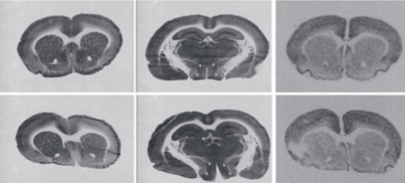
Lithium, a well-known drug for the treatment of bipolar disorder, may also have the ability to reduce neurodegeneration and stimulate cell proliferation. Systemic injection of mitochondrial toxin 3-nitropropionic acid (3NPA) is known to induce a relatively selective, Huntington disease-like brain injury. The aim of this study was to determine the effect of lithium chloride (LiCl) on brain injury caused by 3NPA. Female adult Wistar rats were pre-treated with LiCl (127 mg/kg) 1 day before the first injection of 3NPA (28 mg/kg), and then for 8 days with the same treatment but receiving LiCl 1 hour before 3NPA. Control groups were pre-treated accordingly, with LiCl or with normal saline, but were not treated with 3NPA. Staining for cytochrome c oxidase activity and in situ hybridization autoradiography of synaptotagmin-4 and -7 mRNAs were used to evaluate brain injury caused by 3NPA. There was a significant reduction of body weight in the 3NPA+LiCl group (79%) compared to the 3NPA group (90%, p = 0.031) and both control groups (100%, p = 0.000). Densitometric evaluation of cytochrome c oxidase staining and in situ hybridization autoradiograms revealed that the pre-treatment with LiCl caused an increase in striatal lesion for about 40% (p = 0.049). Moreover, the lesion was observed also in the hippocampus of three animals from the 3NPA+LiCl group and in two animals from the 3NPA group. However, there were no differences between the LiCl and saline group in any of the measured parameters. We concluded that the pre-treatment with a relatively nontoxic dose of LiCl could aggravate brain injury caused by 3NPA.

Downloads

Download data is not yet available.

References

. Diniz BS, Machado-Vieira R, Forlenza OV. Lithium and neuroprotection: Translational evidence and implications for the treatment of neuropsychiatric disorders. Neuropsychiatr Dis Treat. 2013;9:493-500. http://dx.doi.org/10.2147/NDT.S33086.

. Machado-Vieira R, Manji HK, Zarate CA Jr. The role of lithium in the treatment of bipolar disorder: Convergent evidence for neurotrophic effects as a unifying hypothesis. Bipolar Disord. 2009;11 Suppl 2:92-109.

http://dx.doi.org/10.1111/j.1399-5618.2009.00714.x.

. Forlenza OV, de Paula VJ, Machado-Vieira R, Diniz BS, Gattaz WF. Does lithium prevent Alzheimer's disease? Drugs Aging. 2012;29(5):335-42.

http://dx.doi.org/10.2165/11599180-000000000-00000.

. Forlenza OV, De-Paula VJ, Diniz BS. Neuroprotective effects of lithium: Implications for the treatment of Alzheimer's disease and related neurodegenerative disorders. ACS Chem Neurosci. 2014;5(6):443-50. http://dx.doi.org/10.1021/cn5000309.

. Senatorov VV, Ren M, Kanai H, Wei H, Chuang DM. Short-term lithium treatment promotes neuronal survival and proliferation in rat striatum infused with quinolinic acid, an excitotoxic model of Huntington's disease. Mol Psychiatry. 2004;9(4):371-85. http://dx.doi.org/10.1038/sj.mp.4001463.

. Nonaka S, Hough CJ, Chuang DM. Chronic lithium treatment robustly protects neurons in the central nervous system against excitotoxicity by inhibiting N-methyl-D-aspartate receptor-mediated calcium influx. Proc Natl Acad Sci U S A. 1998;95(5):2642-7. http://dx.doi.org/10.1073/pnas.95.5.2642.

. James LF, Hartley WJ, Williams MC, Van Kampen KR. Field and experimental studies in cattle and sheep poisoned by nitro-bearing Astragalus or their toxins. Am J Vet Res. 1980;41(3):377-82.

. Liu X, Luo X, Hu W. Studies on the epidemiology and etiology of moldy sugarcane poisoning in China. Biomed Environ Sci. 1992;5(2):161-77.

. Ming L. Moldy sugarcane poisoning – A case report with a brief review. J Toxicol Clin Toxicol. 1995;33(4):363-7. http://dx.doi.org/10.3109/15563659509028924.

. Ramaswamy S, McBride JL, Kordower JH. Animal models of Huntington's disease. ILAR J. 2007;48(4):356-73. http://dx.doi.org/10.1093/ilar.48.4.356.

. Mirandola SR, Melo DR, Saito A, Castilho RF. 3-nitropropionic acid-induced mitochondrial permeability transition: Comparative study of mitochondria from different tissues and brain regions. J Neurosci Res. 2010;88(3):630-9.

. McCracken E, Dewar D, Hunter AJ. White matter damage following systemic injection of the mitochondrial inhibitor 3-nitropropionic acid in rat. Brain Res. 2001;892(2):329-35. http://dx.doi.org/10.1016/S0006-8993(00)03266-2.

. Szabó A, Papp A, Nagymajtényi L. Effects of 3-nitropropionic acid in rats: General toxicity and functional neurotoxicity. Arh Hig Rada Toksikol. 2005;56(4):297-302.

. Gabrielson KL, Hogue BA, Bohr VA, Cardounel AJ, Nakajima W, Kofler J, et al. Mitochondrial toxin 3-nitropropionic acid induces cardiac and neurotoxicity differentially in mice. Am J Pathol. 2001;159(4):1507-20.

http://dx.doi.org/10.1016/S0002-9440(10)62536-9.

. Milutinovic A, Zorc-Pleskovic R. Glycogen accumulation in cardiomyocytes and cardiotoxic effects after 3NPA treatment. Bosn J Basic Med Sci. 2012;12(1):15-9.

. Glavan G, See RE, Živin M. Differential patterns of synaptotagmin7 mRNA expression in rats with kainate- and pilocarpine-induced seizures. PLoS One. 2012;7(5):e36114. http://dx.doi.org/10.1097/WNR.0b013e3280ef6964.

. Saghafi MM, Pregelj P, Zivin M. Donepezil inhibits diisopropylfluorophosphate-induced seizures and up-regulation of synaptotagmin 4 mRNA. Folia Biol (Praha). 2010;56(6):256-62.

. Glisovic S, Glavan G, Saghafi MM, Zivin M. Upregulation of synaptotagmin IV protein in kainate-induced seizures. Neuroreport. 2007;18(8):831-5.

http://dx.doi.org/10.1097/WNR.0b013e3280ef6964.

. Glavan G, Zivin M. Differential expression of striatal synaptotagmin mRNA isoforms in hemiparkinsonian rats. Neuroscience 2005;135(2):545-54.

http://dx.doi.org/10.1016/j.neuroscience.2005.05.050.

. Glavan G, Schliebs R, Zivin M. Synaptotagmins in neurodegeneration. Anat Rec (Hoboken). 2009;292(12):1849-62. http://dx.doi.org/10.1002/ar.21026.

. Tratnjek L, Zivin M, Glavan G. Up-regulation of Synaptotagmin IV within amyloid plaque-associated dystrophic neurons in Tg2576 mouse model of Alzheimer's disease. Croat Med J. 2013;54(5):419-28.

http://dx.doi.org/10.3325/cmj.2013.54.419.

. Wong-Riley M. Changes in the visual system of monocularly sutured or enucleated cats demonstrable with cytochrome oxidase histochemistry. Brain Res. 1979;171(1):11-28. http://dx.doi.org/10.1016/0006-8993(79)90728-5.

. Zivin M, Milatovic D, Dettbarn WD. Nitrone spin trapping compound N-tert-butyl-alpha-phenylnitrone prevents seizures induced by anticholinesterases. Brain Res. 1999;850(1-2):63-72. http://dx.doi.org/10.1016/S0006-8993(99)02101-0.

. Crespo-Biel N, Camins A, Pallàs M, Canudas AM. Evidence of calpain/cdk5 pathway inhibition by lithium in 3-nitropropionic acid toxicity in vivo and in vitro. Neuropharmacology. 2009;56(2):422-8.

http://dx.doi.org/10.1016/j.neuropharm.2008.09.012.

. Khan A, Jamwal S, Bijjem KR, Prakash A, Kumar P. Neuroprotective effect of hemeoxygenase-1/glycogen synthase kinase-3ß modulators in 3-nitropropionic acid-induced neurotoxicity in rats. Neuroscience. 2015;287(3):66-77. http://dx.doi.org/10.1016/j.neuroscience.2014.12.018.

. Clifford DB, Olney JW, Maniotis A, Collins RC, Zorumski CF. The functional anatomy and pathology of lithium-pilocarpine and high-dose pilocarpine seizures. Neuroscience. 1987;23:953-68. http://dx.doi.org/10.1016/0306-4522(87)90171-0.

. Reddy DS, Kuruba R. Experimental models of status epilepticus and neuronal injury for evaluation of therapeutic interventions. Int J Mol Sci. 2013;14(9):18284-318. http://dx.doi.org/10.3390/ijms140918284.

. Rang HP, Dale MM, Ritter JM, Flower RJ. Antidepressant drugs. Rang and Dales Pharmacology. 6th ed. London, England: Elsevier Churchill Livingstone; 2008. p. 569-73.

. Serdarevic N, Kozjek F, Malesic I. Saliva and serum lithium monitoring in hospitalized patients and possibility to replace serum to saliva. Bosn J Basic Med Sci. 2006;6(4):32-5.

. Serdarevic N, Malesic I, Kozjek F. Comparison of vitros dry slide technology for determination of lithium ions with other methods. Bosn J Basic Med Sci. 2006;6(2):32-6.

. Yamantürk-Çelik P, Unlüçerçi Y, Sevgi S, Bekpinar S, Eroglu L. Nitrergic, glutamatergic and gabaergic systems in lithium toxicity. J Toxicol Sci. 2012;37(5):1017-23.

http://dx.doi.org/10.2131/jts.37.1017.

. Dimitrova M, Petrova E, Gluhcheva Y, Kadiysky D, Dimitrova S, Kolyovska V, et al. Neurodegenerative changes in rat produced by lithium treatment. J Toxicol Environ Health A. 2013;76(4-5):304-10.

http://dx.doi.org/10.1080/15287394.2013.757268.

. Hirvonen MR. Cerebral lithium, inositol and inositol monophosphates. Pharmacol Toxicol. 1991;69(1):22-7. http://dx.doi.org/10.1111/j.1600-0773.1991.tb00403.x.

Lithium chloride could aggravate brain injury caused by 3-nitropropionic acid

Downloads

Additional Files

Published

2016-11-10

How to Cite

1.
Milutinović A. Lithium chloride could aggravate brain injury caused by 3-nitropropionic acid. Biomol Biomed [Internet]. 2016Nov.10 [cited 2023Dec.5];16(4):261-7. Available from: https://www.bjbms.org/ojs/index.php/bjbms/article/view/1206

Issue

Section

Pathology

INTRODUCTION

For a long time, lithium has been successfully used therapeutically for the management of psychiatric disorders, such as bipolar disorder, schizophrenia, and depression. Lithium has also been proposed to possess potential neuroprotective activity by the release of neurotrophic factors, reduction of proinflammatory status, and decreased oxidative stress [1]. It was also shown that lithium has neuroprotective activity against a number of neurotoxic insults in clinical observations [1-4] and animal models [5,6].

The 3-nitropropionic acid (3NPA) is a toxin that is naturally present in leguminous plants used to feed animals and can poison grazing livestock [7]. Human intoxication with 3NPA has occurred in China via ingestion of fungal (Arthrinium and Aspergillus) contaminated sugarcane [8,9]. The 3NPA mimics a downstream process of cell death seen in selective excitotoxic striatal pathology in the Huntington’s disease (HD) brain, namely mitochondrial impairment, where it inhibits the mitochondrial enzyme succinate dehydrogenase [10]. It was found that intoxication with 3NPA causes Ca2+-induced mitochondrial permeability transition [11]. 3NPA may, therefore, be used as an experimental animal model of HD [10]. However, 3NPA-induced injury was observed also in the hippocampus and thalamus [12,13] and in the heart [14,15]. The pre-treatment with lithium chloride (LiCl) in relatively high dose increases neuronal excitability and is, thus, used for lowering the threshold for the induction of seizures by cholinergic seizure-inducing agents in animal models of epilepsy [16,17].

The aim of the study was to determine the effect of a relatively high cumulative dose of LiCl on brain injury caused by 8 daily injections of 3NPA using the cytochrome oxidase (COX) histochemical staining and in situ hybridization of synaptotagmin-4 (Syt4) and synaptotagmin-7 (Syt7) mRNAs expression. COX is the last enzyme in the respiratory electron transport chain of mitochondria located in the mitochondrial membrane. The changes of the histochemical staining intensity of COX activity in the brain are related to the changes in mitochondrial number and activity and may, therefore, be used as an endogenous marker of brain metabolic activity. The changes of the expression of Syt4 and Syt7 are also known to be involved in the pathophysiological mechanisms of neurodegenerative diseases with striatal and/or hippocampal pathology, such as temporal lobe epilepsy [18] and Parkinson’s disease [19] as well as brain ischemia and Alzheimer’s disease [20,21] and are also known to be induced acutely by different excitotoxic seizure-inducing agents [16-18]. Here, we used the autoradiographic method of in situ hybridization that allows visualization and densitometric quantification of the changes of the expression of Syt4 and Syt7 mRNAs for the determination of the effects of lithium on the extent of brain injury induced by 3NPA.

MATERIALS AND METHODS

Animals

We used female Wistar rats weighing from 210 to 288 g at the beginning of the experiment. The study was conducted in compliance with the ethical principles of the European Directive 2010/63/EU, the law on amendments to the animal protection law according to the Federation of European Laboratory Animal Science Associations (FELASA) recommendations. The animals were handled using the “Guide for the care and use of Laboratory Animals”, Institute for Laboratory Animals Research, 2001, and the “National Veterinary Institute Guide for the Care and Use of Laboratory Animals” (National Veterinary Institute, number 323-02-74/00). Animal care was in compliance with standard operating procedures (SOPs) of Animal facility and the European Convention for the Protection of Vertebrate Animals Used for Experimental and other Scientific Purposes (ETS 123).

All efforts were made to minimize animal suffering, and only the number of animals necessary to produce reliable scientific data was used following 3R principles.

Drugs

The LiCl and 3NPA were purchased from Sigma, St. Louis, MO, USA. All drugs were dissolved in 0.9% saline and were administered subcutaneously in a volume of 1 ml/kg. Drug solutions were freshly prepared no more than 30 minutes before injections.

Treatment

Rats were divided into four groups (Table 1):

TABLE 1: The treatment protocol. LiCl (127 mg/kg) and 3NPA (28 mg/kg) were dissolved in 0.9% saline and were administered SC in a volume of 1 ml/kg

  • 3NPA+LiCl group (n = 6): Rats were pre-treated 24 hours prior the first injection of 3NPA (28 mg/kg) with LiCl dissolved in normal saline (127 mg/kg) and they were then treated daily for 8 days, with LiCl (127 mg/kg) 1 hour before the injections of 3NPA (28 mg/kg).

  • 3NPA group (n = 6): Rats were treated in the same way as the 3NPA+LiCl group, but received normal saline pre-treatments instead of pre-treatments with LiCl.

  • LiCl group (n = 3): Rats were treated in the same way as the 3NPA+LiCl group, but received normal saline instead of 3NPA.

  • Saline group (n = 3): Rats were treated in the same way as the 3NPA+LiCl group, but received normal saline instead of the drugs.

The doses of 3NPA and LiCl were selected according to our previous in vivo experiments with these agents and from data published in the literature [15-18].

Measurement of the body weight

The rats were weighted every day for 9 days. The first weighting was performed 1 day before the first application of the substances. The body weight of each animal at the end of experiment was compared to the body weight on the first day of weighting (100%) and expressed in percentage. For each treatment group, the results were expressed as the average percent value ± standard deviation (SD).

Brain sections and staining

After 24 hours of the last injection, the animals were euthanized in CO2 and decapitated (SOP Euthanasia protocol). The brains were rapidly removed and quickly frozen on dry ice. Coronal sections (10 µm) were cut through the striatum and hippocampus using a cryostat and thaw mounted onto microscope slides. The sections were fixed in 4% phosphate-buffered paraformaldehyde, washed in phosphate-buffered saline, dehydrated in 70% ethanol, and stored in 95% ethanol at +4°C until processed for in situ hybridization histochemistry.

Consecutive sections were stained for COX following the diaminobenzidine procedure [22] and for Syt4 and Syt7 mRNAs following the standard procedure for X-ray film autoradiography of in situ hybridization histochemistry described in detail previously [16,17,23]. Briefly, we used 3’ end 35S-labeled oligodeoxyribonucleotide antisense probes (45 bases long) complementary to the rat Syt4 mRNA (bases encoding 1082-1126, sequence 5’-CAG AGG GAG ACC AGA AGT TCA CCC CGT CCA GAA GAC TTC TTA GCA-3’) and rat Syt7 mRNA (bases encoding 300-344, sequence 5’-CCG AGT CTG GCG TGC CCA CCG TCT CCA AGG AGT TCT TGT AGC GTT-3’). GenBank accession numbers used to design the probes were as follows: Syt4 L38247 and Syt7 U20106. For each labeled probe, the control sections were hybridized in the presence of 100-fold excess of unlabeled probe. Air-dried hybridized sections were exposed to X-ray film (Scientific Imaging Film X-Omat AR, Kodak, Rochester, NY) at room temperature for 2-3 weeks and developed using standard darkroom techniques.

Image analysis - measurement of the size of the striatal and hippocampal lesion

The cover-slipped sections that were histochemically stained for COX activity and the autoradiograms of Syt4 and Syt7 mRNA levels were transilluminated in the visual field of a 12-bit digital Photometrics camera CoolSNAP cf (Spectra Services Inc., Ontario, NY, USA) connected to an MCID, M5 Elite image analyzer (Imaging Research Inc., St. Catharines, Ontario, Canada) and visualized as relative optical density (ROD) images. The measurements were performed on three coronal image slices for the striatum and two image slices for the hippocampus.

The boundaries of the striatum were manually outlined by visual observation of the image on the computer screen. For the analysis of autoradiograms, the upper threshold to automatically determine the boundaries of the hypodense areas was set using the MCID software, by setting the upper ROD threshold to the value that excluded the ROD values that were higher than the ROD of the corpus callosum. The size of the lesioned area in the striatum was then evaluated as the percentage of the hypodense area compared to the area representing the whole striatum (100%). For each treatment group, the results were expressed as the average percent value ± SD. The hippocampal lesions on autoradiograms were determined accordingly as the % length of hypodense area observed within the Cornu Ammonis (CA) region of the hippocampal stratum pyramidale (100%). The densitometric analysis of COX histochemistry staining was performed in a similar way; however, the ROD thresholds of hypodense regions were outlined manually, somewhat more arbitrarily, by an experienced observer that was not aware of the treatment protocol.

Statistics

A one-way analysis of variance (ANOVA) followed by Scheffe’s multiple comparison test was used to calculate the differences between the body weights at the end of the 9-day experimental period. Student’s unpaired t-test was used to calculate the statistical differences of the size of striatal/hippocampal lesions between the LiCl and saline pre-treated 3NPA groups. Statistical significance was set at p < 0.05.

RESULTS

The 3NPA-treated and 3NPA+LiCl-treated rats could be distinguished from the LiCl- and saline-treated controls by the hunched posture, declined motor function (locomotor activity, movement pattern, and vacuous chewing movements), reduced resistance at handling, and reduced body weight. One of the 3NPA-treated rats (on the 8th day of the applications) and two of the 3NPA+LiCl-treated rats (one on the 6th day and one on the 8th day of the application of 3NPA) died before the end of the experiment, and they were therefore excluded from the study.

Measurement of body weight

At the end of the experiment, the body weight of treated and control rats measured on the 9th day was compared to the body weight measured 1 day before the first application and expressed in percentage (Figure 1). There was a significant reduction in body weight in the 3NPA+LiCl group (78.7% ± 6.8%, n = 4) compared to the 3NPA group (89.2% ± 3.8%, n = 5, p = 0.031), LiCl (100.1% ± 2.2%, n = 3, p = 0.000), and saline groups (101.5% ± 1.6%, n = 3, p = 0.000). There was also a significant reduction in body weight in the 3NPA group compared to the LiCl (p = 0.041) and saline groups (p = 0.985). There were no differences between LiCl and the saline group (Figure 1).

FIGURE 1: The body weight of the rats that were treated with 3NPA+LiCl, 3NPA, LiCl, and saline. The body weight of the rats that were treated with 3NPA+LiCl (n = 4) and 3NPA (n = 5) and control groups treated with LiCl (n = 3) and saline (n = 3) at the end of the experiment compared to the body weight 1 day before the first application (100%) and expressed in percentage. The bars represent average value ± standard deviation (SD); Statistical significance: One-way ANOVA followed by Scheffe’s multiple comparison test, p < 0.05; *significantly different from all the groups.

Measurement of the lesion in striatum

In the group of rats that were treated with 3NPA+LiCl and with 3NPA the inhibition of COX histochemical staining was typically observed in striatum (Figure 2). The hypodense areas were mostly observed in the central part of striatum. The measurement showed significantly larger lesions in the group treated with 3NPA+LiCl (50.83% ± 34.67%, n = 4) than in the 3NPA group (12.33% ± 10.58%, n = 5, p = 0.049) (Figure 2). Both control groups had no lesions.

FIGURE 2: Effect of LiCl on 3NPA-induced striatal and hippocampal lesion stained with COX, Syt4, and Syt7 mRNAs. The panel demonstrates adjacent coronal/hippocampal sections from the same animal that represents the groups treated with 3NPA+LiCl, 3NPA, LiCl, and saline that were stained for COX activity, or processed for autoradiographic procedure of in situ hybridization of Syt4 and Syt7 mRNAs. The graphs below each column of the images illustrate the results of measurement of the size of striatal (% of hypodense region) and hippocampal (% of hypodense length of stratum pyramidale) lesions. The bars represent the average value ± standard deviation (SD); *statistical significance of the differences between lesions in the groups treated with 3NPA+LiCl (n = 4) and with 3NPA (n = 5) were calculated using unpaired Student’s t-test. Statistical significance was set at p < 0.05. It can be observed that the striatal and hippocampal relative optical densities (RODs) on the images of COX histochemical staining and in situ hybridization autoradiograms of the LiCl and saline groups were apparently unaffected by the treatments.

In the central part of striatum, the hypodense areas were also observed on the sections stained with in situ hybridization autoradiograms (Figure 2). The measurement of the hypodense regions in striatum stained for Syt4 and Syt7 mRNAs showed significantly larger lesions in the group treated with 3NPA+LiCl (52.10% ± 29.60%, 57.00% ± 31.17%, n = 4) than in the 3NPA group (11.34% ± 17.92%, p = 0.037, 14.55% ± 18.76%, n = 5, p = 0.038), respectively (Figure 2).

Measurement of the lesion in hippocampus

In the group of rats that were treated with 3NPA+LiCl and 3NPA, the lesions appeared also in the hippocampus (Figure 2) but not in all animals. In the 3NPA+LiCl group, the lesion was observed in 3 animals, one lacked it, whereas in the group treated with 3NPA, the lesion appeared in 2 animals while 3 lacked it. The hippocampal lesion was seen as a hypodense region of stratum pyramidale, mostly in the CA1 region (Figure 2). The percentage of the length of hypodense stratum pyramidale of the hippocampus in the group treated with 3NPA+LiCl (stained with COX: 62.37% ± 41.70%; Syt4: 53.64% ± 35.77%; Syt7 mRNAs 55.00% ± 31.27%, n = 4) was not significantly higher than the percentage of the length of hypodense stratum pyramidale of hippocampus in the 3NPA group (20.30% ± 38.12%, p = 0.158; 16.37% ± 31.60%, p = 0.141; 17.19% ± 32.90%, p = 0.124, respectively; n = 5) (Figure 2).

DISCUSSION

The present study showed that LiCl in a relatively high, but by itself nontoxic dose, could increase striatal and hippocampal neurodegeneration induced by 3NPA in rats. The pre-treatment with LiCl caused a significant increase in 3NPA-induced striatal and hippocampal lesions. There was also a significant reduction of body weight in the 3NPA+LiCl group as compared to the 3NPA group. On the other hand, we did not find any significant effects of lithium alone on the measured parameters.

These results were somewhat unexpected because lithium is known to possibly have neuroprotective activity against intoxication with 3NPA. In contrast with our results, Crespo-Biel et al. [24] demonstrated that lithium treatment significantly extended survival of 3NPA-treated animals and reduced the weight loss induced by 3NPA. In their experiment, the pre-treatment with lithium also reduced the neurodegenerative changes induced with 3NPA. Lithium was chronically administered for 3 weeks before the treatment with 3NPA by feeding the animals with chow that contained 0.3% LiCl. Our protocol of lithium pretreatment was more similar to the protocol used by Senatorov et al. [5] that found neuroprotective effects of lithium against striatal injury induced by unilateral stereotaxic injection of quinolinic acid. They found that 7 days after quinolinic acid injection, lithium significantly diminished the loss of neurons in the injured striatum. Moreover, a comparable protocol was also used in the recent study by Khan et al. [25] that have demonstrated the neuroprotective effect of LiCl on 3NPA-induced neurotoxicity in rats [25]. In their experiment, all rats survived the 3NPA treatment. They found that LiCl prevented the alteration in body weight and motor impairments and restored the levels of altered oxido-nitrosative stress, the levels of glutathione (GSH) and catalase, reduced the levels of pro-inflammatory markers such as tumor necrosis factor alpha and interleukin 1 beta (IL-1β) and elevated the levels of hemeoxigenase-1 and reduced glycogen synthase kinase-3 beta levels in striatum. These authors administered LiCl in a dose of 25.016 mg/kg and 50.032 mg/kg half an hour before 3NPA for 14 days. They found that LiCl in a dose of 50.032 mg/kg was significantly more effective in protection of striatal injury caused by 3NPA than the 25.016 mg/kg dose [25].

Lithium in a 127 mg/kg dose [16,17,23] is often used to reduce the threshold for the induction of cholinergically induced seizures, such as with muscarinic agonist pilocarpine or irreversible inhibitor of acetylcholinesterase DFP in animal models of seizures. One of the most known experimental models of status epilepticus is the lithium-pilocarpine model. Pretreatment with LiCl seems to potentiate the effect of pilocarpine, since lithium in combination with a relatively low dose of pilocarpine (20-30 mg/kg) induces most consistent status epilepticus in rats [26] compared to pilocarpine alone in a high dose (400 mg/kg). The mortality rate in the lithium-pilocarpine model is very low compared to the high-dose pilocarpine model [27]. It is however not known if the use of epileptic threshold lowering doses of LiCl in these protocols of cholinergically induced seizures may aggravate or protect excitotoxic brain injury following seizure activity.

Lithium is known to have narrow therapeutic range in the serum [28-30]. In a high dose, it could also be neurotoxic. It was shown that lithium, administered intraperitoneally in a dose 763.2 mg/kg after 3 hours, caused clinical signs and death in rats [31]. In acute intoxication with lithium administered intraperitoneally by a single nonlethal dose of 250.16 mg/kg, and subchronically, by 4 administrations of 61.904 mg/kg LiCl over the course of 8 days, the vacuolization of the brain tissue and zones of spongiosis were observed in different brain regions, including cerebral cortex, cerebellum, medulla oblongata, mesencephalon, thalamus, and pons [32]. It was shown that 24 hours after the single intraperitoneal injection of LiCl in the dose range 106-763.2 mg/kg, the dose of LiCl higher than 212 mg/kg caused a remarkable accumulation of lithium in the brain [33]. In our experiment, 127 mg/kg of LiCl was used. However, the group of rats that was administered with LiCl only did not differ from the saline group in behavior, body weight gain, staining for COX activity, and in the amount of Syt4 and Syt7 mRNAs. Considering the above-mentioned neuroprotective effects of LiCl against neurotoxicity induced by 3NPA described in the literature, the results of this study are more in line with the evidence that lithium salts may not just have a narrow therapeutic/toxic ratio, but that therapeutic and toxic effects may be overlapping, depending on the effects that are examined.

CONCLUSION

We concluded that COX staining and in situ hybridization of Syt7 and Syt4 mRNAs autoradiography may be used as good markers for striatal and hippocampal injury induced by 3NPA. The pre-treatment with LiCl in a dose that by itself has no adverse effects on these markers could significantly aggravate the neurotoxicity of 3NPA.

DECLARATION OF INTERESTS

The author declares no conflict of interests.

Acknowledgements

ACKNOWLEDGMENTS

This work was supported by the program grants from the Slovenian Research Agency, ARRS P3-0019 and ARRS P3-0171. I would like to thank Ms. Helena Kupšek and Ms. Ksenja Babić-Benedik for excellent technical assistance.

REFERENCES

  1. , , (). Lithium and neuroprotection: translational evidence and implications for the treatment of neuropsychiatric disorders. Neuropsychiatr Dis Treat. http://dx.doi.org/10.2147/NDT.S33086
  2. , , (). The role of lithium in the treatment of bipolar disorder: convergent evidence for neurotrophic effects as a unifying hypothesis. Bipolar Disord. http://dx.doi.org/10.1111/j.1399-5618.2009.00714.x
  3. , , , , (). Does lithium prevent Alzheimer’s disease?. Drugs Aging. http://dx.doi.org/10.2165/11599180-000000000-00000
  4. , , (). Neuroprotective effects of lithium: implications for the treatment of Alzheimer’s disease and related neurodegenerative disorders. ACS Chem Neurosci. http://dx.doi.org/10.1021/cn5000309
  5. , , , , (). Short-term lithium treatment promotes neuronal survival and proliferation in rat striatum infused with quinolinic acid, an excitotoxic model of Huntington’s disease. Mol Psychiatry. http://dx.doi.org/10.1038/sj.mp.4001463
  6. , , (). Chronic lithium treatment robustly protects neurons in the central nervous system against excitotoxicity by inhibiting N-methyl-D-aspartate receptor-mediated calcium influx. Proc Natl Acad Sci U S A. http://dx.doi.org/10.1073/pnas.95.5.2642
  7. , , , (). Field and experimental studies in cattle and sheep poisoned by nitro-bearing Astragalus or their toxins. Am J Vet Res.
  8. , , (). Studies on the epidemiology and etiology of moldy sugarcane poisoning in China. Biomed Environ Sci.
  9. (). Moldy sugarcane poisoning – a case report with a brief review. J Toxicol Clin Toxicol. http://dx.doi.org/10.3109/15563659509028924
  10. , , (). Animal models of Huntington’s disease. ILAR J. http://dx.doi.org/10.1093/ilar.48.4.356
  11. , , , (). 3-nitropropionic acid-induced mitochondrial permeability transition: comparative study of mitochondria from different tissues and brain regions. J Neurosci Res.
  12. , , (). White matter damage following systemic injection of the mitochondrial inhibitor 3-nitropropionic acid in rat. Brain Res. http://dx.doi.org/10.1016/S0006-8993(00)03266-2
  13. , , (). Effects of 3-nitropropionic acid in rats: general toxicity and functional neurotoxicity. Arh Hig Rada Toksikol.
  14. , , , , , (). Mitochondrial toxin 3-nitropropionic acid induces cardiac and neurotoxicity differentially in mice. Am J Pathol. http://dx.doi.org/10.1016/S0002-9440(10)62536-9
  15. , (). Glycogen accumulation in cardiomyocytes and cardiotoxic effects after 3NPA treatment. Bosn J Basic Med Sci.
  16. , , (). Differential patterns of synaptotagmin7 mRNA expression in rats with kainate- and pilocarpine-induced seizures. PLoS One. http://dx.doi.org/10.1097/WNR.0b013e3280ef6964
  17. , , (). Donepezil inhibits diisopropylfluorophosphate-induced seizures and up-regulation of synaptotagmin 4 mRNA. Folia Biol (Praha).
  18. , , , (). Upregulation of synaptotagmin IV protein in kainate-induced seizures. Neuroreport. http://dx.doi.org/10.1097/WNR.0b013e3280ef6964
  19. , (). Differential expression of striatal synaptotagmin mRNA isoforms in hemiparkinsonian rats. Neuroscience. http://dx.doi.org/10.1016/j.neuroscience.2005.05.050
  20. , , (). Synaptotagmins in neurodegeneration. Anat Rec (Hoboken). http://dx.doi.org/10.1002/ar.21026
  21. , , (). Up-regulation of Synaptotagmin IV within amyloid plaque-associated dystrophic neurons in Tg2576 mouse model of Alzheimer’s disease. Croat Med J. http://dx.doi.org/10.3325/cmj.2013.54.419
  22. (). Changes in the visual system of monocularly sutured or enucleated cats demonstrable with cytochrome oxidase histochemistry. Brain Res. http://dx.doi.org/10.1016/0006-8993(79)90728-5
  23. , , (). Nitrone spin trapping compound N-tert-butyl-alpha-phenylnitrone prevents seizures induced by anticholinesterases. Brain Res. http://dx.doi.org/10.1016/S0006-8993(99)02101-0
  24. , , , (). Evidence of calpain/cdk5 pathway inhibition by lithium in 3-nitropropionic acid toxicity in vivo and in vitro. Neuropharmacology. http://dx.doi.org/10.1016/j.neuropharm.2008.09.012
  25. , , , , (). Neuroprotective effect of hemeoxygenase-1/glycogen synthase kinase-3ß modulators in 3-nitropropionic acid-induced neurotoxicity in rats. Neuroscience. http://dx.doi.org/10.1016/j.neuroscience.2014.12.018
  26. , , , , (). The functional anatomy and pathology of lithium-pilocarpine and high-dose pilocarpine seizures. Neuroscience. http://dx.doi.org/10.1016/0306-4522(87)90171-0
  27. , (). Experimental models of status epilepticus and neuronal injury for evaluation of therapeutic interventions. Int J Mol Sci. http://dx.doi.org/10.3390/ijms140918284
  28. , , , (). . Rang and Dales Pharmacology.
  29. , , (). Saliva and serum lithium monitoring in hospitalized patients and possibility to replace serum to saliva. Bosn J Basic Med Sci.
  30. , , (). Comparison of vitros dry slide technology for determination of lithium ions with other methods. Bosn J Basic Med Sci.
  31. , , , , (). Nitrergic, glutamatergic and gabaergic systems in lithium toxicity. J Toxicol Sci. http://dx.doi.org/10.2131/jts.37.1017
  32. , , , , , (). Neurodegenerative changes in rat produced by lithium treatment. J Toxicol Environ Health A. http://dx.doi.org/10.1080/15287394.2013.757268
  33. (). Cerebral lithium, inositol and inositol monophosphates. Pharmacol Toxicol. http://dx.doi.org/10.1111/j.1600-0773.1991.tb00403.x