Cyclooxygenase-2 inhibition improves antioxidative defense during experimental hypercholesterolemia

Authors

  • Sagheer Ahmed PAPRSB Institute of Health Sciences, University Brunei Darussalam, Jalan Tungku Link Gadong BE 1410, Bander Seri Begawan, Brunei Darussalam. Department of Pharmacy, Kohat University of Science and Technology, Indus Highway, Kohat 26000, Pakistan
  • Saima Gul PAPRSB Institute of Health Sciences, University Brunei Darussalam, Jalan Tungku Link Gadong BE 1410, Bander Seri Begawan, Brunei Darussalam
  • Humera Gul Department of Botany, Abdul Wali Khan University, Nowshera Mardan Road, Mardan- 23200, Pakistan
  • Muhammad Zia-Ul-Haq The Patent Office, 2nd Floor, Kandawala Building, MA Jinnah Road, Karachi-74400, Pakistan
  • Sezai Ercisli Agricultural Faculty, Ataturk University, Kumeevler 60. Blok 25240 Erzurum, Turkey

DOI:

https://doi.org/10.17305/bjbms.2014.2264

Keywords:

Hypercholesterolemia, COX-2, GPX, SOD, TAS, oxidative stress, inflammation

Abstract

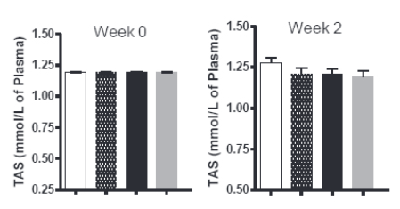
Scientific literature shows that inflammation and oxidative stress contribute in the pathogenesis of atherosclerosis. The oxidative stress is the consequence of an imbalance between the free radical generation and elimination. One source of free oxygen radicals is cyclooxygenase (COX) -2 and, therefore, inhibiting the activity of this enzyme is likely to reduce oxidative stress. In the present study an experimental rabbit model of hypercholesterolemia was developed and the effects of COX-2 inhibitors, nimesulide and celecoxib were observed on the activities of antioxidant enzymes, superoxide dismutase (SOD), glutathione peroxidase (GPx) and total antioxidant status (TAS). Rabbits were divided into four groups-control, saline, nimesulide and celecoxib, with all groups fed a high cholesterol diet, which only received saline. Low activities of SOD, GPx and TAS were measured in the hypercholesterolemic rabbits pretreated with saline. In the same group, a reciprocal relationship was observed between the LDL-cholesterol concentration and the plasma activities of GPx, SOD and TAS. Rabbits in nimesulide and celecoxib group showed significantly higher activities of SOD, GPx and TAS in hypercholesterolemic rabbits compared to saline group (p<0.05). Our study shows that selective and timely use of COX-2 inhibitors would be useful in preventing the onset and development of atherosclerosis by enhancing antioxidant defence system.

Downloads

Download data is not yet available.
Cyclooxygenase-2 inhibition improves antioxidative defense during experimental hypercholesterolemia

Downloads

Additional Files

Published

2014-05-20

How to Cite

1.
Ahmed S, Gul S, Gul H, Zia-Ul-Haq M, Ercisli S. Cyclooxygenase-2 inhibition improves antioxidative defense during experimental hypercholesterolemia. Biomol Biomed [Internet]. 2014May20 [cited 2023Jun.4];14(2):63-9. Available from: https://www.bjbms.org/ojs/index.php/bjbms/article/view/2264

Issue

Section

Biochemistry

INTRODUCTION

Inflammation and oxidative stress are intimately involved in the pathogenesis of atherosclerosis [1]. In hypercholesterolemia, generally recognized as a risk factor of atherogenesis, oxidative stress plays an important role [2]. The tissue concentration of oxygen radicals is limited under normal physiological conditions due to the existence of a delicate balance between the generation of free radicals and the antioxidant defense system [3]. However, if this balance is perturbed in favour of more free radicals, either through an enhanced production or via a reduction in the endogenous antioxidant defence system or both, the body is at risk for free radical-mediated cell damage. In this scenario, an important pathogenic role of these free radicals is a harmful oxidative modification of low-density cholesterol (LDL). The involvement of reactive oxygen species (ROS) in the oxidative modification of LDL is an important element of atherogenesis. These ROS has drawn attention to the anti-oxidative defence of the organism, including the so-called ROS scavengers. Among natural, so-called primary scavengers, antioxidative enzymes of red blood cells and of the serum play a major role [4]. Endothelial damage and increase in polymorphonuclear leukocyte activity which occur during atherogenesis lead to oxidative stress and to an overproduction of reactive forms of oxygen, which in turn exhaust the anti-oxidative pool of the organism. Total antioxidant status (TAS), glutathione peroxidase (GPx) and superoxide dismutase (SOD) are largely responsible for maintaining the redox balance within the body [5]. Patients with ischemic heart disease are detected with the presence of low SOD [6]. The incidence of cardiovascular diseases such as atherosclerosis increases with low levels of GPx [7]. Mitrevky and colleagues reported low TAS levels in patients with myocardial infarction [8]. Low TAS level is also found in patients with atherosclerosis [9], in cancer and rheumatoid arthritis patients [10], in male infertility and diabetic patients [11, 12]. One source of free oxygen radicals is cyclooxygenase (COX)-2 and, therefore, inhibiting the activity of this enzyme is likely to reduce oxidative stress. However, whether COX-2 inhibitors affect the activity of antioxidant enzymes during hypercholesterolemia has not been investigated with any rigor. In the present study an experimental rabbit model of hypercholesterolemia was developed and the effects of COX-2 inhibitors nimesulide and celecoxib were observed on the activities of above-mentioned antioxidant enzymes.

MATERIALS AND METHODS

Animals

Male New Zealand white rabbits weighing 1.5-2 kg were kept in the Animal House of Kohat University of Science & Technology, Kohat, Pakistan for at least 7 days before any experiment. Rabbits were divided into four groups. First group was fed standard rabbit diet (control group), second group was maintained on high cholesterol supplemented diet (1% cholesterol diet, Harlan Teklad, WI, USA) and pre-treated with saline (saline group), third group was maintained on high cholesterol supplemented diet and pretreated with nimesulide (nimesulide group) while fourth group was maintained on high cholesterol supplemented diet and pretreated with celecoxib (celecoxib group).

Procedures

Control and saline groups were injected subcutaneously with 0.5 ml saline per rabbit per day while nimesulide and celecoxib groups were injected with nimesulide (25 mg/ kg) per rabbit per day and celecoxib (25 mg/kg) per rabbit per day throughout the 20 weeks of experimental period. Both nimesulide and celecoxib were dissolved in 1% DMSO (final concentration). Doses of 25 mg/kg for nimesulide and celecoxib administered to the animals were calculated on the basis of recommended daily doses of these drugs in human, and interpolated to rabbits. This protocol was approved by the Animal Committee of Kohat University of Science & Technology. All rabbits were given free access to food and water. Overnight fasting blood was collected from all groups before feeding the normal or high cholesterol diet and pretreatment with COX 2 inhibitor and after 2, 4, 6, 8, 10, 12, 14, 16, 18 and 20 weeks. Blood was taken from the marginal ear vein of these animals and then transferred into siliconized glass tubes. Lipid profile and antioxidant assays were performed within two hours of the blood collection. All the animals used in this study were handled in compliance with the highest ethical guidelines by the National Institute of Health USA, for the care of such animals. Following experiments were carried out on the blood of all rabbits.

Lipid profile

Total cholesterol, HDL and LDL concentrations were determined using routine spectrophotometric assays from kits purchased from RANDOX, UK, and as described by Ukpanukpong et al. [13].

Glutathione peroxidase activity

GPx activity was measured as described by Gul et al. [14]. The assay works by the addition of t-butyl-hydroperoxide which is a substrate for GPx. In this assay the peroxidase reaction is coupled with reductase reaction. Glutathione is used as co-substrate. A change in the absorbance at 340 nm takes place after the reduction of the substrate. This change in the absorbance is reflective of GPX activity.

Superoxide dismutase activity

SOD activity was determined as described by Gul et al. [15]. In this experiment formazane dye is obtained by the addition of the substrate (i.e. 2-(4-iodophenyl)-3-(4-nitrophenol)-5-phenyltetrazoliumchloride (INT)) and after the addition of all other contents of the kit. The basic principle of the method is the generation of superoxide radicals with xanthine and xanthine oxidase. This reaction is inhibited depending upon the concentration and activity of SOD. The absorbance at 505 nm measures the activity of SOD.

Total antioxidant status

TAS assay was performed as described by Gul et al. [14]. This experiment is based on the chemical (2,2’ azino-bis-[3-ethylbenz-thiazoline-6-sulfonic acid] (ABTS)) which is used as a substrate in the reaction. After the addition of plasma and other kit contents, ABTS is added at the very end. As soon as ABTS is added, oxidants in the plasma start oxidizing it to ABTS+. A change in the absorbance at 600 nm shows the degree of inhibition by plasma antioxidants and indicates TAS levels in the plasma. The final plasma anti-oxidant concentration is obtained by multiplying the absorbance of the sample to the specific factor obtained from the standard and the blank and measured in (mmol/L).

Statistical analysis

Statistical analysis was done by one-way analysis of variance (ANOVA) flowed by the New-mal - Keuls test. p<0.05 was considered significant. All the experiments were performed in triplicate.

RESULTS

Lipid profile

There was no significant change in total cholesterol levels in control group throughout the study while in saline group total cholesterol increased significantly to 63.04±17.21 mM and 76.45±13.22 mM at week 10 and 20 respectively (Table 1). In nimesulide group, 51.20±13.67 mM and 43.76±11.45 mM total cholesterol levels were observed at week 10 and 20 respectively and were significantly lower than in the saline group. In celecoxib group 58.05±12.21 mM and 50.09±13.71 mM levels were observed at week 10 and 20 respectively indicating significant lowering compared to saline group at week 20. In saline group, HDL cholesterol decreased to 0.54±0.24mM and 0.52±0.15 mM at week 10 and 20 respectively, in nimesulide group, it increased to 0.85±0.24mM and 0.81±0.20 mM at week 10 and 20 respectively while in celecoxib group, 0.80±0.16 mM and 0.81±0.17 mM of HDL cholesterol was observed at week 10 and 20 respectively. LDL cholesterol increased in the saline group to 61.20±13.21 mM and 72.09±15.26) mM at week 10 and 20 respectively, to 41.27±10.10 mM and 24.47±4.19 mM at week 10 and 20 respectively in nimesulide group while increased to 41.80±13.87 mM and 26.42±3.34 mM at week 10 and 20 respectively in celecoxib group (Table 1). However, this increase in LDL-cholesterol in nimesulide and celecoxib groups was significantly lower (p<0.05) compared to the increase in the saline group at week 10 and 20. All the results are expressed as mean ± S.D.

TABLE 1: Lipid profile of rabbits during hypercholesterolemia. All the values are expressed in mM and as mean(SD)n. *p< 0.05 compared to control group, #p<0.05 compared to saline group.

Total antioxidant status (TAS)

In hypercholesterolemic rabbits treated with nimesulide, there was significant elevation of TAS from 1.19 ± 0.02 to maximum of 1.41 ± 0.10 mmol/L of plasma at week 20 (p<0.05 compared to saline treated hypercholesterolemic rabbits) (See Figure 1 and Table 2). In the control animals fed on the standard diet, TAS remained roughly the same throughout the study with slight increase from 1.190 ± 0.02 at week 0 to a maximum value of 1.28 ± 0.13 mmol/L of plasma at week 12. In hypercholesterolemic rabbits treated with saline, TAS decreased slightly from 1.190 ± 0.02 to 1.57 ± 0.11 mmol/L of plasma at week 16. In hypercholesterolemic rabbits treated with celecoxib, TAS increased from 1.190 ± 0.02 at week 0 to a maximum value of 1.54 ± 0.133 mmol/L of plasma at week 20 (p<0.05 compared to saline treated hypercholesterolemic rabbits). All the results are expressed as mean ± S.D.

FIGURE 1: TAS of rabbits in group A (normal) group B (hypercholesterolemia and saline treated), group C (hypercholesterolemia and nimesulide treated) and group D (hypercholesterolemia and celecoxib treated). *p< 0.05 compared to control group in the same week.
TABLE 2: Table shows TAS of rabbits in group A (normal) group B (hypercholesterolemia and saline treated), group C (hypercholesterolemia and nimesulide treated) and group D (hypercholesterolemia and celecoxib treated) at week 0-20 of the experimental period.

Glutathione peroxidase (GPx)

The cholesterol-rich diet resulted in the elevation of the GPx from 5200 ± 800 to 7200 ± 567 U/L of hemolysate at week 8 in hypercholesterolemic rabbits treated with nimesulide (p<0.05 compared to saline treated hypercholesterolemic rabbits) (See Figure 2 and Table 3). In the control animals fed on the standard diet, there were slight changes in GPx but mostly remained unchanged and increased slightly from 5200 ± 467 to 5412 ± 546 U/L of hemolysate at week 18. In hypercholesterolemic rabbits treated with saline, GPx decreased from 5200 ± 456 at week 0 to 4424 ± 478 U/L of hemolysate at week 18. In hypercholesterolemic rabbits treated with celecoxib, GPx increased from 5200 ± 675 at week 0 to 7362 ± 564 U/L of hemolysate at week 16 (p<0.05 compared to saline treated hypercholesterolemic rabbits). All the results are expressed as mean ± S.D.

FIGURE 2: GPx levels of rabbits in group A (normal) group B (hypercholesterolemia and saline treated), group C (hypercholesterolemia and nimesulide treated) and group D (hypercholesterolemia and celecoxib treated). *p< 0.05 compared to control group in the same week.
TABLE 3: GPx levels of rabbits in group A (normal) group B (hypercholesterolemia and saline treated), group C (hypercholesterolemia and nimesulide treated) and group D (hypercholesterolemia and celecoxib treated).

Superoxide dismutase (SOD)

In hypercholesterolemic rabbits treated with nimesulide, there was significant elevation of the SOD from 179 ± 18 to 234±21 U/L of whole blood at week 16 (p<0.05 compared to saline treated hypercholesterolemic rabbits) (See Figure 3 and Table 4). In the control animals fed on the standard diet. SOD increased slightly from 180 ± 20 to 194 ± 17 U/L of whole blood at week 18. In hypercholesterolemic rabbits treated with saline, SOD decreased from 178 ± 21 at week 0 to 162 ± 17 U/L of whole blood at weeks 4 and 8. In hypercholesterolemic rabbits treated with celecoxib, SOD increased significantly from 179 ± 12 at week 0 to 214 ± 13 U/L of whole blood at week 20 (p<0.05 compared to saline treated hypercholesterolemic rabbits). All the results are expressed as mean ± S.D.

FIGURE 3: SOD levels of rabbits in group A (normal) group B (hypercholesterolemia and saline treated), group C (hypercholesterolemia and nimesulide treated) and group D (hypercholesterolemia and celecoxib treated). *p< 0.05 compared to control group in the same week.
TABLE 4: SOD levels of rabbits in group A (normal) group B (hypercholesterolemia and saline treated), group C (hypercholesterolemia and nimesulide treated) and group D (hypercholesterolemia and celecoxib treated).

DISCUSSION

Various factors, both intrinsic and extrinsic, can influence the activity of antioxidative enzymes [16]. Studies indicate an inverse relationship between cholesterol concentration and antioxidant enzymes including SOD, GPx and TAS [17, 18]. Therefore, people with high LDL-CH can have decreased activity enzymes of antioxidative defence system. This also indicates that such people have an excess of substrate, which is likely to be modified by the oxidative agents [5]. Consistent with previous studies [17, 18], we observed a reciprocal relationship between the levels of LDL cholesterol and the plasma activities of GPx, SOD and TAS in saline treated hypercholesterolemic rabbits (see Table 5). Since the activities of SOD, GPx and TAS correlated negatively with both total cholesterol and LDL cholesterol concentrations in our study, it is also likely that these antioxidative enzymes are modulated by total cholesterol concentration, in particular by LDL cholesterol. Although the involvement of hyperlipidemia in lowering the enzymatic antioxidative pool in persons with atherosclerosis is clear, it is never quantitatively estimated [5]. The conditions favoring ROS overproduction with concomitant exhaustion of enzymatic antioxidative pool and the results presented here point to a major role of hypercholesterolemia in lowering the activities of SOD and GPx and TAS. It is difficult to evaluate to what extent a decrease in the respective values can participate in the initiation of the atherosclerotic cascade. Nevertheless it is evident that enzymatic antioxidative activity is decreased in hypercholesterolemic rabbits which can facilitate and augment the atherosclerotic process. Whether or not the measurements of antioxidative activities of SOD and GPx and TAS can be used in the diagnosis of coronary atherosclerosis in persons with hypercholesterolemia requires further studies. There are some studies, which show a decrease in GPx, SOD and TAS in coronary heart disease population [19] but most of these studies take single measurements. Since atherosclerosis is a chronic process, single measurements of lipids, SOD, GPx, and TAS as well as other risk factors at a particular stage do not allow inferences of their past influence on the disease’s progression [5]. Therefore, we took measurements periodically after 2 weeks up to 20th week. The considerable decrease in the activities of SOD, GPx and TAS in hypercholesterolemic group suggests that a weakening or destruction of the antioxidative barrier of the organism is related to hypercholesterolemia. In nimesulide treated hypercholesterolemic rabbits and to a lesser extent in celecoxib treated hypercholesterolemic rabbits, activities of GPx, SOD and TAS were improved as compared to the saline treated hypercholesterolemic rabbits suggesting antioxidative potential of these COX-2 inhibitors. Our study does not point to the mechanism of antioxidant actions observed with nimesulide and celecoxib. Previous studies, however, show that COX inhibition is associated with improvement in cardiovascular diseases [20, 21]. COX-metabolites influence the force of myocardial contractions through oxidative stress and by affecting changes in Ca2+ cycling [22, 23]. Prostaglandins produced by the action of COX cause induction and growth of inflammatory mediators and their reduction or inhibition by COX-2 inhibitors reduces not only inflammation, but also oxidative stress [24]. Oxidative stress also adversely affects the structure and function of myocardium [25] and COX-2 inhibitors, by causing a reduction in oxidative stress [26] may improve the myocardial contractility. In patients with coronary artery disease, endothelial function was improved by celecoxib mainly through reducing oxidative stress [27]. Selective inhibition of COX-2 is shown to be beneficial in the treatment of myocardial infarction as induction of COX-2 in the ischemic myocardium is followed by oxidative stress [28, 29]. However, further studies are needed to decipher the exact mechanism of these effects of COX-2 inhibitors in improving antioxidative defence system.

TABLE 5: Inverse relationship between LDL cholesterol and antioxidant enzymes in saline-treated hypercholesterolemic rabbits through week 0-20.

CONCLUSION

We conclude that activities of GPx, SOD, and TAS are modulated by total cholesterol concentration, in particular LDL cholesterol. Both nimesulide and celecoxib are able to increase activities of key antioxidant enzymes and there is possibility that these effects of nimesulide and ce-lecoxib may be independent of their COX-2 inhibitory function. Regardless of the mode of action in observed parameters, our study shows that selective and timely use of COX-2 inhibitors would be useful in enhancing the antioxidant defence system during hypercholesterolemia.

DECLARATION OF INTEREST

The authors declare no conflict of interest.

REFERENCES

  1. (). Inflammatory mechanisms: the molecular basis of inflammation and disease. Nutr Rev.
  2. , , , , (). Hypercholesterolemia, oxidative stress and gender dependence in children. Prague Med Rep.
  3. , , , (). Oxidative stress and heart failure. Mol Cell Biochem.
  4. (). Oxygen toxicity, Part II. Defense mechanisms. Postepy Biochem.
  5. (). Activities of red blood cell anti-oxidative enzymes (SOD, GPx) and total anti-oxidative capacity of serum (TAS) in men with coronary atherosclerosis and in healthy pilots. Med Sci Monit.
  6. (). Monitoring of total antioxidant status and activity of superoxide dismutase in patients with ischemic heart disease. Abstracts from MEDLAB, 12th IFCC European Congress of Clinical Chemistry. Poster A94
  7. , (). Total antioxidant status of the patients receiving total parenteral nutrition (TNP). Proceed Nutr Soc. Poster 242A
  8. (). Total antioxidant status and superoxide dismutase after cerebrovascular accident. XVI international Congress of Clinical Chemistry. Poster A106
  9. , (). Lipid peroxides and antioxidants in plasma of healthy subjects and patients with atherosclerosis. Pol. Arch. Med. Wewn.
  10. (). Plasma TAS in various diseases. Abstracts from the MEDLAB; 12th IFCC European Congress of Clinical Chemistry. Poster A11
  11. (). Anti-oxidant therapy: does it have a role in the treatment of human disease? Expert Opin Investig. Drugs.
  12. , , , (). Superoxide scavenging capacity of human seminal plasma. Int J Androl.
  13. , , (). Antioxidant interactions of Pefloxacin, garlic, vitamins C and E on lipid profile level of Albino wistar rats. J Applied Pharm Sci.
  14. , , , (). Investigating the protective effect of Solanum melongena. Asian J Health.
  15. , , , , , (). The antioxidant potential of Brassica rapa L. on glutathione peroxidase, superoxide dismutase enzymes and total antioxidant status. Revista Romănă de Medicina de Laborator.
  16. , , (). Redox regulation by intrinsic species and extrinsic nutrients in normal and cancer cells. Annu Rev Nutr.
  17. , , (). Antioxidative enzyme profiles in fighter pilots. Aviat Space Environ Med.
  18. , , , , , , , , , , (). Effects of D-003, a mixture of high-molecular-weight sugar cane wax acids, on lipid peroxidation markers in older individuals: A randomized, double-blind, placebo-controlled study. Curr Ther Res Clin Exp.
  19. , , , , , (). yperhomocysteinaemia and parameters of antioxidative defence in Tunisian patients with coronary heart disease. Ann Clin Biochem.
  20. , , (). Antiatherosclerotic activity of ibuprofen, a non-selective COX inhibitor-an animal study. Indian J Exp Biol.
  21. , (). Anti-ischemic effects of nimesulide, a cyclo-oxygenase-2 inhibitor on the ischemic model of rabbit induced by isoproterenol. Arch Pharma Res.
  22. , (). Inotropic actions of eicosanoids. Basic Res Cardiol.
  23. , (). Arachidonic acid enhances contraction and intracellular Ca2+ transients in individual rat ventricular myocytes. Am J Physiol.
  24. , , , , , (). Pharmacological and biochemical demonstration of the role of cy-clooxygenase 2 in inflammation and pain. Proc Natl Acad Sci USA.
  25. , (). Oxygen free radicals and excitation-contraction coupling. Antioxid Redox Signal.
  26. , , (). Modulation of oxidant status by meloxicam in experimentally induced arthritis. Pharmacol Res.
  27. , , , , , (). Selective COX-2 inhibition improves endothelial function in coronary artery disease. Circulation.
  28. , , , , (). Inhibition of cyclooxygenase-2 improves cardiac function in myocardial infarction. Biochem Biophys Res Commun.
  29. , (). Role of cyclooxygenase-2 in myocardial infarction and ischaemia. J Coll Physicians Surg Pak.