Effect of met-enkephalin on chromosomal aberrations in the lymphocytes of the peripheral blood of patients with multiple sclerosis

Authors

  • Maida Rakanović-Todić Institute of Pharmacology, Clinical Pharmacology and Toxicology, Faculty of Medicine, University of Sarajevo
  • Lejla Burnazović-Ristić Institute of Pharmacology, Clinical Pharmacology and Toxicology, Faculty of Medicine, University of Sarajevo
  • Slavka Ibrulj Center for Cytogenetics and Molecular Medicine, Faculty of Medicine, University of Sarajevo
  • Nedžad Mulabegović Institute of Pharmacology, Clinical Pharmacology and Toxicology, Faculty of Medicine, University of Sarajevo

DOI:

https://doi.org/10.17305/bjbms.2014.2267

Keywords:

met-enkephalin, chromosomal aberrations, multiple sclerosis, peripheral blood lymphocytes

Abstract

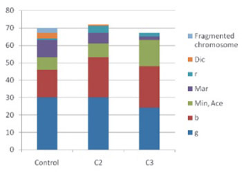
Endogenious opiod met-enkephalin throughout previous research manifested cytoprotective and anti-inflammatory effects. Previous research suggests that met-enkephalin has cytogenetic effects. Reducement in the frequency of structural chromosome aberrations as well as a suppressive effect on lymphocyte cell cycle is found. It also reduces apoptosis in the blood samples of the patients with immune-mediated diseases. Met-enkephalin exerts immunomodulatory properties and induces stabilization of the clinical condition in patients with multiple Sclerosis (MS). The goal of the present research was to evaluate met-enkephalin in vitro effects on the number and type of chromosome aberrations in the peripheral blood lymphocytes of patients with MS. Our research detected disappearance of ring chromosomes and chromosome fragmentations in the cultures of the peripheral blood lymphocytes treated with met-enkephalin (1.2 μg/mL). However, this research did not detect any significant effects of met-enkephalin on the reduction of structural chromosome aberrations and disappearance of dicentric chromosomes. Chromosomes with the greatest percent of inclusion in chromosome aberrations were noted as: chromosome 1, chromosome 2 and chromosome 9. Additionally, we confirmed chromosome 14 as the most frequently included in translocations. Furthermore, met-enkephalin effects on the increase of the numerical aberrations in both concentrations applied were detected. Those findings should be interpreted cautiously and more research in this field should be conducted.


Downloads

Download data is not yet available.
Effect of met-enkephalin on chromosomal aberrations in the lymphocytes of the peripheral blood of patients with multiple sclerosis

Downloads

Additional Files

Published

2014-05-20

How to Cite

1.
Rakanović-Todić M, Burnazović-Ristić L, Ibrulj S, Mulabegović N. Effect of met-enkephalin on chromosomal aberrations in the lymphocytes of the peripheral blood of patients with multiple sclerosis. Biomol Biomed [Internet]. 2014May20 [cited 2023Apr.1];14(2):75-80. Available from: https://www.bjbms.org/ojs/index.php/bjbms/article/view/2267

Issue

Section

Molecular Biology

INTRODUCTION

Met-enkephalin is one of the simplest endogenous opioid peptides within the enkephaline family. Endogenious opioid peptides share amino sequence of tyrosine-glycine-glycine-phenylalanine (aka Opioid motif), and contain one or more copies of met-enkephalin (Tyr-Gly-Gly-Phe-Met) and leu-enkephalin (Tyr-Gly-Gly-Phe-Leu). Opioid receptors (OP) are detected in human phagocytic leukocytes, with a direct binding of naloxone in lymphocytes and thrombocytes [1]. Met-enkephalin binds with high affinity to OP1 (δ) receptors, and with low affinity to OP3 (μ) receptors. Additionally, it specifically binds to receptors on T lymphocytes which are not morphin receptors [1, 2]. As a potential receptor on human lymphocytes a complementary transcript of met-enkephalin is isolated, with a single sequence that matches cytokine receptor γ chain [3]. Multiple sclerosis (MS) is a progressive disease followed by development of the neurological deterioration. Relapsing/remitting form of the disease is highly sensible to immunosuppressive therapy. However, with the extended duration, the response-rate to the treatment tends to decrease as well. As a result of an assumption which claims the existence of the inflammation and of the neurodegenerative phase, patients with MS are recommended for early immunomodulatory treatment [4, 5]. Due to immunomodulatory properties met-enkephalin was applied in clinical studies and it effect on the stabilization of the clinical conditions of MS was documented [6, 7]. It also manifests in vivo citoprotective effects [8]. Met-enkephalin mostly induces immunostimulation when applied in low doses, and immunosuppresion when applied in higher doses. Higher doses of met-enkephalin exerted suppressive effect in experimental treatment of allergic encephalomyelitis [2, 9]. A role of the released cytokines and of Thi cells differentiation disorder are implied in an immuno mediated demyelization [4]. The research of the cerebrospinal fluid in MS patients reveals an increase in the levels of immunoglobulin (lg) and mononuclear pleocytosis. Furthermore the same studies showed the numerous somatic gene mutations in the variable region of the lg Heavy Chain in the cerebrospinal fluid B cells in MS patients [10,11]. Research by Štambuk et al. [3] detected a significant reduction in the frequencies of the structural chromosome aberration in human lymphocytes of the peripheral blood of MS patients.

MATERIALS AND METHODS

The research was conducted at the Neurological Clinic of the Clinical and University Center of Sarajevo, and the Center for Human Genetics of the Faculty of Medicine, University of Sarajevo.

Samples

Blood samples were obtained from seven female patients in relapse in a test tube containing heparin. The eligibility criteria were MS diagnosis as per McDonald Diagnostic Criteria, depicted on existence of objective proofs of at least two lesions (MRI or evoked potentials), or at least two clinically diagnosed symptomatic disease episodes. Patients included in the study were never treated with interferon, and had not received pulse corticosteroid treatment over the past six months.

Tested substance

Met-enkephalin (Biotechnology Laboratories Richmond, USA) was dissolved in distilled water and kept at a temperature of -18°C. Prior to adding to culture, it was kept at a room temperature (18-23°C) up to five minutes. The met-enkephalin concentration per culture 2 (C2) was 1.2 μg/ml and per culture 3 (C3) 120 μg/ml. Control culture (C1) was not incubated with the tested substances.

Research design

Blood samples were cultivated following method described by Moorhead et al. [12], with the incubation of cultures during 72 hours and the application of Colcemid stock solution 25 mcg/ml (0.2 ml) two hours before completion of incubation period. After microscopic analysis of chromosome preparations by standard procedure (Giemsa staining), the identification of rearranged chromosomes was conducted by destaining and applying the G-band technique. The total number of chromosomes included in structural aberrations was determined in the following manner: numerical analysis did not include chromosomes with gaps; its number was separately analyzed. It is deemed that the single chromosome was included when the following structural aberration existed: chromosome/chromatide break, acentric fragment, ring fragment, minute, acentric ring, ring chromosome, marker chromosome. Two chromosomes are deemed included when the following structural aberration existed: dicentric chromosome and translocation.

Statistical analysis

The collected data was statistically processed by computer software SPSS v.11 (Statistical Package for Social Sciences©, March 2004). For the purpose of the hypothesis testing, we used a non-parametric testing for correlated samples, Wilcoxon Signed Ranks Test. Findings from control culture (C1) and cultures incubated with various concentrations of tested substances (C2, C3) were compared.

RESULTS

Female patients included were 34 to 60 years old (41.89±9.17), while the total number of hospitalizations due to MS was from one up to 6 hospitalizations (2.67±1.80). The recorded values of fibrinogen ranged between 9.10-15.90 μmol/L (12.44±2.52).

Our research reviewed 200 mitosis per each tested culture. The total number of chromosomes included in structural aberrations are presented in Table 1. and detected aberrations in Figure 1.

TABLE 1: Descriptive statistics for structural chromosomes aberrations
FIGURE 1: Number of the total detected structural aberrations

Among detected structural aberrations the highest presence of gaps, breaks and marker chromosomes was documented. The ring chromosomes and the chromosome fragmentation were present within the C1 only; while dicentric chromosomes were detected in C1 and C2. When control cultures were compared to incubated ones, no statistically significant differences were recorded either in the number of chromosomes included in structural aberrations (C1 vs C2, p=0.527; C1 vs C3: p=0.089) or in the number of mitosis with aberrations (C1 vs C2, p=1.000; C1 vs C3, p=0.581). The identified chromosomes included in structural chromosome aberrations are displayed in Appendix 1. The frequency of engagement of certain chromosomes in aberrations, expressed in percentages, is shown in Table 2. For the calculation of frequency of associated aberrations with a familiar origin, the following were included: gap, break, translocation, chromosome fragmentation, dicentric and ring chromosomes. The majority of translocated marker chromosomes was detected in C1, while chromosome 14 was mostly included in translocations. Basic descriptive statistics for the numerical aberrations is displayed in Table 3. The number of all detected numerical aberrations is listed in Figure 2. When control cultures were compared to incubated ones, a statistically significant increase in number of numerical aberrations was detected in the incubated cultures (C1 vs C2, p=0.027; C1 vs C3, p=0.039). When observing polyploidy, the most fequent was the presence of endoreduplication, while triploidy and tetraploidy were somewhat rarer. Additionally, hyperdiploidy was detected. Furthermore, after G-banding the most frequent engagement in polysomy was of the X chromosome. Among the detected aneuploidy, the trisomy and tetrasomy of the X chromosome were dominant (Figure 3). When control cultures were compared to incubated ones, a statistically significant increase in the number of poliploidy was detected in the culture treated with a lower concentration of met-enkephalin (C1 vs C2, p=0.034), while no significant difference was documented when controls were compared to cultures treated with higher concentration of met-enkephalin (C1 vs C3, p=0.131). No statistically significant difference existed in the number of endoreduplications (C1 vs C2, p=0.157; C1 vs C3, p=0.334). A statistically significant increase in the number of aneuplody existed in cultures incubated with lower met-enkephalin concentration compared to control cultures (C1 vs C2, p=0.026). No statistically significant difference was revealed when a culture incubated with a higher concentration was compared to control ones (C1 vs C3, p=0.236). Mitotic index was determined as a percent of lymphocytes in mitosis (M1+M2), counted on 300 lymphocytes. No statistically significant difference in mitotic index existed between the control culture and the cultures incubated with various concentrates of substance used for testing (C1 vs C2, p=0.674; C1 vs C3, p=0.753).

APPENDIX 1: Identified chromosomes for cultures not treated with met-enkephalin
TABLE 2: The frequency of associated chromosomes in structural aberrations, expressed in percentages
TABLE 3: Descriptive statistics for numerical chromosome aberrations
FIGURE 2: Number of detected numerical aberrations
FIGURE 3: Structure of detected numerical aberrations

DISCUSSION

Our research detected disappearance of ring chromosomes and chromosome fragmentations in the cultures treated with met-enkephalin. Similar to our results, the study by Štambuk et al. [3] showed disappearance of ring chromosomes and chromosome fragmentation after in vitro treatment of cultures with met-enkephalin (1.2 μg/mL) and incubation period of 48 hours. However, in contrast to their results, our research did not reveal significant effect of met-enkephalin on the reduction of the number of structural aberrations and on disappearance of dicentrical chromosomes. Furthermore, within the five-days cell culturing with incorporated 3H-thymidine, Stambuk et al. [3] documented significant reduction in the number of cells reaching the third stage of mitosis and a significant increase in a number of first metaphase. Chromosomal fragile sites expressed through an increased frequency in gaps and breaks are identified, as well as a presence of conservation of fragile sites throughout evolution [13, 14]. Re et al. [14] suggested that fragile sites via modulated gene expression can participate in the regulation of the cell responsivity rate to oncogenic stress and DNA damage. Ilyin-skikh et al. [15] calculated the expected frequency of chromatic aberrations which are induced by radiation (8.44 up to 2.04 from the first up to 22nd chromosome), while the number of breaks increases with an increase in absolute chromosome length. The longest chromosomes in our research were also most frequently included in aberrations (chromosome 1 in C1 and C2, and chromosome 3 in C3). The frequency of chromosome 9 in structural aberrations was the most prominent. It is difficult to interprete the noticed impact of the met-enkephalin on the number of aberrations in treated cultures. The relationship between the ploidy disorders in malignant cells and an increase in the cell growth potential was suggested [16]. A hypothesis on aneuploidy as chromatic base of cancerogenesis was established [17]. Our research detected dominant engagement of X chromosome in hyperploidy and polysomy. According to data obtained from the Mittleman Data Base on chromatic aberrations in malignant diseases [18], the trisomy of X chromosome is most frequently related to acute lymphoblastic leukemia and lymphoblastic lymphoma. Previous research using met-enkephalin do not point out cancerogenous potential of this peptide; rather it shows quite the opposite [19-22]. When applied with paclitaxel, met-enkephalin enhances the inhibition of tumor growth of squamous cells head and neck carcinoma [19, 20]. Furthermore, throughout the experiment, aneugenic potential of opioids morphine and noskapine was detected [21, 22]. Genotoxic in vitro effects of noskapine were not confirmed in vivo, probably due to fast metabolism and low systemic bioavailability of the medication [21]. Experimental study by Cheng et al. [23] also suggests that met-enkephalin inhibits cell proliferation of various human and animal cells – probably by inducing an expression of inhibitors of cyclin-dependant kinases.

CONCLUSION

In conclusion, although the application of met-enkephalin in the culture of peripheral blood lymphocytes of MS patients did not manifest statistically significant protective effects, it influenced the disappearing of serious structural aberrations such as ring chromosomes and fragmentation of chromosomes. Met-enkephalin showed an impact on the number of numerical aberrations in both treated cultures, which certainly demands further in vivo evaluation.

DECLARATION OF INTEREST

The authors declare no conflict of interest.

Acknowledgements

ACKNOWLEDGEMENTS

The authors want to acknowledge Farmacija d.0.0., Tuzla for providing us with an opportunity to work in this area by donating the tested substances as well as to Ms. Amra Ćatović for technical and other support during the research process.

REFERENCES

  1. , , , , (). Methionine enkefalin: A new cytokine--human studies. Clin Immunol Immunopathol.
  2. (). Enkephalins and immune inflammatory reactions. Acta Neurol (Napoli).
  3. , , , , , (). Cytogenetic Effects of Met-enkephalin (Peptid M) on human lymphocytes. Croat Chem Acta.
  4. , (). Multiple sclerosis and other demyelinating diseases. Harrison's Principles of Internal Medicine.
  5. , , , (). Escalating immunotherapy of multiple sclerosis. Therapeutic Advances in neurological Disorders.
  6. , , , , , (). Peptide M (Lupex(R)) immunotherapy in multiple sclerosis, optic neuritis and uveitis. Int. J. Thymology.
  7. , , , , , , (). Peptid-M (LUPEX”) Effects on the Immune Responseand Clinical Status in Uveitis, Optic Neuritis and Multiple Sclerosis. Uveitis Today, Excerpta Medica ICS1158.
  8. , , , , , , , , (). Protective Effects of Met-enkephalinon Alcohol Induced Gastric Lesions. Croat Chem Acta.
  9. , , , , (). Opioid growth factor suppresses expression of experimental autoimmune encephalomyelitis. Brain Research.
  10. , , , , , (). Clonal Expansion and Somatic Hypermutation of VH genes of B Cells from Cerebrospinal Fluid in Multiple Sclerosis. J Clin Invest.
  11. , , , , , (). Recapitulation of B cell differentiation in the central nervous system of patient with multiple sclerosis. Proc. Natl. Acad. Sci USA.
  12. , , , , (). Chromosome preparations of leukocytes cultured from human peripheral blood. Exp Cell Res.
  13. , (). Fragile sites and human disease. Human Molecular Genetics.
  14. , , , , (). Correlated fragile site expression allows the identification of candidate fragile genes involved in immunity and associated with carcinogenesis. BMC Bioinformatics.
  15. , , (). Chromosome breakage at sites of oncogenes in a population accidentally exposed to radioactive chemical polution. mutagenesis.
  16. , , , (). Deregulation of cell growth and malignant transformation. Croat. Med. J.
  17. , , , , , (). Cancer drug resistance: The central role of the karyotype, Drug Resist. Updat. doi:10.1016/j.drup.2007.02.003
  18. (). Mitelman Database of Chromosome Abberations. Avaliable at http://gap.nci.nih.gov/Chromosomes/Mitelman
  19. , , , , , (). Enhenced growth inhibition of squamous cell carcinoma of the head and neck by combination therapy of pacli-taxel and opioid growth factor. Int J Oncol.
  20. , , , , , (). Opioid growth factor enhances tumor growth inhibition and increases the survival of paclitaxel-treated mice with scuamous cell carcinoma of the head and neck. Cancer Chemother Pharmacol.
  21. (). Genotoxic evaluation of morphine, buprenorphine, pentazocine and noscapine by micronucleus and Comet assay in albino mice. Indian Journal of Pharmacology.
  22. , , , (). Noscapine hydrochloride disrupts the mitotic spindle in mammalian cells and induces aneuploidy as well as polyploidy in cultured human lym-phocytes. Mutagenesis.
  23. , , , (). The OGF-OGFr Axis Utilizes the p16INK4a and p21 W A F 1/CIP1pathway to restrict normal cell proloferation. Molecular biology of the cell.