Morphological aspects of myocardial bridges

Authors

  • Almira Lujinović Department of Anatomy, Faculty of Medicine, University of Sarajevo
  • Amela Kulenović Department of Anatomy, Faculty of Medicine, University of Sarajevo
  • Eldan Kapur Department of Anatomy, Faculty of Medicine, University of Sarajevo
  • Refet Gojak Clinic of Infectious Diseases, Clinical Centre, University of Sarajevo

DOI:

https://doi.org/10.17305/bjbms.2013.2304

Keywords:

human heart, myocardial bridges, morphological aspects

Abstract

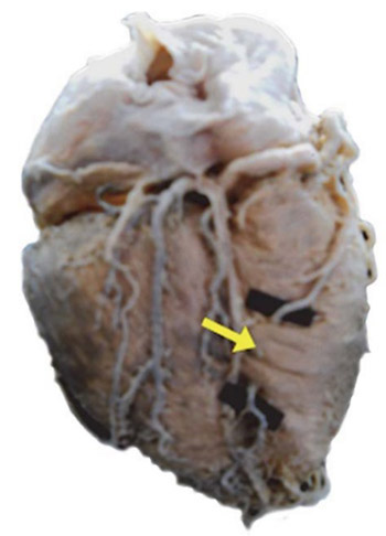
Although some myocardial bridges can be asymptomatic, their presence often causes coronary disease either through direct compression of the “tunnel” segment or through stimulation and accelerated development of atherosclerosis in the segment proximally to the myocardial bridge. The studied material contained 30 human hearts received from the Department of Anatomy. The hearts were preserved 3 to 5 days in 10% formalin solution. Thereafter, the fatty tissue was removed and arterial blood vessels prepared by careful dissection with special reference to the presence of the myocardial bridges. Length and thickness of the bridges were measured by the precise electronic caliper. The angle between the myocardial bridge fibre axis and other axis of the crossed blood vessel was measured by a goniometer. The presence of the bridges was confirmed in 53.33% of the researched material, most frequently (43.33%) above the anterior interventricular branch. The mean length of the bridges was 14.64±9.03 mm and the mean thickness was 1.23±1.32 mm. Myocardial bridge fibres pass over the descending blood vessel at the angle of 10-90 degrees. The results obtained on a limited sample suggest that the muscular index of myocardial bridge is the highest for bridges located on RIA, but that the difference is not significant in relation to bridges located on other branches. The results obtained suggest that bridges located on other branches, not only those on RIA, could have a great contractive power and, consequently, a great compressive force, which would be exerted on the wall of a crossed blood vessel.

Downloads

Download data is not yet available.
Morphological aspects of myocardial bridges

Downloads

Additional Files

Published

2013-11-20

How to Cite

1.
Lujinović A, Kulenović A, Kapur E, Gojak R. Morphological aspects of myocardial bridges. Biomol Biomed [Internet]. 2013Nov.20 [cited 2023Sep.23];13(4):212-7. Available from: https://www.bjbms.org/ojs/index.php/bjbms/article/view/2304

Issue

Section

Anatomy

INTRODUCTION

Myocardial bridges represent an anomaly of coronary artery flow in which the branches flowing subepicardially descend into the myocardium more shallow or deeper, and after a shorter or longer intramyocardial flow, it reappears in the subepicardial tissue. Bundles of myocardial fibres, which in the form of small bridges, pass over the corresponding part of the coronary artery (“tunnel” segment), are marked as the myocardial bridge [1]. It was Reymann who first detected myocardial bridges, as early as in 1737 [2]. Porstmann and Iwig [3] were the first who, in 1960, detected the narrowing of the lumen ramus interventricularis anterior on coronary angiograms and presumed that it was caused by the contraction of myocardial bridge fibres and consequent compression on the “tunnel” segment wall. Data on frequency of myocardial bridges differ a lot and, probably, depend on the method used for their detection. Autopsies ascertained the great frequency of MB, i.e. 34.5%, 52%, 56% and 60% [4-7]. The similar results on the frequency of the MB showed also the more modern method, CT of coronary angiography [8-10], which enables visualization of myocardial bridges and monitoring of their morphological aspects. Angiographic frequency of myocardial bridges is much lower and it ranges from 0.8%, 4.5% to 12% [11-13]. The presence of myocardial bridges can be asymptomatic and some scientists considered them as a benign anomaly with a good long-term prognosis [11]. Yet, in many cases myocardial bridges are connected with heart rhythm disorder [14-16], angina pectoris [17-19], myocardial infarction [20-22] and sudden cardiac death [23, 24], so that their impact on incidence of coronary insufficiency is indisputable. Clinical manifestation of coronary disease, in patients with the myocardial bridge, can appear in two ways: by contraction of myocardial bridge fibres and direct compression of the “tunnel” segment [25-27] or by stimulation and accelerated development of atherosclerosis in the segment proximally to the myocardial bridge [28-30]. The first mechanism leads mainly to coronary insufficiency in young people, particularly in those exposed to the psychophysical exertion, while consequences of the latter mechanism appear most frequently in elderly persons [31]. Incidence and intensity of the coronary disease, in both mechanisms, depends primarily on the length [20-23], localization [31, 32] and thickness of bridges [32-34], what induced us to this research with the aim to detect the frequency of myocardial bridges above certain branches of coronary arteries, to ascertain length and thickness of bridges and if there existed their interrelation. We also wanted to find out if there existed the difference in the value of the myocardial bridge muscle index (MI) between bridges located above the anterior interventricular branch (RIA) and bridges situated above other branches. Our objective was also to detect the angle at which the myocardial bridge fibres pass over the “tunnel” segment of the crossed blood vessel.

MATERIALS AND METHODS

Samples

The research was carried out on 30 human hearts (of persons between 20 and 57 years of age) received from the Department of Anatomy, Faculty of Medicine, Sarajevo. All the persons whose hearts were used in the research suffered a violent death and we are not aware if there were any coronary diseases stated in their history.

Procedures

The hearts were preserved 3 to 5 days in 10% formalin solution. Thereafter, fatty tissue was removed and arterial blood vessels prepared by careful dissection with special reference to the presence of the myocardial bridges. If bridges were detected, their length and thickness were measured by the precise electronic caliper (Black & Decker, 0.00-155.00 mm, Landscheid), while the angle between the myocardial bridge fibre axis and other axis of the crossed blood vessel was measured by a goniometer. The muscle index of the myocardial bridge (MI) was calculated as the product of length and thickness expressed in millimetres.

Statistical analysis

Statistical Package for the Social Sciences (SPSS) was used for statistical data processing. Mean, standard deviation, minimal and maximal values, as well as median were calculated for the length, thickness and MI. Pearson’s correlation coefficient was used for detection of interrelation between length and thickness of the bridges, while Mann Wintney test was used, because of uneven statistic data distribution, for research of the difference in the value of MI between the group of myocardial bridges located above the anterior interventricular branch (RIA) and the group of myocardial bridges located above other branches.

RESULTS

The presence of myocardial bridges was confirmed in 16 hearts (53.33%) out of 30 hearts dissected. In 9 hearts there was detected one bridge in each heart, in 6 hearts two myocardial bridges in each (each of 4 hearts had one bridge above two different branches, while 2 hearts had two bridges each above the same branch). One heart had three bridges located above two branches, so that total number of bridges was 24. Myocardial bridges were found most frequently above the RIA (4.33%); 15 bridges were located above this branch (Figure 1), i.e. 62.5% out of total 24 detected bridges. Frequency of myocardial bridges above other branches (Figure 2) was much lower (Table 1).

FIGURE 1: Myocardial bridge above RIA (RIA-Anterior interventricular branch)
FIGURE 2: Myocardial bridge above Rdd (Rdd) - Right diagonal branch
TABLE 1: The Distribution of the MIC values according to the methods.

The average (mean) length of myocardial bridges amounted to 14.64±9.03 mm, while the average (mean) thickness was 1.23±1.32 mm.Value of myocardial bridge muscle index (MI) ranged from minimum 1.17mm to maximum 110.48mm and amounted to 23.07±30.30 (Table 2). Length and thickness of bridges stand in the correlation which is medium strong and positive r=0.438, p=0.032. With the growth of the bridge length “grows” its thickness as well (Figure 3). The greatest mean length had the myocardial bridges located above Rms and it amounted to M=24.93±7.52 mm, ranging from minimum 19.61 mm to maximum 30.25mm. The average length of bridges located above the RIA was M=14.78±10.20 mm (ranging from minimum 3.44 mm to maximum 39.1 mm) and the two longest bridges 39.10 mm long, 33.91 mm respectively, were located above that branch. The greatest average thickness had the bridges located above the RIA and it amounted to M=1.36±1.51 mm with the broad range from minimum 0.33 mm to maximum 5.68 mm that encompassed the thickest bridge and one from the two thinnest ones. Muscle index of the myocardial bridge (MI) also had the highest average value (mean) in bridges located above the anterior interventricular branch RIA. Although the value of the myocardial bridge muscle index (MI) was distinctly the greatest in myocardial bridges located above the anterior interventricular branch (RIA), Mann-Whitny test did not show any significant difference in the value of that index between the group of bridges located above the RIA and the group of bridges located on another branches (Table 3). In most cases 54,17% (13/24) myocardial bridge fibres passed over the descending blood vessel at the angle of 90 degrees (Table 4).

TABLE 2: Morphological aspects of myocardial bridges
FIGURE 3: Correlation of thickness and length of myocardial bridges. MB-Myocardial bridges
TABLE 3: Difference of the mean values (median) of the MI between bridges located above the RIA and those located on other branches
TABLE 4: The number of myocardial bridges which pass over the certain blood vessel at the quoted angle

The first description of a myocardial bridge originates as early as 1737 [2]. Special interest in myocardial bridges appeared in 20th century, when besides morphologists, also numerous clinicians began to study that phenomenon indicating to their impact on coronary chemodynamics. Although there are numerous data on the presence and frequency of myocardial bridges, they are rather heterogeneous and probably conditioned by the method used for detection of bridges. Researchers who studied the bridges using dissection method report about their high frequency ranging from 30-60% [5-7] what was also confirmed by CT coronary angiography [8-10]. Our findings on myocardial bridges in 53.33% studied cases are in accordance with the results of the quoted authors and confirm rather frequent presence of myocardial bridges above coronary arteries of human hearts. Angiographic frequency of myocardial bridges above coronary arteries is much lower and ranges from 0.8-12% [11-13]. Namely, the myocardial bridges on coronary angiograms are to be detected indirectly, on the basis of the systolic reduction of lumen, i.e. the milky effect, so that many of them remain unnoticed – depending, first of all, on their morphological aspects but also on the presence and intensity of the fixed proximal stenosis, myocardium contractility state, the presence of ascending aorta obstruction [1, 30, 31]. Yet, all results, regardless of the method used, indicate that RIA is the branch above which the myocardial bridges are mostly localized. This is also confirmed by our results which show that 15 of the total 24 detected bridges (i.e. 62.5% out of the total quantity) are located just above that branch. In five hearts, i.e. in 16.67% of the studied material, we detected the presence of the myocardial bridge above the right coronary artery branch flowing along the anterior or diaphragmal wall of the right ventricle. Because this variable branch of the right coronary artery was passing diagonally over the front or the back wall of the right ventricle we named it right diagonal branch (Rdd) like it was called by some other authors [5]. We also detected the presence of two myocardial bridges in each of six hearts: in four hearts above different branches and in two hearts above the same branch, and that above the RIA. One heart had three myocardial bridges located above two branches. Kosinski [36] and Fereira [37] were those who indicated the presence of double and triple bridges. The length of the myocardial bridges amounted to 14.64 mm in average, ranging broadly from 3.44 mm to 39.10 mm, what was in accordance with results reported by Polaček and Kosinski [7,36], who also detected bridges shorter than 5 mm, but significantly differed from the results received by Loukas [4] who reported that the shortest bridge was 12 mm long. We detected very thin myocardial bridges (0.33 mm) but also the ones 5.68 mm thick. As the length stands in positive correlation with the thickness, our confirmed opinion is that the longer bridges are, at the same time, the thicker [28, 31, 32, 38]. The greatest average thickness (median) had the bridges located above the RIA (1.36 mm), and the two longest bridges were also located above that branch, so that it was to be expected that they had the greatest MI. Yet, the discrepancy of the average value (median) of MI, which reflects the contractile force of the myocardial bridge [31,38] between the group of bridges located above the RIA and the group of bridges above other branches is not statistically significant. This fact supports the earlier reports that also myocardial bridges above other branches can cause serious reduction of the lumen and disorder of the chemodynamics leading to serious clinical manifestations of the coronary insufficiency [38-42]. We must underline once again that our results are obtained on a limited human sample and that we did not have the data on a possible history of coronary disease in relation to the persons whose hearts were used in our research, so that the research into the clinically important morphological characteristics of myocardial bridges should continue. Bridge fibres passed most frequently over the crossed blood vessel at the angle of 90 degrees, while a very small angle (10 degrees) was present between the axis of the myocardial fibres and other axis of the crossed blood vessel in bridges located above Rms and Rmd. These findings are in accordance with findings reported by Kosinski [38] and Baptist [43]. Ferriera [37] reports that in the deep type of bridges the RIA is located deeper in the sulcus interventricularis anterior and then it flows toward the right ventricle where it is covered, i.e. surrounded, by bundles of the apical trabecula directed crosswise, aslant and spirally in respect to the descending blood vessel. The author thinks that such a flow and relation of the bridge muscle bundles with the tunnel segment is the main cause of a strong compression and reduction of both systolic and diastolic flow [37]. Besides, the impact of the myocardial bridge on the blood flow depends also on the very structure of the bridge, namely the presence of the connective and fatty tissue [1,30], as well as on the distance of bridge fibres from the adventitia of the crossed blood vessel; all this points towards the need to analyse the myocardial bridges on the ultrasonic level what will probably bee the subject of our further research.

CONCLUSION

The results obtained on a limited sample suggest that the muscular index of myocardial bridge is the highest for bridges located on RIA, but that the difference is not significant in relation to bridges located on other branches. The results obtained suggest that bridges located on other branches, not only those on RIA, could have a great contractive power and, consequently, a great compressive force, which would be exerted on the wall of a crossed blood vessel.

DECLARATION OF INTEREST

There is no conflict of interest to declare.

REFERENCES

  1. , , , (). Myocardial bridges: a review. Prog Cardiovasc Dis.
  2. (). Dissertatio de vasis cordis propriis. Haller, Bibl Anat.
  3. , (). Intramural coronary vessels in the angiogram. Fortschr Geb Rontgenstr Nuklearmed.
  4. , , , , , (). The relationship of myocardial bridges to coronary artery dominance in the adult human heart. J Anat.
  5. (). Krvni sudovi srca. Jugoslovenska medicinska naklada, Zagreb.
  6. , (). The Study of Myocardial Bridges. J. Anat. Soc. India.
  7. , (). Relation of myocardial bridges and loops on the coronary arteries to coronary occlusions. Am Heart J.
  8. , , , , (). Myocardial bridge: evaluation on MDCT. AJR Am J Roentgenol.
  9. , , , , , (). Myo-cardial bridging detection by non-invasive multislice spiral computed tomography: comparison with intravascular ultrasound. Chin Med J (Engl).
  10. , , , , , (). The prevalence and anatomical patterns of intramuscular coronary arteries: a coronary computed tomography angiographic study. J Am Coll Cardiol.
  11. , , , , , (). Isolated myocardial bridges with angiographic milking of the left anterior descending coronary artery: a long-term follow-up study. Am Heart J.
  12. , , , , , (). Myocardial bridges and ischemic heart disease. Eur Heart J.
  13. , , , (). Clinical significance of isolated coronary bridges: benign and frequent condition involving the left anterior descending artery. Am Heart J.
  14. , , , , (). Myocardial bridging as a cause of paroxysmal atrioventricular block. J Am Coll Cardiol.
  15. , , , (). Angiographic evidence of myocardial squeezing accompanying tachyarrhythmia as a possible cause of myocardial infarction. Chest.
  16. , , , , , (). Symptomatic myocardial bridging of coronary artery. Am J Cardiol.
  17. , , (). Myocardial bridge: a clinical review. Catheter Cardiovasc Interv.
  18. , , , (). Coronary events caused by myocardial bridge. Ann Vasc Dis.
  19. , , , , , (). Impact of myocardial bridge on clinical outcome after coronary stent placement. Am J Cardiol.
  20. , , , (). Myocardial infarction to myocardial bridging. Indian Heart J.
  21. , , , , , (). Myocardial bridging of the left anterior descending coronary artery in acute inferior wall myocardial infarction. Clin Cardiol.
  22. , , , , (). Acute myocardial infraction in a young woman with normal coronary arteries and myocardiall bridging. Br J Radiol.
  23. , , , , , (). Sudden death in young competitive athletes:Clinical, demographic, and pathological profiles. JAMA.
  24. , (). Sudden death in athletes: an update. Sports Med.
  25. , , , , , (). Comparison of intravascular ultrasound and angiography in the assessment of myocardial bridging. Circulation.
  26. , , , , , (). Disturbed intracoronary hemodynamics in myocardial bridging: Early normalization by intracoronary stent placement. Circulation.
  27. , , , , , (). Functional characteristics of myocardial bridging. A combined angiographic and intracoronary Doppler flow study. Eur Heart J.
  28. , , , , , (). The significance of myocardial bridge upon atherosclerosis in the left anterior descending coronary artery. J Pathol.
  29. , , , , , (). The effect of myocardial bridging of the coronary artery on vasoactive agents and atherosclerosis localization. J Pathol.
  30. , , , (). Update on myocardial bridging. Circulation.
  31. , , , , (). Significance of the Anatomical Properties of a Myocardial Bridge in Coronary Heart Disease. Circ J.
  32. , , , , , (). Significance of anatomical properties of myocardial bridge on atherosclerosis evolution in the left anterior descending coronary artery. Atherosclerosis.
  33. , , , , (). Intramural LAD: significante of depth of the muscular tunnel. Hum Pathol.
  34. , , , , , (). Correlation between depth of myocardial bridging and coronary angiogeaphy findings. Acta Radiol.
  35. , , (). Morphological description and clinical implications of myocardial bridges: an anatomical study in Colombians. Arq Bras Cardiol.
  36. , (). Myocardial bridges in the human heart: morphological aspects. Folia Morphol (Warsz).
  37. , , , , , (). Myocardial bridges: morphological and functional aspects. Br Heart J.
  38. , , , , , (). Anatomical characteristics of myocardial bridge in patients with myocardial infarction by multi-detector computed tomography. Circ J.
  39. , , , , (). Left circumflex coronary artery bridging. Jpn Heart J.
  40. , , (). Angiographic evidence of right coronary bridging. Cathet Cardiovasc Diagn.
  41. , , , (). Myocardial bridging of left circumflex coronary artery associated with acute myocardial infarction. J Invasive Cardiol.
  42. , , (). Occlusive systolic bridging of circumflex artery. Catheter Cardiovasc Interv.
  43. , (). The relationship between the directions of myocardial bridges and of the branches of the coronary arteries in the human heart. Surg Radiol Anat.