Immunoregulatory cytokines and chronic tonsillitis

Authors

  • Miroljub M. Todorović General Hospital "Danilo I" University of Montenegro
  • Elvir Z. Zvrko Clinic of Otorhinolaryngology and Maxillofacial Surgery, Clinical center of Montenegro, University of Montenegro

DOI:

https://doi.org/10.17305/bjbms.2013.2330

Keywords:

TNF-α, IL-4, IL-6, IFN-γ, tonsillar hypertrophy, recurrent tonsillitis

Abstract

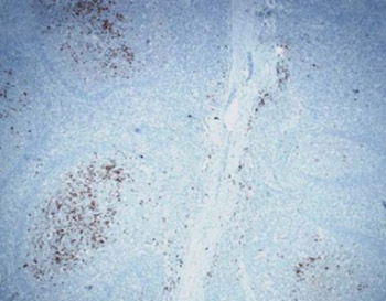
The purposes of the present study were to determine the distribution of cells producing cytokines: tumor necrosis factor α (TNF-α) and interleukin 6 (IL-6) in different morphological sections of tonsils in patients with tonsillar hypertrophy (TH) and recurrent tonsillitis (RT), to analyze the level of production of these cytokines in TH and RT and evaluate the potential of peripheral blood lymphocytes for production of interferon-γ (IFN-γ) and interleukin 4 (IL-4). Analyzed materials consisted of the tonsils after tonsillectomy and blood taken from patients right before tonsillectomy (study group) and blood taken from healthy donors (control group).We used histological and immunohistochemical method, morphometric methods for the quantification of TNF-α and IL- 6 producing cells and immunological methods for determining the concentration of IFN-γ and IL-4. Most of TNF-α producing cells are settled in the subepithelial region (55%). Numerical density of TNF-α producing cells in the crypt epithelium, subepithelial and interfollicular region was significantly higher in RT compared with TH. The concentration of IFN-γ is three times higher in RT then in TH. After the stimulation of peripheral blood lymphocytes in culture there was no significant increase in concentrations of IL- 4. The index of stimulation of IFN-γ was the highest in the RT, and of IL- 4 in TH. The production of Th1-type cytokines (TNF-α and IFN-γ) is higher in RT compared with TH. In both forms of tonsillitis, production of Th1-type cytokines is higher in relation to the production of Th2-type cytokines (IL-6 and IL-4).

Downloads

Download data is not yet available.
Immunoregulatory cytokines and chronic tonsillitis

Downloads

Additional Files

Published

2013-11-20

How to Cite

1.
M. Todorović M, Z. Zvrko E. Immunoregulatory cytokines and chronic tonsillitis. Biomol Biomed [Internet]. 2013Nov.20 [cited 2023Mar.27];13(4):230-6. Available from: https://www.bjbms.org/ojs/index.php/bjbms/article/view/2330

Issue

Section

Immunology

INTRODUCTION

Palatine tonsil is the organ of the first line of host defence against pathogenic agents, but also a place with recurrent chronic inflammatory processes and controversies in treatment. It represents the effectors of systemic humoral and cellular immunity [1-3]. Tonsillar hypertrophy (TH) is characterized by enlarged tonsils, usually with no signs of inflammation or clinical complications in terms of difficulties in swallowing and breathing at the night. While a score of 0 indicate tonsils not extending beyond the pillars, and not obstructing the airway, scores 1+ to 4+ show tonsils that extend and obstruct airway, 1+, 0% to 25%; 2+, 25% to 50%; 3+, 50% to 75%; and 4+, 75% to 100% respectively. The cryptic epithelium of tonsils is thickened and there no clear boundaries with the subepithelial lymphoid tissue. In the epithelium is a keratin mass that fills the intercellular spaces in the form of strips. Beneath the epithelium are enlarged lymph follicles with active hyperplastic germinative centers and thin mantle zone. Interfollicular lymphoid tissue is reduced. Diagnosis of recurrent tonsillitis (RT) was based on a history of >4 tonsillar infections over a period of 12 months, several years in a row, requiring administration of antibiotics [4]. On histological examination there is preserved and occasionally thinned and damaged crypt epithelium with fields of ceratinization [5]. On subepithelial plane, there were numerous, small lymph follicles with clearly limited germinative centers and mantle zone. On extrafollicular plane fibrosis is detected, while in the lumen of tonsillar crypt is cell debris. There is significant difference in the number of lymph follicles in both forms of chronic tonsillitis, but there is a difference in their size [6]. Previous studies have shown that antigenic stimulation of intraepithelial T lymphocytes in the tonsil crypt epithelium produce proinflammatory Th1 type cytokines: interleukin (IL)-2, interferon-γ (IFN-γ), tumor necrosis factor a (TNF-α) and anti-inflammatory Th2 type cytokines: IL-4, IL-5, IL-6, IL-13. Production of Th1 type cytokines is dominant [7]. In tonsillitis production of cytokines starts with Th1-type, including IFN-γ and TNF-α, and later on secretion of Th2 type cytokines [8-10]. An elevated concentration of TNF-α, IL-1 and IL-6 in the tissue is a result of local overproduction because of monocyte-macrophages activation, caused by repeated stimulation by pathogenic agents [7]. Bonanomi et al. [11] have shown that rise of IL-6 after first 24 culture hours, is because of the initial stress. The role of cytokines is known and heterogeneous [12, 13]. Numerous studies related to testing of the immune function in palatine tonsils, i.e. the share in local and systemic immunity, have shown that even damaged tonsils may preserve immune competence [14, 15]. The purposes of the present study were to determine the distribution of cells producing cytokines: TNF-α and IL-6 in different morphological sections of tonsils in patients with TH and RT, to analyze the level of production of these cytokines in TH and RT and to evaluate the potential of peripheral blood lymphocytes for production of IFN-γ and IL-4.

MATERIALS AND METHODS

Patients

The study was conducted at the otorhinolaryngology department of the General Hospital “Danilo I”, Cetinje, Montenegro, and at the Medical Faculty in Niš, Serbia. The study included two groups of patients. The first group consisted of 10 children (6 girls and 4 boys, mean age 9.0 ± 2.7 years) suffering from 2+ or 3+ TH on clinical evaluation and the second group included 13 adults (7 males and 6 females, mean age 23.1 ± 5.2 years) with chronic RT. The control group consisted of 14 healthy adults (6 females and 8 males, mean age 28.3 ± 4.6 years). The control group of healthy children was not formed due to ethical reasons. The study was approved by the Medical board of the General hospital “Danilo I”, Cetinje. Signed written consent was obtained from all patients or their parents.

Samples

Palatine tonsils were obtained by tonsillectomy and classified into groups according to clinical form of tonsillitis. Blood was taken from patients right before tonsillectomy and from healthy, voluntary donors (control group).

Histological methods

Tonsils were taken right after tonsillectomy, repeatedly flushed with saline and immediately fixed for 24 hours in 10% buffered formalin after what the material is brought to a paraffin mold. Histopathological analysis of the material was performed on sections stained with hematoxylin eosin method.

Immunohistochemical method

Streptavidin-biotin immunohistochemical method was performed for the detection of TNF-α and IL-6 producing cells in tonsil tissue. We used the monoclonal antibody antihuman, and as a chromogen, were used 3,3’- diaminobensidin or 3-amino-9-ethylcarbazole. As a primary antibody to IL-6 was used rat monoclonal antibody anti-human, clone MQ2-6A3, and as immunogen recombinant human IL-6, which expresses the COS-7, BD Pharmingen USA manufacturer, catalog number 559 068 in dilution 1:100. As a primary antibody to TNF-α was used the monoclonal mouse anti-human IgG1 class antibody; clone 1E8-G6, and as immunogenic amino-end of human TNF-α, a producer of Santa Cruz, USA, Biotechnology, SC-7317 in dilution 1:100.

Morphometric methods

Quantification of TNF-α and IL-6 producing cells in the tonsils with TH and RT was performed on 5μm thick sections of the tonsils, which were stained by immunohistochemical method LSAB + HRP (LabelledStreptAvidin-Biotin, Horst Readish Peroxidase). Quantification was performed separately in each morphological compartment of tonsils, in crypt epithelium, germinative centers of lymph follicles, and interfollicular regions and subepithelial region. Morphometric analysis was performed on three serial sections for each tonsil (TH-6 tonsils, RT-5 tonsils). Number of TNF-α producing cells was determined on digital images resolution 1280 x 960 pixels, obtained at the NU-2 microscope (Carl Zeis, Jena, Germany) under the x25 objective lens, using a web camera MSI 370. Quantification of the size of lymph follicles and their germinative centers was performed on tonsil sections 4-5 μm thick, stained with hematoxylin-eosin method and included the calculation of several parameters: range (mm2), the mean optical density, volume (mm), circularity, Feret’s diameter (mm) and integrated optical density. These parameters were determined on digital images using a lens and x4 ImageJ program, with manual editing of images using a computer mouse.

Immunological Methods

Mononuclear cells (lymphocytes) were isolated from 10 ml of heparinized (50 IU / ml) blood by applying on a gradient of density 1.077 g/ml (Histopaque 1077, Sigma), and after certain procedures the supernatant is being separated and frozen at -200 until the usage. Concentrations of IFN-γ and IL-4 were detected with immunological testing in the culture of peripheral blood mononuclear cells of people with chronic tonsillitis in unstimulated and lymphocytes stimulated by concavalin (ConA). The concentration of IFN-γ in the supernatant non stimulated and stimulated cells was determined by commercial ELISA test (Biosource International Inc. USA). Test sensitivity was <4.0 pg/ml, value of IFN-γ was calculated by interpolation from the standard curve. The concentration of IL-4 in supernatants nonstimulated and stimulated peripheral mononuclear cells was determined by commercial ELISA (Biosource International Inc., USA). Test sensitivity was <2.0 pg / ml, value of IFN-γ was calculated by interpolation from the standard curve.

Statistical methods

Analysis of the results was performed using the program SigmaStat for statistical data processing and ORIGIN. Results are presented as mean ± standard deviation. Statistical significance among study groups was tested by Student t test and ANOVA test for differences between mean values and the Mann-Whitney rank sum test to test differences between the median.

RESULTS

In all tested tonsils TNF-α producing cells were present but with different distribution and representation in various morphological sections. TNF-α producing cells in TH were primarily localized beneath the crypt epithelium; groups of cells were observed in the germinative center of lymphoid follicle. A few individual cells were seen in the crypt epithelium and inter follicles (Figure 1). TNF-α producing cells in RT were found mainly below crypt epithelium in the form of aggregates, whereas intraepithelial were rare. Groups of cells were presented in the germinative center of lymph follicles. They were found, especially between the lymph follicles and crypt epithelium. Mantle zone usually did not contain TNF-α producing cells (Figure 2). IL-6 producing cells in TH were rarely localized in the subepithelial region, between the lymphoid follicles and crypt epithelium. In RT interfollicular localization of IL-6 producing cells was found and there were a few cells in the subepithelial region. Due to the small number of IL-6 producing cells, morpho-metric analysis was performed only for TNF-α producing cells. We determined the numerical areal density (average number of TNF-α producing cells per mm2 tonsillar tissue) and numerical density (average number of TNF-α producing cells per mm3 tonsillar tissue) (Table 1). ANOVA test showed statistically significant differences in numerical areal density of TNF-α producing cells (Wilks lambda 0.46; Rao R 27.91; p= 0.0001). The t-test found highly statistically significant difference between TH and RT in terms of numerical areal density of TNF-α producing cells in the crypt epithelium and interfollicular regions, while there were no statistically significant differences in germinative centers. TNF-α producing cells had the highest distribution per mm2 in the subepithelial region (TH-56%, RT-52%). Presentation of TNF-α producing cells in the germinative centers of lymph follicles was higher in TH than in RT (TH-34%, RT-25%), while in the interfollicular region was higher in RT than in TH (TH-9%, RT-21%) (Table 2). We found similar percentages of TNF-α producing cells per mm3 in the subepithelial regions (55%) in TH and RT, while the presence of these cells in the germinative centers and in interfollicular regions were significantly different in both forms of chronic tonsillitis (Table 2). For a more precise interpretation of the results of quantification of TNF-α producing cells in certain morphological sections of tonsils, we determined also some morphometric parameters of the lymph follicles and especially for the germinative centers of lymph follicles. Range, volume, diameter and integrated optical density of lymph follicles were significantly higher in TH. Only the optical density of lymph follicles was higher in RT (Table 3). Morphometric analysis of the germinative centers of lymph follicles showed that the examinated parameters, range, volume, diameter and integrated optical density were significantly higher in TH, and there were no differences in terms of optical density and circularity germinative centers of lymph follicles in relation to RT (Table 4). Mean concentrations of IFN-γ in unstimulated peripheral blood lymphocytes in chronic tonsillitis and control group of healthy individuals showed no statistically significant differences, whereas stimulated cells revealed a statistically significant difference (p<0.05) between the RT (306.90 ± 316, 09) and TH (74.10 ± 85.89). The median concentration of IFN-γ by stimulated lymphocytes in the RT group (232.50) was almost five times higher than the median of the tested parameters in the TH group (43.50). The greatest stimulation index was in RT group (16.95), slightly lower in the control group of healthy individuals (13.47), while the lowest was in the TH group (6.07) (Table 5). There was a statistically significant (p < 0.05) value of IL-4 in unstimulated cell supernatants control group (9.71 ± 3.64) compared with the same parameter in the TH group (7.40 ± 2.01), while there were no significant differences in relation to the concentration of IL-4 in the RT group of subjects (9.92 ± 3.84) (Table 6). Mean concentrations of IL-4 in supernatants of stimulated peripheral blood lymphocytes in the control group of healthy subjects (9.50 ± 1.6) and patients with chronic tonsillitis were about the same (TH-8.90±2.81; RT-10.08 ± 3.09). Stimulation index in the control group of healthy individuals (0.97) and patients with RT (1.01) were nearly identical, while the respondents in TH group were slightly higher (1.20) (Table 6).

FIGURE 1: TNF-α producing cells in tonsillar hypertrophy. a) Primarily localized beneath the crypt epithelium; b) Numerous cells in the germinative centers of lymph follicles and severally near the septum. LSAB/ HRPx100
FIGURE 2: TNF-α producing cells in recurrent tonsillitis; a) Below crypt epithelium in groups; b) on the border of germinative centers and mantle zone but in the mantle zone they were not found. LSAB/HRPx100
TABLE 1: Numerical density and numerical areal density of TNF- α producing cells
TABLE 2: Percentage of the presence of TNF- α producing cells per mm2 and mm3 by morphological sections of tonsil
TABLE 3: Morphometric analysis of lymph follicles
TABLE 4: Morphometric analysis of germinative centers in lymph follicles
TABLE 5: The concentration of IFN-γ (pg/ml) in supernatants of peripheral blood lymphocytes
TABLE 6: The concentration of IL-4 (pg/ml) in supernatants of peripheral blood lymphocytes

DISCUSSION

We are confident that there are differences with respect to previous works. We will explain it with the facts. In contrast to our study, Agren and his colleagues, in 1996th, have found that some cytokines, such as IL-8 and IL 1ά are present only in the crypt epithelium and that can not be found in other sections of the morphological tonsils. There was no TNF-and IL-ά 6 producing cells in the crypt epithelium, although they were observed in germ centers of lymph follicles and extrafollicular, they found semiquantitative method, whereas in our study, the largest in sub epithelial tonsillar region. Our result can be considered more valid, because in determining the number of TNF-ά producing cells according to morphological sections of tonsil applied more accurate method of quantification and morphometric measurements. In the literature that we have there is no the quantification of cytokine producing cells using morphometric methods to quantify, but there is data relating to the distribution of cytokine producing cells according to morphological and semiquantitative tonsil sections show their presence [17] Th cells are an important source of cytokine production, as well as other cells such as macrophages and interdigitant cells are involved in the production of cytokines. Recently is discovered subgroup T helper cells - Th 17-producing proinflammatory IL 17 and developmentally, different from Th1 and Th2 cells, which play an important role in antimicrobial immunity. Previous studies shows that palatine tonsils express Th1 and Th2 type cytokines, dominated by Th1 type cytokines [7]. Respond to infection tonsils starts production of Th1 type cytokines, including most important IFN-γ and TNF-α, and later secrete Th2 type cytokines (IL-4, IL-6, etc) [8]. There are significantly increased levels of inflammatory cytokines IL-6 and TNF-α compared to control healthy group [16]. Freshly isolated tonsillar mononuclear cells secrete very small amounts of cytokines other than IL-8, but their production significantly increase after mitogen stimulation of these cells or antigen in culture [8, 17]. Peripheral blood lymphocytes by patients with chronic tonsillitis immediately after isolation show a weak expression of cytokines, except IL-4, while in vitro after mitogen stimulation of peripheral blood T lymphocytes cells secrete significantly more different cytokines [18,19]. We examined the distribution and number of TNF-α producing cells, as representatives of Th1 type cytokines, and for Th2 type representative we analyzed the distribution and number of IL-6 producing cells. Since the tonsils are not only organs of local immunity but also systemic immune responses, we determined the ability of peripheral blood lymphocytes of patients with chronic tonsillitis to produce cytokines. For the representative of Th1-type cytokines was measured the IFN-γ concentration in supernatants of lymphocytes in culture, and for the Th2 cytokine group same parameters were analyzed for IL-4. A comparative analysis of the distribution of TNF-α and IL-6 producing cells in both forms of tonsillitis showed that there was no difference in the localization of these cells in relation to the morphological substrate of chronic tonsillitis with a significant presence of TNF-α in subepithelial region just below crypt epithelium, in the germinative centers and extrafollicular region and individual presence of IL-6 producing cells in extrafollicular and subepithelial region and rarely in the germinative centers. Subepithelial regions of tonsils present position of direct encounters of antigen and immunocompetent cells. This is the “marginal zone” [20]. In contrast to our findings of TNF-α producing cells in the crypt epithelium, Anderson and Anderson [21] argue that there are no cytokine producing cells in the crypt epithelium, but they exist in other morphological sections of tonsil. Also, Argen et al. [17] using semiquantitative method, found that these cells are not in the crypt epithelium and are numerous in the germinative centers. Completely contrary to our results Rostaing et al. [22] considered it is almost impossible to prove intracytoplasmic cytokines in tissue sections of tonsils, but they can only be detected in isolated tonsillar and peripheral blood cells after stimulation in vitro. Also unlike our results, Hoefakker et al. [23] observed that TNF-α producing cells localize exclusively in the mantle zone of lymph follicles and in extrafollicular region. Since we did not get the difference in the distribution of TNF-α producing cells in TH and RT, and to prove the intensity of production of TNF-α we made morphometric measurements, and determined the number of TNF-α producing cells per unit area and per unit volume and in each morphological tonsil compartment. In both forms of tonsillitis 55% of TNF-α producing cells were located in the subepithelial region and 30% in the germinative centers of lymph follicles, 13% in the interfol-licular regions and least about 2% in the crypt epithelium of tonsils. Number of TNF-α producing cells was higher in RT. In addition to the analysis of local cytokine production in tonsillar tissue, we tried to determine whether it has repercussions on the production of cytokines by peripheral blood lymphocytes in patients with chronic tonsillitis. We evaluated the concentrations of IFN-γ and IL-4 in supernatants of lymphocytes in culture, in conditions without stimulation and after stimulation by mitogen phytohaemagglutinin. Unstimulated lymphocytes secrete small amounts of IFN-γ and IL-4 without statistically significant differences between both forms of tonsillitis. Surprisingly, despite the different age of the patients with both forms of tonsillitis there was no difference in cytokine production. If we compare the values of IFN-γ and IL-4 produced by unstimulated peripheral blood lymphocytes in both forms of tonsillitis there were no significant differences, indicating that during chronic inflammatory processes in tonsils usually engages the mechanism of local immune response, as confirmed by our examination of production of TNF-α in the tonsillar tissue. Unstimulated peripheral blood lymphocytes secrete small amounts of tested cytokines, so we tried to determine whether these cells have the potential to secrete additional amounts of cytokines after phytohemagglutinin stimulation. Stimulated lymphocytes significantly increased secretion of IFN-γ within each group of patients. The concentration of IFN-γ was almost three times higher in RT in relation to TH, which is consistent also with results of other authors [17]. Peripheral blood lymphocytes of subjects after mitogen stimulation in culture did not significantly produce IL-4, unlike IFN-γ. The index of stimulation of IFN-γ was highest in RT, and for IL-4 was highest in TH. Considering the results of tests of TNF-α, our findings suggest that Th1-type cytokine secretion is higher in RT. Low production of immunoregulatory cytokines, IL-6 in the tonsils and IL-4 in peripheral blood lymphocyte culture in both form tonsillitis indicates a weaker humoral (Th2) immune response in comparison to the cellular (Th1) immune response. The weaker cytokine production in tonsillar hyperplasia suggests a possible deficit in the activation of the immune system in TH which coincides with the data on a small number of Ig producing cells in tonsils in TH compared with RT.

CONCLUSION

In recurrent tonsillitis production of Th1-type cytokines (TNF-α and IFN-γ) was higher compared with the production of these cytokines in tonsillar hypertrophy. In both forms of tonsillitis the production of Th1-type cytokines (TNF-α and IFN-γ) was higher compared to the production of Th2-type cytokines (IL-6 and IL-4), indicating the dominance of cellular (Th1 type) immune response in relation to humoral (Th2 type) immune response.

DECLARATION OF INTEREST

The authors declare that they have no conflict of interest.

REFERENCES

  1. (). Immunology of tonsils and adenoids: everything the ENT surgeon needs to know. Int J Pediatr Otorhinolaryngol.
  2. (). Mucosal immunity: Some historical perspective on host- pathogen interaction and implication for mucosal vaccines. Immunol Cell Biol.
  3. , , , , , (). Human nasopharyngeal-associated lymphoreticular tissue. Functional analysis of subepithelial and intraepithelial B and T cells from adenoids and tonsills. Am J Pathol.
  4. , , , (). Tonsillar size is an important indicator of recurrent acute tonsillitis. Clin Otolaryngol Allied Sci.
  5. , , (). Scanning electron microscopic characteristics of human tonsillar crypt epithelium. Acta Medica Medianae.
  6. , , , (). Comparison of histology between recurrent tonsillitis and tonsillar hipertrophy. Clin Otolaryngol.
  7. , , , , , (). Structural and immunological characteristics of chronically inflamed adenotonsillar tissue in chidhood. Clin Diagn Lab Immunol.
  8. , , , , (). Cytokines locally produced by lymphocytes removed from the hypertrophic nasopharyngeal and palatine tonsils. Int J Pediatr Otorhinolaryngol.
  9. , , (). Morphology and immunology of the human palatine tonsil. Anat Embriol (Berl).
  10. , , , , , (). Increased cellular proliferation and inflammatory cytokines in tonsils derived from children with obstructive sleep apnea. Pediatr Res.
  11. , , , , , (). Quantitative cytokine gene expression in human tonsils at excision and during histoculture assessed by standardized and calibrated real-time PCR and novel data processing. J Immunol Methods.
  12. (). How the immune system protects the host from infection. Microbes Infect.
  13. , , , , , (). Early commitment of naive human CD4+ T cells to the T follicular helper (Tfh) cell lineage is induced by IL-12. Immunol Cell Biol.
  14. , , (). Detection of cytokine expression in human palatine tonsils. Acta Otolaryngol Suppl.
  15. , , , , , (). Cytokine expression and production by tonsillar lymphocytes. Acta Otolaryngol Suppl.
  16. , , , (). Cytokine evaluation in throat infections. Ann Otol Rhinol Laryngol.
  17. , , , , , (). The production of immunoregulatory cytokines is localized to the extrafollicular area of human tonsils. Acta Otolaryngol.
  18. , , , , , (). Different pattern of cytokine production and mRNA expression by lymphoid and non- lymphoid cells isolated from human palatine tonsil. Int J Clin Lab Res.
  19. , , (). A Study of cytokine in palatine tonsil- cytokine mRNA expression determined by RT-PCR. Nihon Jibiinkoka Gakkai Kaiho.
  20. , , (). Human marginal-zone B cells. Immunol Today.
  21. , , , , , (). Concomitant in vivo production of 19 different cytokines in human tonsils. Immunology.
  22. , , , , , (). Kinetics of intracytoplasmic Th1 and Th2 cytokine production assessed by flow cytometry following in vitro activation of peripheral blood mononuclear cells. Cytometry.
  23. , , , , , (). Immunohistochemical detection of co-localizing cytokine and antibody producing cells in the extrafollicular area of human palatine tonsils. Clin Exp Immunol.