Morphometric measurements of the cranium in congenital bilateral blind males and females

Authors

  • Rengin Kosif Department of Anatomy, Faculty of Medicine, University of Abant Izzet Baysal
  • Ocal Sırmatel Department of Radiology, Faculty of Medicine, University of Abant Izzet Baysal
  • Arzu Canan Department of Radiology, Faculty of Medicine, University of Abant Izzet Baysal

DOI:

https://doi.org/10.17305/bjbms.2013.2331

Keywords:

congenital bilateral blind, human, craniometry

Abstract

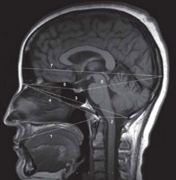
Cranium dimensions differ according to racial, geopraphic, ethnic and nutritional factors. This study will shed light on the question “Is there a difference in the cranial distances measured between congenital bilateral blind people and healthy individuals?” Nine anthropometric measures were performed on the brain MRI midsagittally obtained from male and female congenital bilateral blinds. The anthropometric measures taken included the glabella-opisthocranion, nasion-dorsum sellae, dorsum sellae-opisthocranion, nasion-basion, nasion-opisthion, basion-opisthion, prosthion-basion, basion-vertex and the clival angle. In addition, the supratentorial and infratentorial areas were calculated. Glabella-opisthocranion, nasion-dorsum sellae, nasion-basion, prosthion-basion and clival angle were found to be smaller in the congenital bilateral blind females compared to the healthy group, but these results were not statistically significant. Whereas, other measures out of the basion-opisthion were found to be smaller in the congenital bilateral blind males than in the healthy subjects. Of these results, the distance between glabella-opisthocranion, nasion-dorsum sellae and nasion-basion was significantly smaller compared to the measurements taken from the healthy male group. The infratentorial area was significantly smaller in congenital blind male subjects and the supratentorial area was significantly larger in congenital blind female subjects. This study has revealed that the cranium dimensions of congenital blind people are to different from normal individuals. The most significant difference was in the distance of the nasion-dorsum sellae. The region anterior to the basion and dorsum sellae was prominently smaller. The infratentorial area was significantly smaller in congenital blind male subjects and the supratentorial area was significantly larger in congenital blind female subjects.

Downloads

Download data is not yet available.
Morphometric measurements of the cranium in congenital bilateral blind males and females

Downloads

Additional Files

Published

2013-11-20

How to Cite

1.
Kosif R, Sırmatel O, Canan A. Morphometric measurements of the cranium in congenital bilateral blind males and females. Biomol Biomed [Internet]. 2013Nov.20 [cited 2023Nov.30];13(4):237-41. Available from: https://www.bjbms.org/ojs/index.php/bjbms/article/view/2331

Issue

Section

Anatomy

INTRODUCTION

Measurements of the craniofacial complex are important for studies on human growth, population, variation and some medical problems. The development of cranial proportions is the result of genetic, embriogenetic and environmental factors [1]. The shape and size of the cranium may be affected by genetic disorders, congenital disorders, primer, seconder or metastatic skull tumors and the amount of brain tissue and cerebrospinal fluid present [2,3,4,5]. Craniofacial traits have substantial amounts of genetic variation, but are significantly affected by other non-genetic factors such as sex and year of birth, as has been revealed by the genetic analyses [6]. The brain morphology of the blind people has been investigated previously and it was shown that congential blind subjects may have different brain tissue distribution than do healthy people. Long term living in an environment without visual input can not only induce large changes in the visual cortex but also in other areas [7]. There are distinct differences not only in the visual cortex but also the sensory area, auditory area and motor area [8]. Leporé et al. [9] found that visual regions of the brain were smaller in volume in blind individuals than in sighted ones. However, for non-visual areas, the trend was reversed as these areas grew to be larger in the blind. This, the researchers say, suggests that the brains of blind individuals are compensating for the reduced volume in areas normally devoted to vision. The changes that may occur in the cranium in congenital blind people have not been investigated previously. Traditional anthropometric expressions of the variability in the cranium and face have usually emphasized distances and angles between two or more landmarks [10]. Are they different from any other healthy people? This study aimed to show whether that kind of difference also affect craniometric proportions and to compare standard anthropometric cranial measurements taken from congenital bilateral blind people with those taken from healthy ones.

MATERIALS AND METHODS

Samples and Procedures

For this study, ethic permission was granted to researchers in the Abant Izzet Baysal University. This study was supported by the Abant Izzet Baysal University Scientific Research Committee. Nine anthropometric measures on supratentorial area and two different infratentorial area were performed on the brain via MRI midsagittally obtained from 25 males and 16 females congenital bilateral blind people. Images were obtained with a 1.5 Tesla, Magnetom Symphony (Siemens, Germany) MR device. The subjects’ ages range from 21-61 (37.69±13.78). The results obtained were compared with those of the 27 males and 29 females healthy individuals. The healthy subjects’ ages range from 25-63 (36.72±11.68). The cranial distances measured are shown in Figures 1, 2, 3, 4. The anthropometric measures included the glabella-opisthocranion [11], nasion-dorsum sellae [12], dorsum sellae-opisthocranion [13], nasion-basion [14], nasion-opisthion [15], basion-opisthion [16], prosthion-basion [17], basion-vertex [11] and the clival angle [18]. Glabella (Gl): The most forwardly projecting point in the mid-sagittal plane at the lower margin of the frontal bone. Opisthocranion (Oc): The most posteriorly protruding point on the back of the braincase. Nasion (Na): The middle point of the frontonasal suture. Basion (Ba): The midpoint of the anterior margin of the foramen magnum.

FIGURE 1: Measurements of 7 anthropometric distances. 1: pros-thion-basion 2: basion-opisthion (McRae line), 3:nasion-opisthion, 4: nasion-basion, 5: nasion-dorsum sellae, 6: dorsum sellae-opis-thocranion, 7: glabella-opisthocranion
FIGURE 2: Measurements of the (ba-ver) and (cli-ang). 8: clival angle, 9: basion-vertex
FIGURE 3: Measurement method of supratentorial area and infratentorial area 1
FIGURE 4: Measurement method of infratentorial area 2

Opisthion (Op): The mid-point on the posterior margin of the foramen magnum is termed the opisthion. Prosthion (Pr): The point of the upper alveolar process that projects most anteriorly, a point on the alveolar arch midway between the median upper incisor teeth. Vertex (Ver): The highest point of the skull, the top of the head. Clival angle (Cli-ang): The clival angle, between the ethmoidal plane (the plane of the cribriform plate of the ethmoid bone) and the clival plane (the plane of the intracranial surface of the clivus). Dorsum sellae (DS): A square portion of bone on the body of the sphenoid posterior to the sella turcica or hypophysial fossa.

Surface Area Measurements

  1. Infratentorial area (ITA1) and supratentorial area (STA): (Figure 3) The method has been proposed by Krogness to evaluate the size of the bony posterior fossa with regard to the overall cranial cavity in a more realistic fashion [15]. This method, which omits compensatory tentorial enlargement, better demonstrates retardation of mesodermal osseous growth [15,19]. According to this method, the ITA was outlined by dorsum sellae cranially, internal table of the occipital bone dorsally, the clivus ventrally and the McRae line caudally, whereas, STA was the area between dorsum sellae and the internal tables of the bones in the supratentorial compartment, excluding the sella turcica.

  2. Infratentorial area (ITA2): (Figure 4): In the method of measurement proposed by Nishikawa which permits combine devaluation of the bony andtentorial parts of the posterior fossa [15, 20]. The ITA was marked as the area outlined by tentorial apex-dorsum sellae line, the McRae line occipital bone and clivus. The average values and standard deviations of all measurements were calculated. A two tailed paired t-test was used to compare the measurements. The Pearson correlation test was used for the evaluation of relationships of the measured values with each other and correlation coefficient values were given. A p value less than 0.05 was considered statistically significant.

RESULTS

The means and standard deviations of cranial distances and surface area measurements are shown in Table 1 and 2. The glabella-opisthocranion, nasion-dorsum sellae, nasion-basion, prosthion-basion and clival angle were found to be smaller in the congenital bilateral blind female group compared to the healthy group, but these results were not statistically significant whereas, other measures out of the basion-opisthion were found to be smaller in the congenital bilateral blind males than in the healthy male subjects. Of these results, the distance between the glabella-opisthocranion (173.29±7.04 mm, p=0.016), nasion-dorsum sellae (72.4±13.68 mm, p=0.00025) and nasion-basion (99.72±4.74 mm, p=0.014) was significantly smaller compared to the healthy male group (p< 0.05). The most significant difference was in the distance of nasion-dorsum sellae (p=0.00025) (Table 1). Infratentorial area 2 was significantly smaller in congenital blind males (p=0.049) and supratentorial area was significantly larger in congenital blind females (p=0.036) (Table 2). Surface area measurements were correlated with some anthropometric measurements.

TABLE 1: Means and standard deviations of cranial distances
TABLE 2: Means and standard deviations of surface area measurements

There was no correlation between supraratentorial and infratentorial surface areas and anthropometric measurements in congenital blind females. But with infratentoreal surface area 2, a moderate positive correlation with prosthion-basion distance was recorded (r=0.569, p=0.42). There was no correlation between infratentorial surface areas and anthropometric measurements in congenital blind males. But moderate positive correlation was found between supratentorial area and nasion-basion distance in congenital blind males (r=0.522, p=0.046).

DISCUSSION

In blind individuals, the brain undergoes structural changes and is reorganized by adapting to the absence of sensory inputs. The volume of the regions of the brain that deal with vision has been found to be smaller in blind individals compared to those in sighted subjects. The volume of the regions of the brain that are not involved in vision has been found to be bigger to compensate for this condition. It was found that frontal lobes were significantly enlarged in blind subjects compared to sighted subjects [9]. Compared with the normal controls, the regions expanded in the blind men’s brain located at Brodmann 19, 31 where one finds the occipital lobe, cuneus, superior occipital gyrus, limbic lobe, cingulate gyrus. The contracted regions are located at Brodmann 17, 18 where one finds the occipital lobe and lingual gyrus [7]. There are distinct differences not only in the visual cortex but also the sensory area, auditory area and motor area [8]. According to our knowledge, this is the first craniometric study of congenital bilateral blind people. This study has revealed that the cranium dimensions of congenital blind people are different. The most significant difference was in the distance of nasion-dorsum sellae (p=0.00025). The region anterior to the basion and dorsum sellae was prominently smaller. No meaningfull change occurred at the region behind the dorsum sellae. Unilateral clinical anophthalmia (absence or extreme hypoplasia of the eyeball) always results in hypoplasia of the ipsilateral bony orbit, as there is no stimulus inducing growth of the orbital socket [21]. Our findings supported this study. The reason for why the front part of the dorsum sellae is shorter in sighted people, can be explained by undevelopment of structures of orbita. Cotton found vertex-basion height 135.66±6.56 mm; glabella-opisthocranion length 181.94±6.40 mm and basion-opisthion distance 36.5±2.36 mm in his study [22]. Cameron was found, female nasion-basion length 94.8± 4.51 mm, glabella-occipital length 174±7.54 mm, male nasion-basion length 100± 4.39 mm, glabella-occipital length 182±7.98 mm in his study [23]. For congenital bilateral blind people and healthy ones, the glabella-opistho-cranion distance was found smaller in this study (Table 1). Naderi was found distance between the basion and opisthion 34.7±2.3 mm [24]. In our study this measurement was found to be larger but not significantly. Ursi et al. [25] reported that sella-nasion distances are significantly larger in males in sexual dimorphism. In our study this measurement is larger, but not significantly so. Cistulli found nasion-dorsum sellae 64.1± 0.4 mm in normal males and that distances were significantly increased this distance in the Marfan sendrom male [26]. In our study, the distance of nasion-dorsum sellae was found to be significantly smaller in congenital bilateral blind male. Guyot found to be clival angle 124.14± 10.04° in his study [27]. In our study, the clival angle was found to be smaller in both blind and sighted people (Table 1). The STA was larger in the congenital blind males and females but significantly larger in the congenital blind female in this study. Erdogan et al reported that a large supratentorial area means that there is no atrophy in white matter [28]. It was indicated that small STA signals brain atrophy [29]. It has been reported that supratentorial and infratentorial volume were lesser in multiple sklerosis subjects and supratentorial volume was smaller in autism [30,31]. This study has shown that brain atrophy did not occur in congenital bilateral blind people. Infratentorial area 2 was significantly smaller in congenital blind males when compared to sigted males (p=0.049). In the same patient group, we found the volume of cerebellum to be smaller, but not significantly so [32]. The findings are consistent with in congenital blind males. The supratentorial area and nasion-basion distance was correlated in congenital blind males. This finding shows expansion the front side of basion is correlated with an expanding supratentorial area. Small infratentorial area that would be able to early findings at infratentorial tumors as medullo-blastoma in this people. Supratentorial area was significantly larger in congenital blind females when compared to sigted females (p=0.036). The diagnosis of supratentorial tumors may be delayed for this reason in congenital blind females. Widespread hypertrophy was observed in non-occipital white matter, particularly in the prefrontal and frontal areas in early and late-onset blind adults [9]. In this study, the supratentorial area was significantly larger in congenital blind females (p=0.036) but infratentorial area was not significantly different in congenital blind females. The study is that we have done in the same patient group, we found the volume of cerebellum to be significantly smaller [32].

CONCLUSION

As a result cranial measurements of congential bilateral blind people are different from sighted people, this difference is clear in the front of basion and dorsum sellae. Brain atrophy did not occur in congenital bilateral blind people in supratentorial area because of larger than sigthed people. When the infratentorial area 2 was evaluated, significant atrophy had occurred in congenital bilateral blind males. The normative data described here form a basis for quantitative analysis of MRI from congenital bilateral blind individuals. We hope this study will provide additional new knowledge the studies in craniometric, morphologic, clinic and antropometric areas of research. To our knowledge, this is the first study showing whether congential blindness affects cranial dimensions. Although found to be statistically significant, the differences are small. Thus, further studies with more subjects and using more precise measurement methods are necessary to confirm our findings.

DECLARATION OF INTEREST

The authors declare no conflict of interest.

Acknowledgements

ACKNOWLEGMENT

This study was supported by the Abant Izzet Baysal University Scientific Research Committee.

REFERENCES

  1. , , , (). Large forehead: A novel sign of undiagnosed coeliac disease. Dig Liver Dis.
  2. , (). Skull deformities. Pediatr Clin North Am.
  3. , , , , (). Large osteolytic skull tumor presenting as a small subcutaneous scalp lesion. J Am Board Fam Med.
  4. , , (). Basicranial influence on overall cranial shape. J Hum Evol.
  5. (). Rönning. Growth of the cranial vault: influence of intracranial and extracranial pressures. Acta Odontol Scand.
  6. , , , , , (). Heritability of human cranial dimensions: comparing the evolvability of different cranial regions. J Anat.
  7. , , , (). Deformation-based morphometric study on blind men's brain structures. 7th Asian-Pacific Conference on Medical and Biological Engineering IFMBE Proceedings. DOI 10.1007/978-3-540-79039-6
  8. , , (). Voxel-based morphometric study on chinese blind men's brain structure. Journal of Beijing Institute of Technology.
  9. , , , , , (). Brain structure changes visualized in early- and late-onset blind subjects. Neuroimage.
  10. (). Morphometric analysis of cartesian coordinates of the human skull. Am J Phys Antropol.
  11. , , , , , (). Cranial sutures and craniometric points detected on MRI. Surg Radiol Anat.
  12. , (). Glabellar approach: simplified midline anterior skull base approach. Minim Invasive Neurosurg.
  13. , , , , , (). The relationship between craniofacial morphology and obstructive sleep apnea in whites and in African-Americans. Am J Respir Crit Care Med.
  14. , , , , , (). Three-dimensional imaging and osteometry of adult human skulls using helical computed tomography. Surg Radiol Anat.
  15. , , (). Morphometric measurements of the cranium in patients with Chiari type I malformation and comparison with the normal population. Acta Neurochir (Wien).
  16. , , , (). Age, sex and body mass index in relation to calvarial diploe thickness and craniometric data on MRI. Forensic Sci Int.
  17. (). Computer-graphic facial reconstruction Elsevier Academic Press.
  18. , , , , , (). An anthropometric study of relationships between the clival angle and craniofacial measurements in adult human skulls. Surg Radiol Anat.
  19. , (). Size of posterior fossa in Chiari type I malformation in adults. Acta Neurochir (Wien).
  20. , , , , (). Pathogenesis of Chiari malformation a morphometric study of the posterior cranial fossa. J Neurosurg.
  21. , , , , (). Expansion of the socket and orbit for congenital clinical anophthalmia. Plast Reconst Surg.
  22. , , , , , (). Cranial sutures and craniometric points detected on MRI. Surg Radiol Anat.
  23. (). Craniometric studies. American Journal of Physical Anthropology.
  24. , , , , , (). Morphometric analysis of human occipital condyle. Clin Neurol Neurosurg.
  25. , , , (). Sexual dimorphism in normal craniofacial growth. Angle Orthod.
  26. , , (). Relationship between craniofacial abnormalities and sleep-disordered breathing in Marfan's syndrome. Chest.
  27. , , , , , (). An anthropometric study of relationships between the clival angle and craniofacial measurements in adult human skulls. Surg Radiol Anat.
  28. , , , (). A novel index to estimate the corpus callosum morphometry in adults:callosal/supratentorial- supracallosal area ratio. Diagn Interv Radiol.
  29. , , (). The relationship of brain and cervical cord volume to disability in clinical subtypes of multiple sclerosis: a three-dimensional MRI study. Acta Neurol Scand.
  30. , , , , (). Three dimensional MRI estimates of brain and spinal cord atrophy in multiple sclerosis. J Neurol Neurosurg Psychiatry.
  31. , , , , (). Reduced Central White Matter Volume in Autism: Implications for Long-Range Connectivity. Psychiatry Clin Neurosci.
  32. , , (). Comparison of cerebellar volume between the people who are bilateral congenital blind with healthy ones. International Journal Of Morphology.