Electrophysiological Effects of Bosentan in Rats with Induced Cerebral Ischemia-Reperfusion

Authors

  • Bekir Akgun Department of Neurosurgery, Faculty of Medicine, Firat University,
  • Metin Kaplan Department of Neurosurgery, Faculty of Medicine, Firat University,
  • Caner F. Demir Department of Neurology, Faculty of Medicine, Firat University
  • Aysel Sari Department of Chemistry, Faculty of Science, Firat University
  • Hasan H. Ozdemir Department of Neurology, Faculty of Medicine, Firat University
  • Said M. Berilgen Department of Neurology, Faculty of Medicine, Firat University

DOI:

https://doi.org/10.17305/bjbms.2013.2358

Keywords:

Bosentan, cerebral ischemia-reperfusion, electroencephalography, rat

Abstract

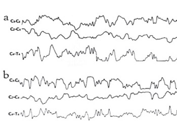
We examined the effect of bosentan, an ETA and ETB receptor antagonist, on EEG, an indicator of neuronal activity, in rats with experimentally induced cerebral ischemia-reperfusion. The rats were divided into three groups with seven rats in each group. Before the procedures, the EEGs of all rats were recorded for ten minutes. 30 mg/kg bosentan in 2 cc physiological serum was administered to the first group, and the second and third groups were injected with 2 cc physiological serum intraperitoneally. After the administration, the right and the left common carotid arteries of the animals in Groups 1 and 2 were clipped for 10 minutes using aneurysm clippings. The rats in the third group received only a subcutaneous incision. Ten minutes after the clips were removed in the first and second groups and after the incision in the third group, EEG recordings were repeated for 10 minutes. All the rats were decapitated and MDA values in the brain tissue were measured for evaluation of the efficiency of induced cerebral ischemia. Induced cerebral ischemia was performed effectively because the MDA levels in Groups 1 and 2 were elevated, compared to the levels in Group 3 (p<0.05). After the application of the Cerebral Ischemia-Reperfusion Technique, the EEG showed minimal slowing in the rats in Group 1, and generalized diffuse slowing in the rats in Group 2 compared to pre-ischemic findings. Bosentan may reduce the damage induced by ischemia on neuronal electrophysiology, likely through its vasodilation effect on cerebral vessels.

Downloads

Download data is not yet available.
Electrophysiological Effects of Bosentan in Rats with Induced Cerebral Ischemia-Reperfusion

Downloads

Additional Files

Published

2013-08-20

How to Cite

1.
Akgun B, Kaplan M, F. Demir C, Sari A, H. Ozdemir H, M. Berilgen S. Electrophysiological Effects of Bosentan in Rats with Induced Cerebral Ischemia-Reperfusion. Biomol Biomed [Internet]. 2013Aug.20 [cited 2023Sep.23];13(3):175-9. Available from: https://www.bjbms.org/ojs/index.php/bjbms/article/view/2358

Issue

Section

Pharmacology

INTRODUCTION

Endothelin-1 (ET-1) is known to have potent effects on vasoconstriction, mitosis, and cell proliferation. The ET-1 concentrations are increased in the tissues and plasma of patients with pulmonary arterial hypertension (PAH) due to a reduction in clearance in the pulmonary vascular bed [1]. Besides, ET-1 is considered to be a central pathogenic factor in connective tissue diseases (CTDs) such as systemic sclerosis (SSc), leading to vasoconstriction, fibrosis and inflammation. Bosentan, dual Endothelin receptor antagonist, which has been proved to be effective in the treatment of PAH and SSc. The efficiency of bosentan in improving exercise capacity has been demonstrated in patients with idiopathic PAH or PAH associated with CTD.

Despite the potential for treatment-related teratogenicity and hepatotoxicity, long-term data indicate that bosentan is generally well tolerated at the approved dosages [1, 2]. Endogenous endothelins (ETs) are known to have significant effects on hypoperfusion following cerebral ischemia [3]. Therefore, recent studies have focused on the effects of bosentan, an Endothelin A and B (ETA-ETB) receptor antagonist, on cerebral ischemia-reperfusion damage [4, 5]. Cell membranes contain phospholipids that are vulnerable to oxidation and rich in unsaturated fatty acids. Ischemic stroke is caused by the obstruction of blood flow to the brain, resulting in energy failure that initiates a complex series of metabolic events, ultimately causing neuronal death. Free radicals that are released in cerebral ischemia by hydrolysis of membrane phospholipids were reported to have harmful effects. Free radicals bring about a chain reaction that breaks down the double bonds of unsaturated fatty acids in the presence of oxygen. As a result, cell membrane stability is disturbed, permeability is affected, and the ability to form membrane potential is impaired [6, 7]. Malondialdehyde (MDA) results from the reaction of thiobarbituric acid (TBA) and the products that arise from lipid peroxidation. As they are very short-lived, the direct measurement of free radicals is extremely difficult. Measured MDA levels provide information about the damage induced by free radicals [6, 8]. Therefore, in the present study we preferred to determine MDA values in the brain tissue in order to examine the effectiveness of experimentally-induced cerebral ischemia. Electroencephalography (EEG) is a useful method for monitoring the neuronal function in ischemic stroke patients [9]. It was shown that bioelectrical brain activity disturbances occurred during the acute stroke period even after the improvement of neurological status [10]. Therefore EEG has been used in some neurovascular surgical interventions for the evaluation of ischemia [11]. Therefore, we evaluated the effects of bosentan administration on cerebral electrophysiology by examining the EEG activities of the animals that induced cerebral ischemia-reperfusion.

MATERIALS AND METHODS

The present study was carried out in the Experimental and Surgical Animals Research Laboratory of our University after the approval of the Ethics Board was obtained.

Animals

A total of 21 Wistar Albino female rats, weighing 300 to 350 grams, were randomly allocated to three groups. All the rats were anesthetized using 100 mg/kg ketamine (i.m.) injection. The body temperature of the animals was measured with a rectal thermometer and kept at around 37°C. Before the procedures, the EEGs of all rats were recorded for about ten minutes. The study groups were then subjected to the following procedures: Group 1 (n=7): Anesthetized rats were injected with i.p. 30 mg/kg bosentan in 2 cc physiological serum. An hour later, the Cerebral Ischemia-Reperfusion technique was performed on the rats. Then the EEG of the rats was recorded for 10 minutes. Finally, the rats were decapitated and MDA levels in the brain tissue were measured. Group 2 (n=7): Anesthetized rats were administered i.p. 2 cc physiological serum. Procedures were conducted as in Group 1. The same sequence of Cerebral Ischemia-Reperfusion technique, followed by EEG recording and brain tissue MDA measurement after decapitation were carried out in this group as well. Group 3 (n=7): Anesthetized rats were administered i.p. 2 cc physiological serum, but Cerebral Ischemia-Reperfusion technique was not conducted. The rats in this group received only a midline incision. After the incision was made, an EEG recording was performed for 10 minutes. Finally, the rats in this group were decapitated and the MDA levels in the brain tissue were measured. Application of the “CerebralIschemia - Reperfusion Technique” After the anesthesia application, rats were fixed in a supine position on the operation table and a midline strip was shaved on the neck. After disinfecting the operation site, a midline incision was made. A superficial micro-dissection was followed by deep micro-dissection proceeding toward the common carotid arteries. When the trachea became visible, the paratracheal muscles were dissected to reach the common carotid arteries. Yaşargil aneurysm clips were placed on the right and the left common carotid arteries. The clips were kept closed for 10 minutes.

EEG

Recordings were performed in C3-C2, C2-C4, C4-T4 derivations with intrascalp electrodes by a “Nihon Kohden” analogous EEG device.

Measurement of Tissue MDA levels

Lipid peroxidation in injured brain tissue was estimated by the thiobarbituric acid reaction method for MDA described by Ohkawa et al. to give a red species absorbing at 535 nm [12]. The MDA results were expressed as nmol/g wet tissue. 0.2 ml of 10% (weight/volume) tissue homogenate was added to 0.2 ml of 8.1% sodium dodecyl sulfate and a 1:5 aqueous solution of thiobarbituric acid. The mixture was diluted to 4.0 ml with distilled water heated in an oil bath at 95°C for 60-min. After cooling with tap water, 1.0 ml of distilled water and 5.0 ml of a mixture of N-butanol and pyridine (15:1 volume : volume) were added and the mixture was shaken. After centrifugation at 4000 rpm for 10 min, the organic layer was taken and its absorbance at 532 nm was measured spectrophotometrically. Tetramethoxy propane was measured as an external standard, and the level of lipid peroxides was expressed nmol MDA per gram wet weight.

Statistical analysis

While the study data were evaluated for the statistical significancy, a One Way Anova test and post hoc TUKEY test were used. Comparison of definitive statistical methods (Average, Standard Deviation, Percentage) and Quantitative data, and in the case of more than two groups, for the inter-group comparison of parameters, and an LSD test was utilized to detect which group was causing the difference. P values less than 0.05 were accepted as significant.

RESULTS

The post-procedural EEGs of the rats in Group 1 showed minimal slowing, in comparison to the pre-procedural EEGs (Figure 1a, b).

FIGURE 1: EEGs of Group 1; rats were injected with i.p. 30 mg/kg bosentan in 2 cc physiological serum. An hour later, the Cerebral Ischemia-Reperfusion technique was performed on the rats. (a) before the application of bosentan, (b) after the application of bosentan.

When pre-and post-procedure EEGs were examined, generalized and diffuse slowing was established in Group 2 after the procedure (Figure 2a, b). Pre- and post-procedural EEGs of the rats in Group 3 were the same (Figure 3a, b).

FIGURE 2: EEGs of Group 2; rats were administered i.p. 2 cc physiological serum. An hour later, the Cerebral Ischemia-Reperfusion technique was performed on the rats. (a) before cerebral ischemia, (b) after cerebral ischemia.
FIGURE 3: EEGs of control group (Group 3; rats were administered i.p. 2 cc physiological serum, but Cerebral Ischemia-Reperfusion technique was not conducted.) (a) before the procedure, (b) after the procedure.

When MDA levels were compared, no significant difference was established between Groups 1 and 2 (p≥0.05). However, MDA levels in Groups 1 and 2 were found elevated, compared to the levels in Group 3 (p<0.05). This data showed that induced cerebral ischemia was performed effectively in our study. Table 1 presents the MDA levels according to groups.

TABLE 1: Cerebral tissue MDA levels of groups (Group 1; were injected with i.p. 30 mg/kg bosentan in 2 cc physiological serum and an hour later, the Cerebral Ischemia-Reperfusion technique was performed. Group 2; were administered i.p. 2 cc physiological serum and an hour later, the Cerebral Ischemia-Reperfusion technique was carried out. Group 3; were administered i.p. 2 cc physiological serum, but Cerebral Ischemia-Reperfusion technique was not conducted.)

DISCUSSION

In order for the ordinary human brain to function and to maintain its functioning, it needs to continuously take some substances from blood. Most notable among these substances are oxygen (O2) and glucose. Ischemia occurs when the O2 and glucose input required for the maintenance of the metabolism in a tissue is curtailed or cut [13-15]. Lack of an adequate cellular energy supply plays a major role in the development of ischemic injury. Under normal circumstances, the energy need of the brain is met by the ATP formed as a result of aerobic glycolysis. In the case of ischemia-induced lack of oxygen, due to impairment of mitochondrial electron transport and oxidative phosphorylation, ATP cannot be synthesized, resulting in an energy deficit. At the onset of ischemia, oxygen deficiency leads to the activation of anaerobic glycolysis for a short period. Accumulation of lactic acid and H+ as a result of the anaerobic metabolism causes acidosis inside and outside the cell. This gives rise to the factors that have a part ischemic injury, such as lactic acid and low pH protein denaturation, loss of enzyme function, and free radical formation. ATP deficiency brings about a gradual decrease in the synthesis procedures of macromolecules such as phospholipids, polysaccharides, and proteins [13-16]. Endothelins are a family of vasoconstrictor peptides that have a potent activity in cerebral blood vessels in particular and which, despite having been originally isolated from endothelial cells, are produced by neurons, glial cells, vascular smooth muscle cells and monocytes [17, 18]. Lampl et. al. [18] reported an increase in the cerebrospinal fluid and plasma ET-1 level in the early post-ischemic stroke period. The development of reperfusion injury after ischemia rests heavily upon the shifts in oxidant-antioxidant balance [16, 19]. In the light of this information, we aimed to assess the efficiency of bosentan for inhibition of vasoconstruction effects of endothelins. In ischemic stroke, the EEG shows slow wave activity, periodical lateralized epileptiform discharges, as well as a decrease in amplitude when there is cortex involvement [20]. Cerebral blood flow is 50 ml/min/100gr brain tissue in a normal human. When it decreases to below 25 ml/min/100gr, decelerations are observed in EEG waves [21]. In comparison of pre-ischemic and post-ischemic EEG findings, these disturbances were observed in our study. The efficiency of our cerebral ischemia procedure was proven when the EEG findings and MDA levels were assessed together. Pre- and post-procedural EEG recordings of the rats in Group 3 were the same. The EEGs of the rats in Group 2 showed a generalized, diffuse slowing after the procedure, in comparison to the pre-procedural recording. However, the EEGs of rats in Group 1, which were administered intraperitoneal bosentan revealed minimal slowing after the procedure, relative to the recording before the procedure, although carotid occlusion was performed for the same period of time using the same clips. EEG results demonstrated that bosentan administration had favorable electrophysiological effects on the ischemic cerebral tissue. The concerned effect likely resulted from the vasodilation caused by bosentan in the cerebral vascular structures. In addition, bosentan may contribute to the cortical circulation, especially through its effects on collateral circulation via pial anastomoses. These findings suggest that bosentan may be used for brain protection before neurosurgical procedures that can cause cerebral ischemia. In particular, it can be preferred in neurovascular operations such as carotid endarterectomies and aneurism surgeries in which temporary clippings were carried out.

CONCLUSION

In conclusion, as a result of the evaluation of the EEG results in rats with induced ischemia-reperfusion, it is suggested that bosentan, which is the antagonist of the ETA-ETB receptors of endothelins (ETs), which are from the family of vasoconstrictor peptides and known to have significant effects in hypoperfusion following cerebral ischemia, may reduce neuronal damage. Bosentan may be used for brain protection and prevention of ischemic complications, before the neurosurgical interventions that can cause cerebral ischemia. Further experimental and clinical studies are needed to identify the effects of bosentan in cerebral ischemia.

DECLARATION OF INTEREST

There is no conflict of interest in this paper.

REFERENCES

  1. , , , , , (). Beneficial and adverse effects of bosentan treatment in korean patients with pulmonary artery hypertension. Korean Circ J.
  2. , (). Bosentan: a review of its use in pulmonary arterial hypertension and systemic sclerosis. Am J Cardiovasc Drugs.
  3. , , , , (). Study of endothelin-1 in acute ischemic stroke. Rom J Intern Med.
  4. , , , , , (). High-dose bosentan in the prevention and treatment of subarachnoid hemorrhage-induced cerebral vasospasm: an open-label feasibility study. Neurocrit Care.
  5. , , (). Safe treatment of pulmonary hypertension with bosentan in a patient with moyamoya disease and cerebral ischemia. J Child Neurol.
  6. , (). Phospholipase A(2), reactive oxygen species, and lipid peroxidation in CNS pathologies. BMB Rep.
  7. , (). Phospholipase A2, reactive oxygen species, and lipid peroxidation in cerebral ischemia. Free Radic Biol Med.
  8. , , , , , (). Early biochemical effects after unilateral hypoxiaischemia in the immature rat brain. Int J Dev Neurosci.
  9. , , , , , (). Quantitative EEG correlates of low cerebral perfusion in severe stroke. Neurocrit Care.
  10. , , , , , (). Clinical-electrophysiol characteristics of the cognitive sphere in patients in the acute period of the first cerebral ischemic stroke. [Article in Russian] Zh Nevrol Psikhiatr Im S S Korsakova.
  11. , , , , , (). Predictors of clamp-induced electroencephalo graphicchanges during carotid endarterectomies. Clin Neurophysiol.
  12. , , (). Assay for lipid peroxides in animal tissues by thiobarbituric acid reaction. Anal Biochem.
  13. (). Pathophysiology and treatment of focal cerebral ischemia. Part I: Pathophysiology. J Neurosurg.
  14. (). Mitochondria and neuronal death/survival signaling pathways in cerebral ischemia. Neurochem Res.
  15. , , , , , (). Initial hyperlac- tatemia in the ED is associated with poor outcome in patients with ischemic stroke. Am J Emerg Med.
  16. , , (). Lactacidosis modulates gluta- thione metabolism and oxidative glutamate toxicity. J Neurochem.
  17. (). Endothelins in health and disease. Eur J Intern Med.
  18. , , , , , (). Endothelin in cerebrospinal fluid and plasma of patients in the early stage of ischemic stroke. Stroke.
  19. (). Reactive oxygen radicals in signaling and damage in the ischemic brain. J Cereb Blood Flow Metab.
  20. , , (). Rapid EEG changes indicate reperfusion after tissue plasminogen activator injection in acute isch- aemic stroke. Clin Neurophysiol.
  21. (). Serebral Kan Akimi ve Fizyolojisi. Temel Norosirurji. Turkish