Induction of apoptosis by grape seed extract (Vitis vinifera) in oral squamous cell carcinoma

Authors

  • Amirala Aghbali Department of Oral and Maxillofacial Pathology, Faculty of Dentistry, Tabriz University of Medical Sciences
  • Sepideh Vosough Hosseini Department of Oral and Maxillofacial Pathology, Faculty of Dentistry, Tabriz University of Medical Sciences
  • Abbas r Delaza Drug Applied Research Center, Tabriz University of Medical Sciences
  • Nader Kalbasi Gharavi Department of Oral and Maxillofacial Pathology, Faculty of Dentistry, Tabriz University of Medical Sciences
  • Fatemeh Zare Shahneh Immunology Research Center, Tabriz University of Medical Sciences
  • Mona Orangi Immunology Research Center, Tabriz University of Medical Sciences
  • Ali Bandehagh Drug Applied Research Center, Tabriz University of Medical Sciences
  • Behzad Baradaran Drug Applied Research Center, Tabriz University of Medical Sciences

DOI:

https://doi.org/10.17305/bjbms.2013.2360

Keywords:

cytotoxicity, apoptosis, Anticancer, oral squamous cell carcinoma, grape seed extract

Abstract

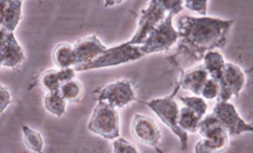
Development of novel therapeutic modalities is crucial for the treatment of oral squamous cell carcinoma (OSCC). Recent scientific studies have been focused on herbal medicines as potent anti-cancer drug candidates. This study is the first to investigate the cytotoxic effects and the mechanism of cell death induced by grape seed extract (GSE) in oral squamous cell carcinoma (KB cells). MTT (3-(4,5-dimetylthiazol-2-yl)-2,5 diphenyltetrazolium bromide) and trypan blue assays were performed in KB cells as well as human umbilical vein endothelial cells (HUVEC) were used to analyze the cytotoxic activity of GSE. Furthermore, the apoptosis-inducing action of the extract was determined by TUNEL, DNA fragmentation and cell death analysis. Statistical significance was determined by analysis of variance (ANOVA), followed by Duncan’s test at a significance level of P≤0.05. The results showed apoptotic potential of GSE, confirmed by significant inhibition of cell growth and viability in a dose- and time- dependent manner without inducing damage to non-cancerous cell line HUVEC. The results of this study suggest that this plant contains potential bioactive compound(s) for the treatment of oral squamous cell carcinoma.

Downloads

Download data is not yet available.
Induction of apoptosis by grape seed extract (Vitis vinifera) in oral squamous cell carcinoma

Downloads

Additional Files

Published

2013-08-20

How to Cite

1.
Aghbali A, Vosough Hosseini S, Delaza A r, Kalbasi Gharavi N, Zare Shahneh F, Orangi M, Bandehagh A, Baradaran B. Induction of apoptosis by grape seed extract (Vitis vinifera) in oral squamous cell carcinoma. Biomol Biomed [Internet]. 2013Aug.20 [cited 2023Dec.11];13(3):186-91. Available from: https://www.bjbms.org/ojs/index.php/bjbms/article/view/2360

Issue

Section

Pharmacology

INTRODUCTION

Oral squamous cell carcinoma (OSCC) is one of the most common oral cancers with increasing risk in younger people less than 40 years of age, especially in developed countries. Principal methods for treatment of OSCC are radiotherapy and surgery but recurrences are common with further resistance to therapy [1-3]. Nowadays complementary and alternative medicine (CAM) provides treatments for various cancers using herbal medicine derivatives from natural products. Recent scientific studies have been focused on herbal medicine as potent anti-cancer drug candidates [4]. Taxol from Taxus brevifolia L, as a plant-derived compound, is extensively used in the clinical practice. Cytotoxic effects of medicinal plant extracts can be mediated through the induction of apoptosis [5]. Apoptosis, programmed cell death, occurs in physiological and pathological conditions and is characterized by morphological alterations such as cell shrinkage, membrane blebbing, chromatin condensation and DNA fragmentation [6, 7]. Morphological hallmarks of apoptosis in the nucleus are chromatin condensation and nuclear fragmentation. Apoptosis plays a key role in eliminating damaged or abnormal cells without harming normal cells [8]. Grapes (Vitis vinifera) are indigenous to southern Europe and Western Asia cultivated worldwide. The grape vine is rich in flavonoids, polyphenols, anthocyanin, proan-thocyanidins, procyanidins and trans-resveratrol [9]. The seeds and the leaves of the grape vine are used in herbal medicines, whilst fruit is consumed as a dietary supplement. Recent studies have revealed that grape seed extract (GSE) has antioxidant and free radical scavenging, anti-diabetic, cardioprotective, hepatoprotective, anti-carcinogenic, anti-microbial, and anti-viral activities [10-12]. In the present study, cytotoxic effects of grape seed extract (GSE) were investigated for the first time and its possible cell death induction potential on oral squamous cell carcinoma (KB cell line) was determined. Furthermore, the possible side effects of the extracts on human umbilical vein endothelial cells (HUVEC), as normal cells, were evaluated.

MATERIALS AND METHODS

Reagents

Grape (Vitis vinifera) plant was collected from Eastern Azerbaijan Province, Iran, in April 2012. A voucher specimen was deposited at the Herbarium of Faculty of Pharmacy, Tabriz University of Medical Sciences. KB cell (oral squamous cell carcinoma cell line) and HUVEC (human umbilical vein endothelial cells) were purchase from the National Cell Bank of Iran (Pasteur Institute, Tehran, Iran). RPMI-1640 medium, fetal bovine serum (FBS), penicillin, and streptomycin were purchased from Sigma (Germany), dimethylsulfoxide (DMSO) and trypan blue from Merck (Germany), trypsin/EDTA solution from Gibco (U.K), and 3-(4,5-dimetylthiazol-2-YL)- 2,5-diphenyltetrazolium bromide (MTT reagent), Cell Death Detection ELISA plus kit, and In Situ Cell Death Detection Kit, POD from Roche Diagnostics GmbH (Germany) and DAB (3, 3’-diaminobenzidine).

Preparation of extracts

The grape seeds from Vitis Vinifera were washed, dried and ground to powder using a blender. Extractions were performed in a Soxhlet apparatus with methanol. Dry powder of the grape seeds were extracted with N-hexane, Dichloromethane and methanol in Soxhlet extractor, the obtained extract dried in a rotary evaporator in low power and 45°C temperature, then dried powders were stored in a covered glass container in refrigerator until usage, so the mentioned extract was completely dry and did not contain any methanol and its cytotoxic effects are because of the pure extract itself. The grape seed extract (GSE) was concentrated by a rotary evaporator (Heildolph, Germany) at about 45°C and then dried under very low pressure. The dried extracts were stored at −20°C; 20 mg of each extract were dissolved in 100 μL of DMSO and diluted with RPMI-1640 medium to the defined concentrations as indicated. Then, test solutions were sterilized using 0.22-μm syringe filters (Nunc, Denmark) and used as stock solution for further experiments.

Cell culture

The KB and HUVEC cells were grown in RPMI-1640 medium supplemented with 10% FBS, 100 U/mL penicillin and 100 μg/mL streptomycin. The cells were incubated in a humidified incubator containing 5% CO2 at 37°C. After reaching an 80% confluence, the cells were rinsed with PBS/0.5% EDTA and harvested from 25 cm2 flasks using 0.25% trypsin/EDTA solution. Then, the cells were sub-cultured into 75-cm2 flasks, 96-well plates or 6-well plates (Nunc, Denmark) according to experiments. All applied measurements were performed in triplicate.

MTT assay

Cytotoxicity of GSE was assessed on KB cells as well as HUVEC using the MTT reagent according to manufacturer’s protocol. The cells were seeded in 96-well plates (104 cells/ well) and incubated for 24 h at 37°C with 5% CO2. The cells were treated with different concentrations of solvent extracts (50, 100, 200, 300, 400, 500, 600 μg/mL) and 0.2% (v/v) DMSO as a negative control. After 24 h of treatment, 50 μL of MTT reagent (2 mg/mL) labeling reagent was added to each well. The plates were incubated at 37°C with 5% CO2 for 4 hours. Then, 200 μL of the solubilizing solution was added to each well, followed by incubation overnight at 37°C to dissolve formazan crystals. Finally, absorbance was read using an ELISA plate reader (Bio Teck, Germany) at a wavelength of 570 nm [13]. The percentages of cytotoxicity and cell viability were calculated using the following equations: % cytotoxicity = 1- [means absorbance of treated cells/mean absorbance of negative control], % viability = 100 - % cytotoxicity.

Trypan blue assay

Cell membrane integrity was evaluated using trypan blue dye exclusion. Trypan blue passes through the membranes of dead cells and thus stains them, unlike live cells. KB cells (104 cells/well) in 96-well plates were exposed against different concentrations of GSE and 0.2% (v/v) DMSO for 24 h. After medium was removed from the wells, cells were washed with 200 μL of PBS. The cells were detached by adding 100 μL of 0.5% trypsin/EDTA. RPMI-1640 medium supplement with 10% FBS (50 μL) and 0.5% trypan blue (50 μL) was added to each well, and the plates were incubated for 5 min. Then, 20-μL aliquot was removed and placed on a Neubauer hemacytometer. Finally, the number of viable cells was counted under light microscopy. The number of viable cells was calculated according to the following formula: [The unstained cell count × the dilution of the cell suspension × 104]= viable cells.

The percentage of viability was calculated as: [viable cells/the total cell count] x 100= viability %

Assessment of apoptosis

Apoptotic cells death were measured using the Cell Death Detection ELISA Plus kit, which quantifies histone-associated DNA fragments (mono- and oligo-nucleosomes). KB cells (104 cells/well) were treated against different concentrations of GSE and 0.2% (v/v) DMSO at 37°C for 48 hrs. The procedure was performed according to manufacturer’s protocol. Briefly, the culture supernatants and lysate of cells were prepared and incubated in the microtiter plate coated with anti-histone antibody. After color development, the results were measured spectrophoto-metrically using an ELISA plate reader at 405 nm [14].

TUNEL assay

DNA fragmentation was detected by terminal deoxytransferase (TdT)-mediated dUTP nickend labeling (TUNEL) with the In Situ Cell Death Detection Kit, POD (Roche Diagnostics GmbH, Germany) as described by manufacturer’s protocol. Briefly, (1.5×10⁵) KB cells were sub-cultured into 6-well plates and incubated for 24 h at 37°C and 5% CO2. The cells were treated with GSE at concentrations required for 50% inhibition of growth of KB cells (IC50) for 24 h. Negative control cells were treated with the same final concentration of DMSO present in treated wells [0.2% (v/v)]. After treatment, the cells were fixed with 4% (w/v) paraformaldehyde in PBS (pH 7.4) for 1 hour at room temperature and rinsed twice with PBS. Then, the fixed cells were incubated with blocking solution (3% H2O2 in methanol) for 10 min and rinsed with PBS. The cells were then incubated in permeabilizing solution (0.1% Triton X-100 in 0.1% sodium citrate) for 2 min on ice. Subsequently, 50 μL of reaction mixture containing TdT enzyme and nucleotide were added to the cells and incubated for 1 h at 37°C. After washing three times with PBS, the slides were incubated with 50 μL converter-POD sterptavidin HRP solution for 30 min, and rinsed three times with PBS. Finally, the cells were incubated with DAB. Substrate solution and the stained cells were analyzed using invert microscopy [15].

DNA Fragmentation assay

Biochemically, apoptosis is characterized by the activation of a nuclear endonuclease that cleaves DNA into multimers of 180-200 base pairs and can be visualized as an oligosomal ladder by standard agarose gel electrophoresis. KB cells were seeded in 6-well plates and kept in a CO2 incubator. KB cells were treated by GSE in IC50 concentrations for 24 hours. At the end of incubation period, the cells were centrifuged for 1000 rpm for 3 min at 14°C. The pellet was suspended in a lysis buffer (10 mM Tris-HCI, pH=8.0, 10 mM NaCl, I0 mM EDTA, 20 mg/mL Proteinase K, 10% SDS), and incubated at 37°C. The pellet was dissolved in TE buffer (0.1 M

Tris-HCl, pH=8.0, 10 mM EDTA). DNA samples were elec-trophoretically separated on 1.8% agarose gel, containing ethidium bromide (0.4 μg/mL). DNA was visualized by a UV transilluminator [16]. Untreated cells were used as control.

Statistical analysis

Data are presented as means ± SEM. Statistical significance was carried out by ANOVA, followed by Duncan’s test. Statistical significance was defined at p<0.05. IC50 values were derived from prohibit analysis. SPSS 20 program was used for statistical analysis of data.

RESULTS

The cytotoxic effects of grape seed extract (GSE) on the growth of oral squamous cell carcinoma (KB cell line) were determined by MTT and trypan blue assays as shown in Figures 1 and 2. As shown in Figure 1, the cells treated with hydroalcoholic extract exhibited significant decline in viability in comparison to the untreated control cells, whereas no cytotoxic activity was observed on normal human umbilical vein endothelial cells (HUVEC). Moreover, treatment of KB cells with GSE showed cell growth inhibition in a time- and dose-dependent manner. The most significant cytotoxicity was achieved in the higher concentration and longer time of the GSE treatment on KB cells. Data analysis of cytotoxicity assay showed that IC50 (dose required for 50% inhibition) of GSE on KB cells was 245.984 μg/mL for 24 hours. Direct counting for viable cells using the trypan blue exclusion test showed that 39% of GSE-treated KB cells with the highest concentration (600 μg/mL) absorbed the dye at 24 h (Figure 2). The results showed that dose-dependent apoptosis of KB cells occurred under the incubation with different concentration of GSE.. The highest apoptotic effect of GSE was 69.56%. One of the hallmarks of apoptotic KB cells (treated with GSE)was the presence of nucleosomal DNA fragments, confirmedby TUNEL assay. As shown in Figure 3, after the treatment of KB cells with 24 hIC50 concentration of GSE, the apoptotic cells produced dark brown-stained nuclei, whereas the non-apoptotic cells were not stained; a similar observation was noted in the negative control cells treated with 0.2% (v/v) DMSO. To confirm the effect of extracts on the induction of apoptosis in KB cells, the GSE was examined on the internucleosomal DNA fragmentation as a characteristics feature of apoptosis. As shown by agarose gel electrophoresis in Figure 4, increased DNA fragmentation was apparent in KB cells after treatment with 250 μg/mL (near to IC50) of GSE. Fragmented DNA was clearly observed in KB cells, whereas untreated cells did not exhibit ladders. Therefore, it is possible that GSE causes apoptosis in KB cells.

FIGURE 1: Effects of GSE on proliferation of KB and HUVEC cells for 24 h.
FIGURE 2: Effects of GSE on KB and HUVEC cells viability for 24 h.
FIGURE 3: Nuclei morphological changes during GSE-induced apoptosis in KB cells detected by TUNEL assay. For KB cells, (a) shows negative control (without treatment) and (b) and (c, d) treated with extract (IC50) for 24 h (n: 3). (b), (c) and (d) indicate representative apoptotic cells with nuclei morphological changes.
FIGURE 4: DNA laddering: DNA fragmentation on 1.8% agarose gel electrophoresis; Lane 1, 1000 bp marker; Lanes 2 and 3, untreated cells; Lanes 4 and 5, GSE-treated cells.

DISCUSSION

Oral squamous cell carcinoma (OSCC) poses an important health concern all over the world. Standard treatment plan for oral squamous cell carcinoma depends on the stage of the disease. Early-stage tumors are treated primarily by surgery or radiotherapy, with both modalities resulting in similar local control and survival rates. More advanced carcinomas often require multi-modality therapy with surgery, radiation and chemotherapy, which can result in very high morbidity [17]. Treatment modalities of oral SCC have numerous side effects. Radical surgery can result in disfigurement and functional impairment. Common side effects of radiotherapy include mucositis, oral candidiasis, loss of taste and xerostomia, which may be permanent due to the detrimental effect of radiation on salivary glands [18]. Osteoradionecrosis of bones within the radiation field (most commonly the mandible) may occur as a result of damage to the bone vasculature and osteocytes and is one of the most serious complications of radiotherapy [19]. Current chemotherapeutic drugs exert many cytotoxic effects on normal cells, resulting in adverse systemic and cytotoxic effects and development of resistance to therapy in OSCC patients. Despite continuing research and advances in treatment, the clinical outcomes for HNSCC have not improved significantly over the last several decades, with the overall 5-year survival rate as low as 50% [20-22]; therefore, development of new treatment modalities is crucial for prevention and to reduce mortality. Herbal medicines (plant-derived compounds), in combination with routinely implemented treatment modalities, such as radiotherapy and surgery, can reduce morbidity and mortality in OSCC patients. Recent studies focus on herbal medicines as potent anti-cancer drug candidates. Previous studies have revealed the anticancer activities of herbal extracts against oral squamous cell carcinoma cell lines. These include Tamoxifen in combination with Cisplatin, 5-Fluorouracil, Cordycepin, Scutellariabaicalensis, Quercetin and Artemisinin [5]. In a study by Ghashmet et al, time-and dose-dependent inhibitory effect of anti-proliferative activity of local honey (Tualang) on OSCC and HOS cell lines was investigated by MTT assay, light and fluorescent microscope, and Annexin V-FITC Apop-tosis Detection Kit [23]. In a study by Hseu et al, ethanol (70%) extracts of Alpiniapricei rhizome could induce G2/M phase of the cell cycle arrest and apoptosis in KB cells [24]. Grapes (Vitisvinifera) are rich in flavonoids, polyphenols, anthocyanin, proanthocyanidins, trans-resveratrol, and procyanidins. Recent studies have revealed that GSE has antioxidant and free radical scavenging activities, and anti-diabetic, cardioprotective, hepatoprotective, anti-carcinogenic, antimicrobial, and anti-viral effects. Dinicola et al showed that GSE has anti-proliferative and apoptotic effects on CaCO2 and HCT-8 colon cancer cell lines [25]. Sun et al. [26] indicated that GSEs have the ability to inhibit HNSCC cell invasion by targeting the expression of EGFR and activation of NF-κ B as well as inhibiting the epithelial-to-mesenchymal transition In the present study, for the first time the cytotoxic effects of the grape seed extract was investigated and their possible effects on cell death properties in KB cell line were determined. Furthermore, the probable side effects of the grape seed extract on HUVEC were evaluated. The results showed that grape seed extract inhibited the growth of oral squamous cell carcinoma through the induction of apoptosis. The IC50 values strongly indicated that the GSE had a potent cytotoxic effect on KB cells. The crucial results of our study addressed to selectivity of GSE effects on KB vs. HUVEC cells (effect was observed on KB, but HUVEC cells) indicate possibility for its promising use for its promising use for the treatment oral squamous cell carcinoma. The different sensitivities of human oral squamous cell carcinoma and normal cells to GSE suggested that used GSE may be used as a natural chemotherapeutic drug. Many chemotherapeutic agents can trigger the apoptosis of cancer cells. Morphological changes in apoptosis are chromatin condensation and nuclear fragmentation. Apoptotic DNA fragmentation is a key feature of apoptosis, characterized by the activation of endogenous endonucleases with subsequent cleavage of chromatin DNA into internucleosomal fragments of approximately 180-200 base pairs (bp) [27, 28]. The results of our study showed induction of apoptosis in the cancer cells as a result of treatment with the GSE. Cell death assay, TUNEL and DNA fragmentation analyses were used to reveal if induction of apoptosis was achieved by GSE. It was shown that DNA fragmentation happened in the late stage of apoptosis. Therefore, results of our in vitro study have shown an anticancer potential of the GSE and selectivity to inhibit growth and induce apoptosis in KB, unlike HUVEC cells. However, additional experiments should be done to test grape seed extracts effect on other cancer cell lines in vitro and in vivo (animal studies) conditions to establish their therapeutic ability as well as its adverse effects.

CONCLUSION

The present study demonstrated the in vitro cytotoxic and apoptotic activity of GSE in human KB cell line, possibly suggesting a new potential chemotherapeutic agent for the treatment of oral squamous cell carcinoma.

Acknowledgements

ACKNOWLEDGEMENTS

This study was supported by a grant from the Drug Applied Research Center of Tabriz University of Medical Sciences.

DECLARATION OF INTEREST

The authors declare that there is no conflicting interest.

REFERENCES

  1. , (). Recent advances in oral oncology 2007: epidemiology, aetiopathogenesis, diagnosis and prognostication. Oral Oncol.
  2. (). Global epidemiology of oral and oropharyngeal cancer. Oral Oncol.
  3. , (). Oral squamous cell carcinoma: Etiology, pathogenesis and prognostic value of genomic alterations. Indian J Cancer.
  4. , (). Morphologic and biochemical hallmarks of apoptosis. Cardiovasc Res.
  5. , , , (). Cancer chemoprevention by natural products: How far have we come?. Pharm Res.
  6. , (). Morphological features of cell death. News Physiol Sci.
  7. , , , , , (). Cell death modalities: classification and pathophysiological implications. Cell Death Differ.
  8. (). Apoptosis: a review of programmed cell death. Toxicol Pathol.
  9. , , , (). Grape seed proantho-cyanidins induce apoptosis throughp53, Bax, and Caspase 3 pathways. Neoplasia.
  10. , , , , (). Grape seed extract inhibits in vitro and in vivo growth of human colorectal carcinoma cells. Clin Cancer Res.
  11. , , , , , (). The cytotoxic effects of a novel IH636 grape seed proanthocy- anidin extract on cultured human cancer cells. Mol Cell Biochem.
  12. , , , , , (). The importance of polymerization and galloylation for the antiproliferative properties of procyanidin-rich natural extracts. FEBS J.
  13. (). Rapid colorimetric assay for cellular growth and survival: application to proliferation and cytotoxicity assays. J Immunol Methods.
  14. , (). Enzyme-linked immunosorbent assay (ELISA) for the specific detection of apoptotic cells and its application to rapid drug screening. J Immunol Methods.
  15. , , , , (). Initiation of DNA fragmentation during apoptosis induces phos- phorylation of H2AX histone at serine 139. J Biol Chem.
  16. , (). A rapid and sensitiveassay for the detection of DNA fragmentation during early phases of apoptosis. Nucleic Acids Res.
  17. (). Integrating systemic agents into multimodality treatment of locally advanced head and neck cancer. Ann Oncol.
  18. , , , , , (). Oral sequelae of head and neck radiotherapy. Crit Rev Oral Biol Med.
  19. , , , (). Osteoradionecrosis of the jaws: clinical characteristics and relation to field of irradiation. J Oral Maxillofac Surg.
  20. (). Survival times in end-stage head and neck cancer. Eur J Surg Oncol.
  21. , , , (). Head and neck cancer. N Engl J Med.
  22. , , , , , (). Competing causes of death and second primary tumors in patients with locoregionally advanced head and neck cancer treated with chemoradiotherapy. Clin Cancer Res.
  23. , , , , (). Antiproliferative effect of Tualang honey on oral squamous cell carcinoma and osteosarcoma cell lines. BMC Complement Altern Med.
  24. , , (). Alpinia pricei rhizome extracts induce cell cycle arrest in human squamous carcinoma KB cells and suppress tumor growth in nude mice. Evid Based Complement Alternat Med 2011.
  25. , , , , , (). Antiproliferative and Apoptotic Effects Triggered by Grape Seed Extract (GSE) versus Epigallocatechin and Procyanidins on Colon Cancer Cell Lines. Int J Mol Sci.
  26. , , , (). Grape seed proanthocy-anidins inhibit the invasiveness of human HNSCC Cells by targeting targeting EGFR and reversing the epithelial-to-Mesenchymal Transition. PLoS ONE.
  27. , , (). Apoptosis: a link between cancer genetics and chemotherapy. Cell.
  28. , (). The role of apoptosis in cancer development and treatment response. Nat Rev Cancer.