Dexmedetomidine reduces ventilator-induced lung injury (VILI) by inhibiting Toll-like receptor 4 (TLR4)/nuclear factor (NF)-κB signaling pathway

Authors

  • Hongli Chen Department of Anesthesiology, Qilu Hospital of Shandong University, Jinan, Shandong, China
  • Xiaotong Sun Department of Anesthesiology, Qilu Hospital of Shandong University, Jinan, Shandong, China
  • Xiaomei Yang Department of Anesthesiology, Qilu Hospital of Shandong University, Jinan, Shandong, China
  • Yonghao Hou Department of Anesthesiology, Qilu Hospital of Shandong University, Jinan, Shandong, China
  • Xiaoqian Yu Department of Anesthesiology, Qilu Hospital of Shandong University, Jinan, Shandong, China
  • Yang Wang Department of Anesthesiology, Qilu Hospital of Shandong University, Jinan, Shandong, China
  • Jianbo Wu Department of Anesthesiology, Qilu Hospital of Shandong University, Jinan, Shandong, China
  • Dejie Liu Department of Anesthesiology, Qilu Hospital of Shandong University, Jinan, Shandong, China
  • Huanliang Wang Department of Anesthesiology, Qilu Hospital of Shandong University, Jinan, Shandong, China
  • Jingui Yu Department of Anesthesiology, Qilu Hospital of Shandong University, Jinan, Shandong, China
  • Wenbo Yi Department of Anesthesiology, Qilu Hospital of Shandong University, Jinan, Shandong, China

DOI:

https://doi.org/10.17305/bjbms.2018.2400

Keywords:

Dexmedetomidine, ventilator-induced lung injury, VILI, Toll-like receptor 4, TLR4, Nuclear factor κB, NF-κB, inflammatory cytokine, α2-adrenoceptor

Abstract

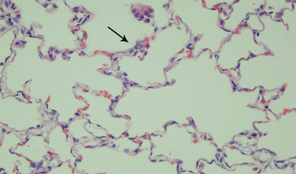
Mechanical ventilation (MV) may lead to ventilator-induced lung injury (VILI). Previous research has shown that dexmedetomidine attenuates pulmonary inflammation caused by MV, but the underlying mechanisms remain unclear. Our study aims to test whether dexmedetomidine has a protective effect against VILI and to explore the possible molecular mechanisms using the rat model. Thirty adult male Wistar rats weighing 200-250 g were randomly assigned to 5 groups (n = 6): control, low tidal volume MV (LMV), high tidal volume (HVT) MV (HMV), HVT MV + dexmedetomidine (DEX), HVT MV + dexmedetomidine + yohimbine (DEX+Y). Rats were euthanized after being ventilated for 4 hours. Pathological changes, lung wet/dry (W/D) weight ratio, lung myeloperoxidase (MPO) activity, levels of inflammatory cytokines (i.e., interleukin [IL]-1β, tumor necrosis factor alpha [TNF-α], and IL-6) in the bronchoalveolar lavage fluid (BALF) and lung tissues, expression of Toll-like receptor 4 (TLR4) and nuclear factor (NF)-κB, and activation of NF-κB in lung tissues were measured. Compared with HMV, DEX group showed fewer pathological changes, lower W/D ratios and decreased MPO activity of the lung tissues and lower concentrations of the inflammatory cytokines in the BALF and lung tissues. Dexmedetomidine significantly inhibited the expression of TLR4 and NF-κB and activation of NF-κB. Yohimbine partly alleviated the effects of dexmedetomidine. Dexmedetomidine reduced the inflammatory response to HVT-MV and had a protective effect against VILI, with the inhibition of the TLR4/NF-κB signaling pathway, at least partly via α2-adrenoceptors.

Downloads

Download data is not yet available.

Author Biographies

Hongli Chen, Department of Anesthesiology, Qilu Hospital of Shandong University, Jinan, Shandong, China

Department of Anesthesiology

Xiaotong Sun, Department of Anesthesiology, Qilu Hospital of Shandong University, Jinan, Shandong, China

Department of Anesthesiology

Xiaomei Yang, Department of Anesthesiology, Qilu Hospital of Shandong University, Jinan, Shandong, China

Department of Anesthesiology

Yonghao Hou, Department of Anesthesiology, Qilu Hospital of Shandong University, Jinan, Shandong, China

Department of Anesthesiology

Xiaoqian Yu, Department of Anesthesiology, Qilu Hospital of Shandong University, Jinan, Shandong, China

Department of Anesthesiology

Yang Wang, Department of Anesthesiology, Qilu Hospital of Shandong University, Jinan, Shandong, China

Department of Anesthesiology

Jianbo Wu, Department of Anesthesiology, Qilu Hospital of Shandong University, Jinan, Shandong, China

Department of Anesthesiology

Dejie Liu, Department of Anesthesiology, Qilu Hospital of Shandong University, Jinan, Shandong, China

Department of Anesthesiology

Huanliang Wang, Department of Anesthesiology, Qilu Hospital of Shandong University, Jinan, Shandong, China

Department of Anesthesiology

Jingui Yu, Department of Anesthesiology, Qilu Hospital of Shandong University, Jinan, Shandong, China

Department of Anesthesiology

Wenbo Yi, Department of Anesthesiology, Qilu Hospital of Shandong University, Jinan, Shandong, China

Department of Anesthesiology

References

Petrucci N, De Feo C. Lung protective ventilation strategy for the acute respiratory distress syndrome. Cochrane Database Syst Rev 2013;(2):CD003844. https://doi.org/10.1002/14651858.CD003844.pub4.

Loring SH, Talmor D. Inhomogeneous computed tomographic densities in lungs in acute respiratory distress syndrome: Stress multipliers leading to ventilator-induced injury? Am J Respir Crit Care Med 2014;189(2):123-4.

https://doi.org/10.1164/rccm.201312-2189ED2189ED.

Slutsky AS, Ranieri VM. Ventilator-induced lung injury. N Engl J Med 2013;369(22):2126-36. https://doi.org/10.1056/NEJMra1208707.

Scheiermann J, Klinman DM. Suppressive oligonucleotides inhibit inflammation in a murine model of mechanical ventilator induced lung injury. J Thorac Dis 2016;8(9):2434-43. https://doi.org/10.21037/jtd.2016.08.18.

Ko YA, Yang MC, Huang HT, Hsu CM, Chen LW. NF-κB activation in myeloid cells mediates ventilator-induced lung injury. Respir Res 2013;14(1):69. https://doi.org/10.1186/1465-9921-14-69.

Cruickshank M, Henderson L, MacLennan G, Fraser C, Campbell M, Blackwood B, et al. Alpha-2 agonists for sedation of mechanically ventilated adults in intensive care units: A systematic review. Health Technol Assess 2016;20(25):v-xx, 1-117. https://doi.org/10.3310/hta20250.

Li B, Li Y, Tian S, Wang H, Wu H, Zhang A, et al. Anti-inflammatory effects of perioperative dexmedetomidine administered as an adjunct to general anesthesia: A meta-analysis. Sci Rep 2015;5:12342. https://doi.org/10.1038/srep12342.

Schoeler M, Loetscher PD, Rossaint R, Fahlenkamp AV, Eberhardt G, Rex S, et al. Dexmedetomidine is neuroprotective in an in vitro model for traumatic brain injury. BMC Neurol 2012;12:20. https://doi.org/10.1186/1471-2377-12-20.

Chen C, Zhang Z, Chen K, Zhang F, Peng M, Wang Y. Dexmedetomidine regulates inflammatory molecules contributing to ventilator-induced lung injury in dogs. J Surg Res 2014;187(1):211-8. https://doi.org/10.1016/j.jss.2013.09.018.

Medzhitov R, Janeway C Jr. Innate immune recognition: Mechanisms and pathways. Immunol Rev 2000;173:89-97. https://doi.org/10.1034/j.1600-065X.2000.917309.x.

Takeda K, Akira S. Toll receptors and pathogen resistance. Cell Microbiol 2003;5(3):143-53. https://doi.org/10.1046/j.1462-5822.2003.00264.x.

Takeuchi O, Hoshino K, Kawai T, Sanjo H, Takada H, Ogawa T, et al. Differential roles of TLR2 and TLR4 in recognition of gram-negative and gram-positive bacterial cell wall components. Immunity 1999;11(4):443-51. https://doi.org/10.1016/S1074-7613(00)80119-3.

Guijarro-Munoz I, Compte M, Alvarez-Cienfuegos A, Alvarez-Vallina L, Sanz L. Lipopolysaccharide activates Toll-like receptor 4 (TLR4)-mediated NF-κB signaling pathway and proinflammatory response in human pericytes. J Biol Chem 2014;289(4):2457-68. https://doi.org/10.1074/jbc.M113.521161.

Wong ET, Tergaonkar V. Roles of NF-κB in health and disease: Mechanisms and therapeutic potential. Clin Sci (Lond) 2009;116(6):451-65. https://doi.org/10.1042/CS20080502.

Hu G, Malik AB, Minshall RD. Toll-like receptor 4 mediates neutrophil sequestration and lung injury induced by endotoxin and hyperinflation. Crit Care Med 2010;38(1):194-201. https://doi.org/10.1097/CCM.0b013e3181bc7c17.

Lucas K, Maes M. Role of the Toll Like receptor (TLR) radical cycle in chronic inflammation: Possible treatments targeting the TLR4 pathway. Mol Neurobiol 2013;48(1):190-204. https://doi.org/10.1007/s12035-013-8425-7.

Peri F, Calabrese V. Toll-like receptor 4 (TLR4) modulation by synthetic and natural compounds: An update. J Med Chem 2014;57(9):3612-22. https://doi.org/10.1021/jm401006s.

Li H, Su X, Yan X, Wasserloos K, Chao W, Kaynar AM, et al. Toll-like receptor 4-myeloid differentiation factor 88 signaling contributes to ventilator-induced lung injury in mice. Anesthesiology 2010;113(3):619-29. https://doi.org/10.1097/ALN.0b013e3181e89ab2.

Villar J, Cabrera NE, Casula M, Flores C, Valladares F, Diaz-Flores L, et al. Mechanical ventilation modulates TLR4 and IRAK-3 in a non-infectious, ventilator-induced lung injury model. Respir Res 2010;11:27. https://doi.org/10.1186/1465-9921-11-27.

Vaneker M, Joosten LA, Heunks LM, Snijdelaar DG, Halbertsma FJ, van Egmond J, et al. Low-tidal-volume mechanical ventilation induces a toll-like receptor 4-dependent inflammatory response in healthy mice. Anesthesiology 2008;109(3):465-72. https://doi.org/10.1097/ALN.0b013e318182aef1.

Gajic O, Dara SI, Mendez JL, Adesanya AO, Festic E, Caples SM, et al. Ventilator-associated lung injury in patients without acute lung injury at the onset of mechanical ventilation. Crit Care Med 2004;32(9):1817-24. https://doi.org/10.1097/01.CCM.0000133019.52531.30.

Dhanireddy S, Altemeier WA, Matute-Bello G, O'Mahony DS, Glenny RW, Martin TR, et al. Mechanical ventilation induces inflammation, lung injury, and extra-pulmonary organ dysfunction in experimental pneumonia. Lab Invest 2006;86(8):790-9. https://doi.org/10.1038/labinvest.3700440.

Sen V, Guzel A, Sen HS, Ece A, Uluca U, Soker S, et al. Preventive effects of dexmedetomidine on the liver in a rat model of acid-induced acute lung injury. Biomed Res Int 2014;2014:621827. https://doi.org/10.1155/2014/621827.

Chen Y, Miao L, Yao Y, Wu W, Wu X, Gong C, et al. Dexmedetomidine ameliorate CLP-induced rat intestinal injury via inhibition of inflammation. Mediators Inflamm 2015;2015:918361. https://doi.org/10.1155/2015/918361.

Hayden MS, Ghosh S. NF-κB in immunobiology. Cell Res 2011;21(2):223-44. https://doi.org/10.1038/cr.2011.13.

Hayden MS, Ghosh S. Regulation of NF-κB by TNF family cytokines. Semin Immunol 2014;26(3):253-66. https://doi.org/10.1016/j.smim.2014.05.004.

Hayden MS, Ghosh S. NF-κB, the first quarter-century: Remarkable progress and outstanding questions. Genes Dev 2012;26(3):203-34. https://doi.org/10.1101/gad.183434.111.

Ghosh G, Wang VY, Huang DB, Fusco A. NF-κB regulation: Lessons from structures. Immunol Rev 2012;246(1):36-58. https://doi.org/10.1111/j.1600-065X.2012.01097.x.

Hoesel B, Schmid JA. The complexity of NF-κB signaling in inflammation and cancer. Mol Cancer 2013;12:86. https://doi.org/10.1186/1476-4598-12-86.

Camp SM, Ceco E, Evenoski CL, Danilov SM, Zhou T, Chiang ET, et al. Unique Toll-like receptor 4 activation by NAMPT/PBEF induces NFκB signaling and inflammatory lung injury. Sci Rep 2015;5:13135. https://doi.org/10.1038/srep13135.

Dexmedetomidine reduces ventilator-induced lung injury (VILI) by inhibiting Toll-like receptor 4 (TLR4)/nuclear factor (NF)-κB signaling pathway

Downloads

Additional Files

Published

2018-05-20

How to Cite

1.
Chen H, Sun X, Yang X, Hou Y, Yu X, Wang Y, Wu J, Liu D, Wang H, Yu J, Yi W. Dexmedetomidine reduces ventilator-induced lung injury (VILI) by inhibiting Toll-like receptor 4 (TLR4)/nuclear factor (NF)-κB signaling pathway. Biomol Biomed [Internet]. 2018May20 [cited 2023Dec.1];18(2):162-9. Available from: https://www.bjbms.org/ojs/index.php/bjbms/article/view/2400

Issue

Section

Pharmacology

INTRODUCTION

Mechanical ventilation (MV) is an irreplaceable therapeutic measure for some critically ill patients [1]. Nevertheless, there are several disadvantages of MV associated with the abnormal stretch and strain that may induce an iatrogenic lung injury known as ventilator-induced lung injury (VILI) [2,3]. Ample evidence has demonstrated that, in addition to mechanical damage, upregulation of inflammatory molecules and excessive inflammatory response play a crucial role in the occurrence and development of VILI [4,5].

Dexmedetomidine, a selective α2-adrenoceptor agonist, exhibits a variety of pharmacological effects, including sedation, antianxiety, analgesia and sympatholytic effect [6]. Dexmedetomidine is widely used in patients subjected to MV. Many reports showed that dexmedetomidine has an anti-inflammatory effect on specific organs probably by inhibiting the release of inflammatory cytokines [7,8]. Recently, a few studies demonstrated that dexmedetomidine can attenuate pulmonary inflammation in animal models of VILI, but the underlying molecular mechanisms have not yet been explained [9].

Toll-like receptor (TLR) 4, a member of the TLR family of proteins that are expressed in different lung cells, is a type of pattern recognition receptor [10,11]. TLR4 was originally recognized as the main sensor for pathogen-associated molecular patterns [PAMPs] (e.g. lipopolysaccharides) [12], activating nuclear factor kappa-light-chain-enhancer of activated B cells (NF-κB) and then inducing the production of inflammatory cytokines and chemokines [13-15]. Recently, significant advances have been made to illustrate that TLR4 may also recognize damage-associated molecular patterns [DAMPs], e.g. high-mobility group box 1 and heat shock proteins, released from damaged tissue [16,17]. Current studies suggest that TLR4 is involved in lung injury caused by mechanical stretch [18-20]. Along with the activation of TLR4/NF-κB signaling pathway, the production of inflammatory cytokines is induced by MV, which plays a major role in the subsequent development of inflammatory response.

Our study aims to test whether treatment with dexmedetomidine has a protective effect against VILI, induced by high-tidal volume MV (HVT-MV) in a rat model, and to explore the function of the TLR4/NF-κB signaling pathway in the anti-inflammatory process of dexmedetomidine in the VILI model.

MATERIALS AND METHODS

Animals

Adult male Wistar rats (200-250 g) were provided by Shandong University Experimental Animal Center (Jinan, China). All animals were fed in uncrowded cages with standard laboratory chow at a room temperature of 22°C (humidity: 45-55%) until the day of the experiment. All animal studies and the handling of the animals were approved by the Medical Ethics Committee for Experimental Animals of Shandong University, China (number ECAESDUSM 2012029) and were in accordance with National Institutes of Health guidelines.

Experimental protocol

The rats were anesthetized with an intraperitoneal (IP) injection of 50 mg/kg body weight sodium pentobarbital and underwent tracheotomy. They were then randomly assigned to 5 groups (n = 6): 1) control group with spontaneous breathing without MV for 4 hours; 2) MV with a low VT of 8 ml/kg (LMV); 3) MV with a HVT of 20 ml/kg (HMV); 4) MV with a HVT of 20 ml/kg and dexmedetomidine treatment (DEX); 5) pretreatment with yohimbine (a competitive α2-adrenoceptor antagonist) and MV with a HVT of 20 ml/kg and dexmedetomidine treatment (DEX+Y). In MV groups, all rats were ventilated for 4 hours with a respiratory rate of 50 times/minute. The rats in DEX group were given a loading dose of dexmedetomidine (Hengrui Medicine Co, Ltd., China), 1 µg/kg intravenously (IV) over 15 minutes before MV, and then a maintenance IV dose of 1 µg/kg/hour during MV. The rats in DEX+Y group were pretreated with yohimbine (Sigma-Aldrich, USA), 0.1 mg/kg IV over 10 minutes before dexmedetomidine was given, and after that, they were treated as DEX group. The rats in control, LMV, and HMV groups received an equal volume of normal saline. The rectal temperature was maintained at 37°C using a heating lamp.

At the end of each experiment, the lungs were rapidly removed and immediately injected with 2 ml of phosphate-buffered saline (PBS) through the trachea 3 times, to collect bronchoalveolar lavage fluid (BALF), followed by centrifugation at 400 g for 5 minutes. Then, the left lung lobe was collected for determining the lung wet/dry (W/D) weight ratio, after removing the blood and water from the lung surface. The right upper lung lobe was immersed in 4% paraformaldehyde to create slices for pathological and immunohistochemical analysis; the remaining lung tissue was immediately stored in liquid nitrogen and frozen at -80°C, until assayed.

Histopathological analysis

The tissues were immersed in 4% paraformaldehyde overnight to be fixed. Then the tissues were embedded in paraffin and sectioned (3-4µm slices). The sections were stained with hematoxylin and eosin (H&E) and then evaluated with a microscope by a pathologist who was blinded to the groups. The levels of lung injury were recorded as normal (0), mild (1), moderate (2), or severe (3), according to the following histological parameters: alveolar edema, intra-alveolar infiltration of inflammatory cells, congestion, and diffuse alveolar hemorrhage. All slides were observed and imaged under a Nikon eclipse 80i light microscope (Nikon Corporation, Japan).

W/D ratio measurements

The wet and dry weights of the left lung lobe were measured after dissection at the end of each experiment, and after drying in a ventilated oven at 70°C for 24 hours. Then, we calculated the W/D ratio by dividing the wet weight by the dry weight.

Myeloperoxidase (MPO) activity assay

The activity of MPO in the lung tissues was assayed using a MPO assay kit (Jiancheng Bioengineering Institute of Nanjing, China), according to the manufacturer’s protocols. Briefly, the lung tissue samples were stored in 1 ml ice-cold sample buffer. After homogenization and subsequent centrifugation of homogenates at 16,000 rpm for 30 minutes at 4°C, the MPO activity was measured over 25 seconds at 450 nm, by a Varioskan Flash spectral scanning multimode reader (Thermo Scientific, USA).

Enzyme-linked immunosorbent assay (ELISA)

The levels of inflammatory cytokines including tumor necrosis factor alpha (TNF-α), interleukin (IL)-1β, and IL-6 in the BALF were detected using ELISA kits (Wuhan Xinqidi Biological Technology, China), according to the manufacturer’s instructions.

Quantitative reverse transcription polymerase chain reaction (qRT-PCR)

The relative gene expression of TNF-α, IL-1β, IL-6 and TLR4 in the lung tissue homogenates was measured by qRT-PCR. We extracted total RNA from the lung tissues using RNeasy Mini kits (Aidlab Biotechnologies, Beijing, China), following the manufacturer’s instructions. After cDNA synthesis by the ReverTra Ace qPCR RT Kit (TOYOBO, Japan), the quantitative expression of genes was determined using a Bioer real-time PCR system (Bioer Technology, China) in a 10-ml reaction as follows: 1 µl cDNA, 3.8 µl nuclease-free water, 0.1 µl forward and reverse primers and 5 µl mix (Vazyme Biotech, China). The sense and antisense primers, listed in Table 1, were obtained from GBI (Shanghai, China). The relative mRNA levels of the genes of interest were normalized to glyceraldehyde 3-phosphate dehydrogenase (GAPDH).

TABLE 1: Primer sequences for quantitative reverse transcription polymerase chain reaction

Western blot analysis

The lung tissues were immersed in radioimmunoprecipitation assay (RIPA) buffer and then homogenized with an electric homogenate machine to obtain supernatant fluid of homogenates after centrifugation at 12,000 rpm for 20 min at 4°C. A BCA Protein Assay Kit (ComWin Biotech, China) was used to measure the protein concentration of TLR4 and NF-κB in supernatants, according to the manufacturer’s instructions. The supernatants added to equal volume of sample buffer were boiled 10 minutes at 100°C. Then, we loaded the supernatants containing 20 µg of proteins onto 10% sodium dodecyl sulfate polyacrylamide gel electrophoresis (SDS-PAGE), and the proteins were transferred to a polyvinylidene difluoride (PVDF) membrane (Millipore, USA). The membranes were blocked for 1 hour at room temperature in blocking buffer containing 1 g nonfat dry milk per 20 ml buffer. After washing in Tween-Tris-buffered saline, the membranes were incubated overnight at 4°C with rabbit anti-TLR4 antibody (dilution 1:1000, Abcam, USA), anti-NF-κB p65 antibody (dilution 1:2000, Cell Signaling Technology, USA), anti- phosphor(p)-NF-κB p65 (dilution 1:1000, Abcam, USA), or anti-GAPDH antibody (dilution 1:2000, Proteintech Group, USA). The next day, all membranes were incubated with goat anti-rabbit or goat anti-mouse immunoglobulin G (IgG) antibody (dilution 1:2000, ZSGB-BIO, China) for 1 hour at room temperature and then washed in Tween-Tris-buffered saline. The membranes were imaged by a BioRad ChemiDoc gel documentation system after visualization with enhanced chemiluminescence [ECL] (ComWin Biotech, China).

Immunohistochemistry

NF-κB activation was observed by immunohistochemical analysis. The slices were blocked with 3% bovine serum albumin (BSA) and incubated with anti-NF-κB p65 antibody (1:200, Abcam, USA) at 4°C overnight. The DAB detection kit (Gene Tech, China) was used to stain the sections. Counterstaining was performed with H&E stain. All slides were observed and imaged under a Nikon eclipse 80i light microscope.

Statistical analysis

The results were analyzed with IBM SPSS Statistics for Windows, Version 20.0. (IBM Corp., Armonk, NY). The differences between the groups were determined with one-way ANOVA. Two-tailed Student’s t-test followed by a Bonferroni correction was performed for paired samples. All data were presented as mean ± standard error of the mean (SEM). A value of p < 0.05 was considered statistically significant.

RESULTS

Dexmedetomidine attenuates histopathological injury in the rat lungs

The histopathological changes of the injury were estimated by H&E staining and under an optical microscope. Structural changes in the rat lungs and pulmonary edema were induced more severely in HMV compared to control group (Figure 1). Furthermore, lung injuries were significantly less severe in the rats treated with dexmedetomidine compared to HMV group, and the effects of dexmedetomidine were partly inhibited by yohimbine (Figure 1). Similar results were reflected by the histological lung injury scores (HMV vs. Control, p = 0.0125; DEX vs. HMV, p = 0.0265; DEX+Y vs. DEX, p = 0.0335; Figure 1F).

FIGURE 1: Hematoxylin and eosin (H&E) staining of rat lung tissue (magnification, 400×). Compared with control group (A) and MV group with a low VT of 8 ml/kg (LMV) (B), rats ventilated with HVT of 20 ml/kg (HMV group) (C) showed a marked increase in inflammatory cell infiltration, alveolar edema, and alveolar hemorrhage in the lungs. Dexmedetomidine treatment (DEX group) reduced the histopathological injury in the lungs induced by HVT of 20 ml/kg (D) and yohimbine (DEX+Y group) inhibited partly the effects of dexmedetomidine (E). The histopathological scores for the levels of lung injury (F). The arrows in the figure indicate alveolar edema. The data are presented as mean ± SEM. *p < 0.05 vs. control;#p < 0.05 vs. HMV;Dp < 0.05 vs. DEX.

Dexmedetomidine reduced the lung W/D ratio and MPO activity

The pulmonary edema was also evaluated by the W/D ratio. The lung W/D ratios in HMV group were higher compared to control group (p = 0.0103, Figure 2A). Dexmedetomidine significantly reduced the lung W/D ratios compared to HMV group (p = 0.0258, Figure 2A), and yohimbine pretreatment offset the effects of dexmedetomidine (p = 0.0460, Figure 2A).

The MPO activity, which reflects the degree of neutrophil recruitment, was increased significantly in HMV compared with control group (p = 0.0205, Figure 2B). In relation to HMV group, dexmedetomidine significantly reduced the MPO activity (p = 0.0268) and yohimbine partly reversed the effect of dexmedetomidine on the rat lungs treated with HVT-MV (p = 0.0438, Figure 2B).

FIGURE 2: The wet/dry (W/D) weight ratios and myeloperoxidase (MPO) activity of rat lung tissues. The values of W/D ratio (A) and MPO activity (B) in different groups are presented as mean ± SEM. *p < 0.05 vs. control; #p < 0.05 vs. HMV; Dp < 0.05 vs. DEX. Control (C) group: spontaneous breathing without MV for 4 hours; LMV: MV with a low VT of 8 ml/kg; HMV: MV with a HVT of 20 ml/kg; DEX: MV with a HVT of 20 ml/kg and dexmedetomidine treatment; DEX+Y: pretreatment with yohimbine (a competitive α2-adrenoceptor antagonist) and MV with a HVT of 20 ml/kg and dexmedetomidine treatment.

Dexmedetomidine reduced the concentration of inflammatory cytokines in BALF

The concentrations of IL-1β, TNF-α and IL-6 in the BALF from rats were measured to determine the level of inflammatory changes in the lungs induced by MV. We found that HVT-MV treatment significantly increased the production of IL-1β, TNF-α, and IL-6 compared to control group (p < 0.01 or p < 0.05, Figure 3). Dexmedetomidine significantly reduced the levels of the inflammatory cytokines induced by HVT-MV (p < 0.01 or p < 0.05, Figure 3). Yohimbine pretreatment partly reversed the effects of dexmedetomidine (p < 0.05, Figure 3).

FIGURE 3: The concentrations of inflammatory cytokines interleukin (IL)-1β (A), tumor necrosis factor alpha [TNF-α] (B), and IL-6 (C) in bronchoalveolar lavage fluid (BALF) from rats. The cytokine concentrations in the BALF were measured using an ELISA kit. The data are presented as mean ± SEM. *p < 0.05, **p < 0.01 vs. control; #p < 0.05, # #p < 0.01 vs. HMV;Dp < 0.05 vs. DEX. Control (C) group: spontaneous breathing without MV for 4 hours; LMV: MV with a low VT of 8 ml/kg; HMV: MV with a HVT of 20 ml/kg; DEX: MV with a HVT of 20 ml/kg and dexmedetomidine treatment; DEX+Y: pretreatment with yohimbine (a competitive α2-adrenoceptor antagonist) and MV with a HVT of 20 ml/kg and dexmedetomidine treatment.

Dexmedetomidine decreased mRNA levels of inflammatory cytokines in rat lung tissues

The mRNA levels of inflammatory cytokines (IL-1β, TNF-α, and IL-6) in the rat lung tissues were determined by qRT-PCR. Consistent with the results for inflammatory cytokines in the BALF, the mRNA levels of IL-1β, TNF-α, and IL-6 were significantly higher in HMV compared to control group (p < 0.05, Figure 4). Dexmedetomidine decreased those mRNA levels, while yohimbine pretreatment reversed the effects of dexmedetomidine to some extent (p < 0.05, Figure 4).

FIGURE 4: The mRNA levels of interleukin (IL)-1β (A), tumor necrosis factor alpha [TNF-α] (B), and IL-6 (C) in rat lung tissues determined by quantitative reverse transcription polymerase chain reaction in different groups. Data are presented as mean ± SEM. *p < 0.05 vs. control; #p < 0.05 vs. HMV;Dp < 0.05 vs. DEX. Control (C) group: spontaneous breathing without MV for 4 hours; LMV: MV with a low VT of 8 ml/kg; HMV: MV with a HVT of 20 ml/kg; DEX: MV with a HVT of 20 ml/kg and dexmedetomidine treatment; DEX+Y: pretreatment with yohimbine (a competitive α2-adrenoceptor antagonist) and MV with a HVT of 20 ml/kg and dexmedetomidine treatment.

Dexmedetomidine reduced the mRNA and protein levels of TLR4 in rat lung tissues

The mRNA and protein expression of TLR4 in the rat lung tissues was determined by qRT-PCR and Western blotting, respectively. TLR4 mRNA and protein levels were significantly higher in HMV compared to control group (p < 0.05, Figure 5). Dexmedetomidine decreased those levels compared to HMV group, while the effects of dexmedetomidine were reversed to some extent with yohimbine pretreatment (p < 0.05, Figure 5).

FIGURE 5: Toll-like receptor (TLR4) mRNA (A) and protein (B) levels in rat lung tissues, as determined by quantitative reverse transcription polymerase chain reaction and Western blotting, respectively. The protein expression of TLR4 was quantified by measuring the band intensity and displayed as fold increase relative to glyceraldehyde 3-phosphate dehydrogenase (GAPDH). Data are presented as mean ± SEM. *p < 0.05 vs. control; #p < 0.05 vs. HMV; Dp < 0.05 vs. DEX. Control (C) group: spontaneous breathing without MV for 4 hours; LMV: MV with a low VT of 8 ml/kg; HMV: MV with a HVT of 20 ml/kg; DEX: MV with a HVT of 20 ml/kg and dexmedetomidine treatment; DEX+Y: pretreatment with yohimbine (a competitive α2-adrenoceptor antagonist) and MV with a HVT of 20 ml/kg and dexmedetomidine treatment.

Dexmedetomidine inhibited NF-κB activation in rat lung tissues

Compared with control group, the NF-κB activation was greater in HMV group and dexmedetomidine reduced the activation of NF-κB induced by HVT-MV (Figure 6A-E). In DEX+Y group, yohimbine partly reversed the effects of dexmedetomidine (Figure 6E).

FIGURE 6: Activation of nuclear factor kappa-light-chain-enhancer of activated B cells (NF-κB) in rat lung tissues analyzed by immunohistochemical staining in different groups. The slices are shown in the micrographs (magnification, 400×). Compared with control (A) and LMV group (B), greater NF-κB activation was observed in HMV group (C). Treatment with dexmedetomidine reduced the NF-κB activation in DEX group (D), while yohimbine partly inhibited the effect of dexmedetomidine in DEX+Y group (E). Western blotting was performed to evaluate the protein expression of phospho(p)-NF-κB p65 (F). The level of p-NF-κB p65 was quantified by measuring the band intensity and displayed as fold increase relative to NF-κB p65. Data are presented as mean ± SEM. *p < 0.05 vs. control; #p < 0.05 vs. HMV; Dp < 0.05 vs. DEX. Control (C) group: spontaneous breathing without MV for 4 hours; LMV: MV with a low VT of 8 ml/kg; HMV: MV with a HVT of 20 ml/kg; DEX: MV with a HVT of 20 ml/kg and dexmedetomidine treatment; DEX+Y: pretreatment with yohimbine (a competitive α2-adrenoceptor antagonist) and MV with a HVT of 20 ml/kg and dexmedetomidine treatment.

In addition, we analyzed the expression of p-NF-κB p65 by Western blotting, which can reflect the activation level of NF-κB. The expression of p-NF-κB p65 was increased in HMV and inhibited in DEX group (p < 0.05, Figure 6F). Similarly as in the other experiments, yohimbine partly inhibited the effects of dexmedetomidine on the rats treated with HVT-MV (p < 0.05, Figure 6F).

DISCUSSION

Mechanical ventilation is essential in general anesthesia and is a life-saving therapy for patients with acute respiratory failure, e.g., acute lung injury (ALI) and acute respiratory distress syndrome (ARDS) [1]. However, excessive ventilation has the potential to exacerbate a pre-existing lung injury or even to injure a healthy lung, which is referred to as VILI [2,3,21]. Recent research has found that VILI is mainly characterized by pathological changes, vascular permeability increase, pulmonary edema, influx of various inflammatory cells, and upregulation of pulmonary inflammatory cytokines [22]. In this study, we successfully established the rat VILI model using HVT-MV, based on previous studies [9,18].

As a widely used anesthetic adjuvant in clinical setting, dexmedetomidine has a high affinity for the α2-adrenoceptor and is approved for sedation in critically ill patients, due to its sedative effects without respiratory depression [6]. Many studies also showed the anti-inflammatory effect of dexmedetomidine by reducing the levels of inflammatory cytokines in some specific cases [7,23,24]. Based on previous research [9], we set the dose of dexmedetomidine at 1 µg/kg/hour which is the equivalent of the clinical dose in humans. Our data showed that dexmedetomidine significantly decreased the high levels of inflammatory cytokines (i.e. IL-1β, IL-6, and TNF-α) and reduced the MPO activity, W/D ratios, and pathological changes in the rats treated with HVT-MV. Moreover, the pretreatment with yohimbine partly attenuated the effects of dexmedetomidine. In concert with previous studies, our results suggest that dexmedetomidine reduces the lung injury induced by HVT-MV probably by inhibiting the excessive inflammatory response, and these protective effects may, at least partly, be mediated by α2-adrenoceptors.

TLRs, as transmembrane proteins, comprise a family of signal transduction molecules expressed in many cell types [10,11]. TLR4 is considered an important mediator in inflammation and immune responses. In addition to PAMPs [12], emerging evidence suggests that TLR4 can also recognize endogenous ligands termed DAMPs [16,17]. Recently, significant advances have been made to demonstrate that mechanical stretch may induce the release as well as increase of some endogenous ligands for TLR4; TLR4 could be activated after binding to these ligands and promote the occurrence and development of VILI [18,19]. Here we analyzed the expression levels of TLR4 in the lung tissues of different groups of rats with induced VILI, using qRT-PCR and Western blotting. The results indicated that TLR4 plays a role in the development of VILI in the absence of infection, and that dexmedetomidine could reduce the expression of TLR4 in the VILI model. Moreover, yohimbine partly attenuated the effects of dexmedetomidine on TLR4, suggesting that the α2-adrenoceptor is involved in the inhibition of TLR4 expression by dexmedetomidine.

NFκB, an important nuclear transcription factor, has a crucial role in immune and inflammatory responses [25-27]. Activated NF-κB translocates to the nucleus and binds to DNA, regulating the genes encoding a wide array of inflammatory molecules (e.g. IL-1β, TNF-α, and IL-6) [28]. Excessive activation of the NF-κB pathway plays a pivotal role in the development of many inflammatory diseases, including VILI [5,29]. Data from immunohistochemistry and Western blotting in our study showed that NF-κB was activated by HVT-MV and inhibited significantly by dexmedetomidine.

TLR4-mediated signaling pathways lead to the activation of NF-κB [13,30]. The TLR4/NF-κB signaling pathway is important for investigation due to its central role in the signal transduction network [25,26] and because it governs the expression of inflammatory cytokines and chemokines [28]. Our data showed that dexmedetomidine could reduce the expression of TLR4, activation of NF-κB, and production of inflammatory cytokines in the rat VILI model. These results, in combination with the aforementioned relationship between TLR4 and NF-κB, and NF-κB and upregulation of inflammatory cytokines, indicate that the inhibition of the TLR4/NF-κB signaling pathway may be the mechanism through which dexmedetomidine ameliorated inflammatory responses in the VILI induced by HVT-MV.

CONCLUSION

Overall, we showed that dexmedetomidine reduced the inflammatory response in the lungs induced by HVT-MV and had a protective effect against VILI. Furthermore, we suggested that the inhibition of the TLR4/NF-κB signaling pathway by dexmedetomidine was, at least partly, via α2-adrenoceptors. Our results are the first evidence of TLR4 inhibition by dexmedetomidine in a VILI model with no presence of infection. However, how dexmedetomidine affects the expression of TLR4 is still unclear and further research is warranted. Our findings will facilitate future investigation of therapeutic approaches for VILI and other inflammatory diseases.

Acknowledgements

ACKNOWLEDGMENTS

Our thanks are due to Professor Jingxin Li for the assistance with the experiments and to Dr. Tao Meng for the valuable discussion.

This work was supported by the National Natural Science Foundation of China (81570044, 81470264) and Shandong Provincial Natural Science Foundation of China (ZR2014HP050, ZR2015PH036, ZR2012HM027).

DECLARATION OF INTERESTS

The authors declare no conflict of interests.

REFERENCES

  1. , (). Lung protective ventilation strategy for the acute respiratory distress syndrome. Cochrane Database Syst Rev. https://doi.org/10.1002/14651858.CD003844.pub4
  2. , (). Inhomogeneous computed tomographic densities in lungs in acute respiratory distress syndrome: Stress multipliers leading to ventilator-induced injury?. Am J Respir Crit Care Med. https://doi.org/10.1164/rccm.201312-2189ED2189ED
  3. , (). Ventilator-induced lung injury. N Engl J Med. https://doi.org/10.1056/NEJMra1208707
  4. , (). Suppressive oligonucleotides inhibit inflammation in a murine model of mechanical ventilator induced lung injury. J Thorac Dis. https://doi.org/10.21037/jtd.2016.08.18
  5. , , , , (). NF-κB activation in myeloid cells mediates ventilator-induced lung injury. Respir Res. https://doi.org/10.1186/1465-9921-14-69
  6. , , , , , (). Alpha-2 agonists for sedation of mechanically ventilated adults in intensive care units: A systematic review. Health Technol Assess. https://doi.org/10.3310/hta20250
  7. , , , , , (). Anti-inflammatory effects of perioperative dexmedetomidine administered as an adjunct to general anesthesia: A meta-analysis. Sci Rep. https://doi.org/10.1038/srep12342
  8. , , , , , (). Dexmedetomidine is neuroprotective in an in vitro model for traumatic brain injury. BMC Neurol. https://doi.org/10.1186/1471-2377-12-20
  9. , , , , , (). Dexmedetomidine regulates inflammatory molecules contributing to ventilator-induced lung injury in dogs. J Surg Res. https://doi.org/10.1016/j.jss.2013.09.018
  10. , (). Innate immune recognition: Mechanisms and pathways. Immunol Rev. https://doi.org/10.1034/j.1600-065X.2000.917309.x
  11. , (). Toll receptors and pathogen resistance. Cell Microbiol. https://doi.org/10.1046/j.1462-5822.2003.00264.x
  12. , , , , , (). Differential roles of TLR2 and TLR4 in recognition of gram-negative and gram-positive bacterial cell wall components. Immunity. https://doi.org/10.1016/S1074-7613(00)80119-3
  13. , , , , (). Lipopolysaccharide activates Toll-like receptor 4 (TLR4)-mediated NF-κB signaling pathway and proinflammatory response in human pericytes. J Biol Chem. https://doi.org/10.1074/jbc.M113.521161
  14. , (). Roles of NF-κB in health and disease: Mechanisms and therapeutic potential. Clin Sci (Lond). https://doi.org/10.1042/CS20080502
  15. , , (). Toll-like receptor 4 mediates neutrophil sequestration and lung injury induced by endotoxin and hyperinflation. Crit Care Med. https://doi.org/10.1097/CCM.0b013e3181bc7c17
  16. , (). Role of the Toll Like receptor (TLR) radical cycle in chronic inflammation: Possible treatments targeting the TLR4 pathway. Mol Neurobiol. https://doi.org/10.1007/s12035-013-8425-7
  17. , (). Toll-like receptor 4 (TLR4) modulation by synthetic and natural compounds: An update. J Med Chem. https://doi.org/10.1021/jm401006s
  18. , , , , , (). Toll-like receptor 4-myeloid differentiation factor 88 signaling contributes to ventilator-induced lung injury in mice. Anesthesiology. https://doi.org/10.1097/ALN.0b013e3181e89ab2
  19. , , , , , (). Mechanical ventilation modulates TLR4 and IRAK-3 in a non-infectious, ventilator-induced lung injury model. Respir Res. https://doi.org/10.1186/1465-9921-11-27
  20. , , , , , (). Low-tidal-volume mechanical ventilation induces a toll-like receptor 4-dependent inflammatory response in healthy mice. Anesthesiology. https://doi.org/10.1097/ALN.0b013e318182aef1
  21. , , , , , (). Ventilator-associated lung injury in patients without acute lung injury at the onset of mechanical ventilation. Crit Care Med. https://doi.org/10.1097/01.CCM.0000133019.52531.30
  22. , , , , , (). Mechanical ventilation induces inflammation, lung injury, and extra-pulmonary organ dysfunction in experimental pneumonia. Lab Invest. https://doi.org/10.1038/labinvest.3700440
  23. , , , , , (). Preventive effects of dexmedetomidine on the liver in a rat model of acid-induced acute lung injury. Biomed Res Int. https://doi.org/10.1155/2014/621827
  24. , , , , , (). Dexmedetomidine ameliorate CLP-induced rat intestinal injury via inhibition of inflammation. Mediators Inflamm. https://doi.org/10.1155/2015/918361
  25. , (). NF-κB in immunobiology. Cell Res. https://doi.org/10.1038/cr.2011.13
  26. , (). Regulation of NF-κB by TNF family cytokines. Semin Immunol. https://doi.org/10.1016/j.smim.2014.05.004
  27. , (). NF-κB, the first quarter-century: Remarkable progress and outstanding questions. Genes Dev. https://doi.org/10.1101/gad.183434.111
  28. , , , (). NF-κB regulation: Lessons from structures. Immunol Rev. https://doi.org/10.1111/j.1600-065X.2012.01097.x
  29. , (). The complexity of NF-κB signaling in inflammation and cancer. Mol Cancer. https://doi.org/10.1186/1476-4598-12-86
  30. , , , , , (). Unique Toll-like receptor 4 activation by NAMPT/PBEF induces NFκB signaling and inflammatory lung injury. Sci Rep. https://doi.org/10.1038/srep13135