Effects of carbamazepine on serum parathormone, 25- hydroxyvitamin D, bone specific alkaline phosphatase, C-telopeptide, and osteocalcin levels in healthy rats

Authors

  • Hale Maral Kir Departments of Biochemistry, School of Medicine, Kocaeli University
  • Şebnem Garip Department of Biochemistry, Middle East Technical University
  • Deniz Şahin Departments of Physiology, School of Medicine, Kocaeli University
  • Berrin Öztaş Departments of Biochemistry, School of Medicine, Kocaeli University

DOI:

https://doi.org/10.17305/bjbms.2012.2445

Keywords:

carbamazepine, bone-specific alkaline phosphatase, C-telopeptide, 25-hydroxyvitamin D, osteocalcin

Abstract

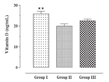
It is still not completely clear whether carbamazepine causes alterations in vitamin D status and in bone metabolism. The objective of this study was to investigate the effects of carbamazepine on serum levels of 25-hydroxyvitamin D and on biomarkers of bone formation and resorption in healthy rats. Levels of calcium, 25-hydroxyvitamin D, parathormone, C-telopeptide, bone specific alkaline phosphatase and osteocalcin were measured in 3 groups of rats consisting of controls (n=10), isotonic saline solution group (n=10) and carbamazepine group (n=10). Mean calcium levels were found to be significantly lower in healthy controls in comparison to isotonic saline solution and carbamazepine groups (10.0±0.24, 10.81±0.16, 10.93±0.22 mg/dL, respectively, p<0.05). Mean levels of 25-hydroxyvitamin D, were found to be significantly higher in control group compared to isotonic saline solution group (25-hydroxyvitamin D; 25.91±1.12, 19.99±0.99 ng/mL, respectively, p<0.01). Mean levels of parathormone and osteocalcin were found to be significantly higher in control group compared to isotonic saline solution group and carbamazepine group. Parathormone levels were measured as 3.46±0.83, 1.08±0.08, 0.94±0.02 pg/mL, respectively (p<0.01). Osteocalcine levels were measured as 1.66±0.001, 1.32±0.002, 1.32±0.001 ng/mL, respectively (p<0.001). A significant difference in terms of mean serum bone specific alkaline phosphatase and C-telopeptide levels among groups was not observed. The main outcome of this prospective study in healthy rats showed no change in biochemical parameters of bone turnover during treatment with carbamazepine.

Downloads

Download data is not yet available.
Effects of carbamazepine on serum parathormone, 25- hydroxyvitamin D, bone specific alkaline phosphatase, C-telopeptide, and osteocalcin levels in healthy rats

Downloads

Additional Files

Published

2012-11-20

How to Cite

1.
Maral Kir H, Garip Şebnem, Şahin D, Öztaş B. Effects of carbamazepine on serum parathormone, 25- hydroxyvitamin D, bone specific alkaline phosphatase, C-telopeptide, and osteocalcin levels in healthy rats. Biomol Biomed [Internet]. 2012Nov.20 [cited 2023Sep.23];12(4):240-4. Available from: https://www.bjbms.org/ojs/index.php/bjbms/article/view/2445

Issue

Section

Biochemistry

INTRODUCTION

Epileptic seizures result from excessive discharge in a population of hyperexcitable neurons [1]. Antiepileptic drugs (AEDs) are increasingly used for the treatment of several non-epileptic neurological conditions and psychiatric disorders [2]. Most of the patients with epilepsy require a long-term and sometimes lifelong therapy with AEDs and, therefore, they are exposed to the potential undesirable metabolic side effects of medical treatment [3]. The reported effects of AEDs on bone include rickets, osteomalacia, osteoporosis, and fractures [4]. Most of the published studies and evidence involve patients receiving AEDs that induce the cytochrome P450 enzyme system (phenobarbital, phenytoin, and carbamazepine) are most commonly associated with abnormalities in bone [4-7]. Markers of bone formation, which include alkaline phosphatase and osteocalcin, have been assessed in patients receiving AEDs [8-10]. Carbamazepine (CBZ), which has been mostly used in epilepsy treatment for 20 years, is also used for the treatment of neuropsychiatric disorders, neuropathy and depression in recent years [11-13]. Up to now, either epileptic animals or patients have been used in the studies. Therefore, it is not clear whether the effects in question are due to carbamazepine treatment only, or whether seizures also contribute to this situation. In these studies the action mechanism of the anti-epileptic drug on bone metabolism can not be detected. In this present study, the effects and action mechanism of carbamazepine on bone metabolism of healthy rats were investigated in order to detect the drug-related side effects only. Thus we measured serum osteocalcin (OC), parathormone (PTH), C-telopeptide (CTX), bone specific alkaline phosphatase (BAP) and 25-OH vitamin D (25-OH-VD) levels in healthy rats receiving carbamazepine and investigated whether side effects on bone are a result of the drug alone.

MATERIALS AND METHODS

Animals

30 male, 6 monthly Wistar rats provided by the Experimental Medical Research Unit (DETAB, Kocaeli University Medical School, Kocaeli) were used in the study. The Kocaeli University Medical School Animal Ethic Committee approved the experimental design. Animals were maintained on a 12-hour light-dark cycle and provided with standart laboratory rat chow and tap water ad libitum. The animals were divided into three groups. Group 1 rats (n=10) were control group. Group 2 rats (n=10) were injected with physiological saline. Group 3 rats (n=10) were injected with carbamazepine.

Procedures

Injections were done between 09:00-11:00 am in all groups. 75 mg/kg carbamazepine in isotonic saline solution not exceeding 1 mL in volume was injected intra-peritoneally once a day over 5 weeks in sterile conditions. According to previous studies carried out with various anti-epileptic drugs, a 5-week anti-epileptic treatment is sufficient for observing the side effects on the bone structure and metabolism of rats [14-16]. Body weight of animals were followed up in the course of 5 week drug therapy. Blood samples obtained from the animals at the end of 5 weeks were centrifuged at 4 °C, 3000 rpm for 15 minutes. Serum samples were stored at -80 °C until biochemical analysis was completed. Serum calcium levels were measured with Abbot Aeroset autoanalyser using Aeroset kit (Abbot Lab. Abbot Park, IL 60064, USA). Serum osteocalcin, parathormone, C-telopeptide, bone specific alkaline phosphatase and 25-OH vitamin D levels were measured with ELISA method using microarray device. OC and PTH levels were measured with DRG ELISA kit (DRG International, Inc. USA), BAP and 25-OH-VD levels were measured with IDS ELISA kit (IDS Ltd. Fountain Hills, USA), whereas CTX levels were measured with Uscn ELISA kit (Uscn Life Science Inc. Wuhan). Analyses of all the samples, standards, and controls were run in duplicate.

Statistical analysis

Statistical analysis was performed by using the SPSS for windows (version 13.0) statistical package (SPSS Inc., Chicago, IL, USA). The values were expressed as mean ±SEM. Differences between the groups were analyzed using the one way ANOVA and Bonferroni multiple comparisons tests. p values of < 0.05 were considered statistically significant.

RESULTS

Mean serum calcium levels were found to be significantly lower in Group 1 compared to Group 2 and 3 (10.0±0.24, 10.81±0.16, 10.93±0.22 mg/dL, respectively, p<0.05) (Table 1), (Figure 1). Mean serum 25-OH-VD levels were found to be significantly higher in Group 1 compared to Group 2 (25.91±1.12, 19.99±0.99 ng/mL, respectively, p<0.01) (Table 1), (Figure 2). PTH levels were found significantly higher in Group 1 compared to Group 2 and 3 (3.46±0.83, 1.08±0.08, 0.94±0.02 pg/mL, respectively, p<0.01) (Table 1), (Figure 3). Mean serum OC levels were significantly higher in Group 1 in comparison to other groups (1.66±0.001, 1.32±0.002, 1.32±0.001 ng/mL, respectively, p<0.001) (Table 1), (Figure 4). A significant difference was not found between groups in terms of mean serum BAP and CTX levels (Table 1). The intra- and inter-assay coefficient of variation for pooled plasma were 4.7 % and 3.5 % for OC, 5.3 % and 4.6 % for 25-OH-VD, 6.5 % and 5.8 % for BAP, 8.3 % and 7.6 % for CTX, 3 % and 5.1 % for PTH, respectively.

TABLE 1: Plasma calcium, 25-OH-VD, PTH, CTX, BAP and OC levels of groups
FIGURE 1: Calcium levels of the three groups. *Significantly lower than those of the Group II and Group III (respective values: p < 0.05, p <0.05).
FIGURE 2: 25-OH vitamine D levels of the three groups. **Significantly higher than those of the Group II (respective values: p < 0.01).
FIGURE 3: Parathormon levels of the three groups. ***Significantly higher than those of the group II and group III (respective values: p < 0.01, p <0.01).
FIGURE 4: Osteocalsin levels of the three groups. ****Significantly higher than those of the group II and group III (respective values: p < 0.001, p <0.001).

DISCUSSION AND CONCLUSION

The main outcome of this prospective study in healthy rats showed no change in biochemical parameters of bone turnover during treatment with CBZ. Bone is a complex dynamic tissue that responds to external and internal forces. Accordingly, body weight, exercise, and calcium homeostasis can alter bone structure and architecture. These changes are complex and involve osteoclast and osteoblast dynamics, vitamin D, calcium, and phosphorous homeostasis as well as connective tissue arrangements [17, 18]. Increased risk for bone disease including changes in bone turnover, osteoporosis, alterations in bone quality, and most importantly fracture. Although carbamazepine is an enzyme inducing AED, data on the issue is not conclusive [8]. In a study by Nissen-Meyer et al. [19], researchers emphasized the importance of using animal models in order to put forward the effects of anti-epileptic drugs on bone. They stated that animal tests were more beneficial for testing anti-epileptic drug-related bone fragility because such studies allow the elimination of possible alterations resulting from life style and genetic and individual disease characteristics. The 1,25 dihydroxy vitamin D (1,25 (OH)2D) has its effect on the classic target organs bone, intestine and kidney and stimulates calcium transport from these organs to the blood. The production of 1,25 (OH)2D is stimulated by PTH. There is a negative feedback through calcium which decreases PTH and a direct negative feedback from 1,25 (OH)2D to PTH [20]. While vitamin D deficiency leads to osteoporosis, vitamin D insufficiency-subsequent form of vitamin D deficiency-leads to osteomalacia [21]. Some studies show that, biochemical abnormalities in patients receiving carbamazepine include hypocalcemia, hypophosphatemia, reduced levels of active vitamin D metabolites, elevated PTH levels, and elevated markers of bone resorption and formation [6, 18, 22, 23, 24, 25]. However, reduction was not detected in serum calcium and 25-OH-VD levels and elevation of PTH levels was not detected in the group receiving anti-epileptic drug in our study. Alkaline phosphatase is the most commonly used marker of bone formation [26-28]. Because serum total alkaline phosphatase is derived from bone, liver, and other sources, it lacks sensitivity and specificity in evaluating bone disease. In reports that measured the isoenzymes, the increase in total alkaline phosphatase was attributed mainly to the bone fraction [4, 27, 28]. ALP increases have been seen in both children and adults receiving AEDs [29-31]. In contrast to the aforementioned studies, a statistically significant elevation was not found in BAP levels in the rats receiving carbamazepine treatment in the present study. Osteocalcin, or bone-gla protein, is a small noncollagenous protein that is specific for bone tissue and dentin and is synthesized predominantly by osteoblasts. High serum levels of osteocalcin with AED treatment are described [3, 6, 29, 32]. Levels of osteocalcin, the marker of bone formation, was also not elevated in the carbamazepine receiving group. In one study, low serum calcium concentrations were considered to be the long term outcome of treatment with anti-epileptic drugs [29]. Studies report that alterations in markers of bone formation and resorption are observed particularly in patients receiving multi-drug therapy [3]. Verrotti et al. [25] reported as the result of the study carried out by administering carbamazepine to epilepsy patients that bone turnover may increase despite normal vitamin D levels. Some another studies reported abnormalities in the bone metabolism of patients treated with AEDs independently of vitamin D deficiency [6, 25, 33, 34]. These data suggest that AEDs might alter the skeleton via other mechanisms, e.g. direct effects on bone cells, intestinal calcium transport and resistance to parathyroid hormone, rather than monocausally by decreased serum 25-OHVD [17, 35, 36]. In some recent studies, genetic variation was shown to have some effects as reducing bone mineral density and active vitamin D metabolism in epilepsy patients receiving antiepileptic drugs [37, 38]. In conclusion, a statistically significant alteration has not been observed in biomarkers of bone formation and resorption in healthy rats receiving carbamazepine. Studying the effects of carbamazepine in healthy animals enabled elimination of some confounding factors affecting bone metabolism (e.g. renal disease, hyperparathyroidism, cancer, gastrointestinal disorders, liver insufficiency, diabetes mellitus, drug use as corticosteroids and diet) [3]. However based on the studies reporting that side effects concerning bone may arise in the long term, we consider that animal models studying long term effects of carbamazepine could be built up in future studies.

DECLARATION OF INTEREST

The authors declare that they have no conflict of interest related to the publication of this manuscript.

REFERENCES

  1. , (). Cellular biology of epileptogenesis. Lancet Neurol.
  2. , (). Antiepileptic drugs: indications other than epilepsy. Epileptic Disord.
  3. , , , , , , , , , , , (). Effects of antiepileptic drug therapy on vitamin D status and biochemical markers of bone turnover in children with epilepsy. Eur J Pediatr.
  4. , , (). Bone disease associated with antiepileptic drugs. Cleve Clin J Med.
  5. , , , , , (). Effect of carbamazepine and valproate on bone mineral density. J Pediatr.
  6. , , , (). Increased bone turnover in epileptic patients treated with carbamazepine. Ann Neurol.
  7. (). The Association Between Antiepileptic Drugs and Bone Disease. Epilepsy Curr.
  8. (). Bone health in people with epilepsy: Is it impaired and what are the risk factors?. Seizure.
  9. , (). Effect of topiramate on bone resorption in adults. Am Epilepsy Soc Abstr.
  10. , , , , (). Measurement of bone mineral density in patients on levetiracetam monotherapy. Am Epilepsy Soc Abstr.
  11. , , , , (). Comparison of carbamazepine and lithium in treatment of bipolar disorder: a systematic review of randomized controlled trials. Hum Psychopharmacol.
  12. (). Sydenham's chorea, and its complications affecting the nervous system. Brain Dev.
  13. , , , , , , , , (). Chronic coadministration of carbamazepine together with imipramine produces antidepressant-like effects in an ACTH-induced animal model of treatment-resistant depression: involvement of 5-HT(2A) receptors?. Pharmacol Biochem Behav.
  14. , , , , , , , (). Effects of chronic administration of zonisamide, an antiepileptic drug, on bone mineral density and their prevention with alfacalcidol in growing rats. J Pharmacol Sci.
  15. , , , (). Effects of phenytoin and/or vitamin K2 (menatetrenone) on bone mineral density in the tibiae of growing rats. Life Sci.
  16. , , , , , (). Phenytoin-induced bone loss and its prevention with alfacalcidol or calcitriol in growing rats. Calcif Tissue Int.
  17. (). Pathophysiology of bone loss in patients receiving anticonvulsant therapy. Epilepsy Behav.
  18. , (). Bone consequences of epilepsy and antiepileptic medications. Semin Pediatr Neurol.
  19. , , , , , (). How can antiepileptic drugs affect bone mass, structure and metabolism? Lessons from animal studies. Seizure.
  20. (). Vitamin D physiology. Prog Biophys Mol Biol.
  21. (). Diagnosis of disorders of vitamin D-metabolism and osteomalacia. Clin Lab.
  22. , (). Calcium metabolism in patients with epilepsy. Br Med J.
  23. , , , , , (). A comparative study of the relative influence of different anticonvulsant drugs, UV exposure and diet on vitamin D and calcium metabolism in out-patients with epilepsy. Q J Med.
  24. , , , , , (). Effect of antiepileptic drugs on bone density in ambulatory patients. Neurology.
  25. , , , , (). Increased bone turnover in prepubertal, pubertal, and postpubertal patients receiving carbamazepine. Epilepsia. 58(9): 1348-1353
  26. , (). Disturbance of calcium metabolism by anticonvulsant drugs. Br Med J.
  27. , , (). Isoenzymes of alkaline phosphatase in epileptic patients receiving carbamazepine monotherapy. J Clin Pathol.
  28. , , , , (). Bone metabolism alteration on antiepileptic drug therapy. Indian J Pediatr.
  29. , , , , , , , , (). Bone mass and turnover in women with epilepsy on antiepileptic drug monotherapy. Ann Neurol.
  30. , , , , , , (). Effect of carbamezapine and valproic acid on bone mineral density, IGF-I and IGFBP-3. J Pediatr Endocrinol Metab.
  31. , , , (). Evaluation of bone mineral density in children receiving carbamazepine or valproate monotherapy. Acta Paediatr Taiwan.
  32. , , , , , , , (). Decreased bone mass and increased bone turnover with valproate therapy in adults with epilepsy. Neurology.
  33. , , , , , , (). Antiepileptic drug-induced bone loss in young male patients who have seizures. Arch Neurol.
  34. , , , (). Normal vitamin D and low free estradiol levels in women on enzyme-inducing antiepileptic drugs. Epilepsy Behav.
  35. , , , , (). Long-term anticonvulsant therapy leads to low bone mineral density--evidence for direct drug effects of phenytoin and carbamaze-pine on human osteoblast-like cells. Exp Clin Endocrinol Diabetes.
  36. , , , , , , , (). Bone mineral density measured by dual-energy x-ray absorptiometry and novel markers of bone formation and resorption in patients on antiepileptic drugs. J Bone Miner Res.
  37. , , , , , , , , , (). BsmI vitamin D receptor's polymorphism and bone mineral density in men and premenopausal women on long-term antiepileptic therapy. Eur J Neurol.
  38. , , , , , , (). Steroid and xenobiotic receptor and vitamin D receptor crosstalk mediates CYP24 expression and drug-induced osteomalacia. J Clin Invest.