Anti-fibrotic effect of Aliskiren in rats with deoxycorticosterone induced myocardial fibrosis and its potential mechanism

Authors

  • Likun Ma Department of Cardiology, Anhui Provincial Hospital
  • Jinsheng Hua Department of Cardiology, Anhui Provincial Hospital
  • Lifeng He Department of Cardiology, Anhui Provincial Hospital
  • Qian Li Department of Cardiology, Anhui Provincial Hospital
  • Junling Zhou Department of Cardiology, Anhui Provincial Hospital
  • Jiangtao Yu Department of Cardiology, Zentralklinik Bad Berka GmbH

DOI:

https://doi.org/10.17305/bjbms.2012.2498

Keywords:

Aliskiren, deoxycorticosterone, myocardial fibrosis, renin- angiotensin - aldosterone system

Abstract

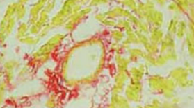
The objective of our study was to investigate the effect of Aliskiren, a renin inhibitor, on the deoxycorticosterone (DOCA) induced myocardial fibrosis in a rat model and its underlying mechanism. A total of 45 Sprague-Dawley (SD) rats underwent right nephrectomy and were randomly assigned into 3 groups: control group (CON group: silicone tube was embedded subcutaneously); DOCA treated group (DOC group: 200 mg of DOCA was subcutaneously administered); DOCA and Aliskiren (ALI) treated group (ALI group: 200 mg of DOCA and 50 mg/kg/d ALI were subcutaneously and intragastrically given, respectively). Treatment was done for 4 weeks. Sirius red staining was employed to detect the expression of myocardial collagen, and the myocardial collagen volume fraction (CVF) and perivascular collagen volume area (PVCA) were calculated. Radioimmunoassay was carried out to measure the renin activity (RA) and content of angiotensin II (Ang II) in the plasma and ventricle. Western blot assay was done to detect the expressions of extracellular signal-regulated kinase 1/2 (ERK1/2), phosphorylated ERK1/2 (PERK1/2) and matrix metalloproteinase 9 (MMP-9). In the DOC group and ALI group, the CVF and PVCA were significantly increased; the RA and Ang II levels in the plasma and ventricle were remarkably lowered when compared with the CON group. The RA and Ang II levels in the ventricle of the ALI group were significantly lower than those in the DOC group. Moreover, the expressions of ERK1/2, PERK1/2 and MMP9 were the lowest in the CON group, but those in the ALI group were significantly reduced as compared to the DOC group. ALI can inhibit the DOCA induced myocardial fibrosis independent of its pressure-lowing effect, which may be related to the suppression of RA and Ang II production, inhibition of ERK1/2 phosphorylation and MMP9 expression in the heart.

Downloads

Download data is not yet available.
Anti-fibrotic effect of Aliskiren in rats with deoxycorticosterone induced myocardial fibrosis and its potential mechanism

Downloads

Additional Files

Published

2012-05-20

How to Cite

1.
Ma L, Hua J, He L, Li Q, Zhou J, Yu J. Anti-fibrotic effect of Aliskiren in rats with deoxycorticosterone induced myocardial fibrosis and its potential mechanism. Biomol Biomed [Internet]. 2012May20 [cited 2023Sep.23];12(2):69-73. Available from: https://www.bjbms.org/ojs/index.php/bjbms/article/view/2498

Issue

Section

Pharmacology

INTRODUCTION

Myocardial fibrosis is a common pathological feature shared by several heart diseases at the end stage. To prevent or even reverse myocardial fibrosis has been a key goal in the prevention and treatment of severe cardiovascular events including heart failure, arrhythmia and sudden cardiac death. Studies have demonstrated that renin - angiotensin - aldosterone system (RAAS) plays important roles in the regulation of myocardial collagen metabolism and the occurrence of myocardial fibrosis [1, 2]. Evidence shows Aliskiren (ALI), a new rennin inhibitor, can not only lower the blood pressure, but improve the myocardial fibrosis and subsequent remodeling via its anti-inflammatory and anti-oxidative effects [3, 4]. Currently, the anti-fibrotic effect of ALI and its potential mechanism are less studied. The present study aimed to investigate the cardioprotective effect of ALI on the deoxycorticosterone (DOCA) induced myocardial fibrosis and its potential mechanism in a rat model.

MATERIALS AND METHODS

Reagents

ALI (Novartis, Switzerland), DOCA (Sigma, USA), primary antibodies against β-actin, extracellular signal kinase 1/2 (ERK1/2), phosphorylated ERK1/2 (PERK1/2) and metalloproteinase-9 (MMP-9) (Santa Cruz, USA), secondary antibodies, two - quinolinecarboxylic acid (BCA) (Beijing Zhongshan Golden-Bridge Biotech, China), electrochemiluminescence (ECL) kit (Pierce), Sirius red (Beijing Haide Biotech, China) and radioimmunoassay kit (Beijing Yuanzi Biotech, China) were used in the present study.

Grouping and modelling

A total of 45 Sprague-Dawley (SD) male rats weighing 187~245 g were purchased from the Animal Center of Anhui Medical University. The investigation conforms with the Guide for Care and Use of Laboratory Animals published by the US National Institutes of Health (NIH Publication, 8th edition). Animals were randomly assigned into 3 groups (n=15 per group): control (CON) group, DOCA (DOC) group and ALI (ALI) group. Following anesthesia with intraperitoneal 10% chloral hydrate (300 mg/kg), right nephrectomy was done in these animals. One week after surgery, animals were given access to 1% NaCl and received following treatments. In the CON group, silicone tube was embedded subcutaneously in the left lower abdomen. In the DOC group, 200 mg of DOCA were embedded in the left lower abdomen. In the ALI group, 200 mg of DOCA were embedded in the left lower abdomen and animals were intragastrically treated with ALI at 50 mg/kg/d for 4 weeks. In addition, animals in the CON group and DOC group were also intragastrically treated with normal saline for consecutive 4 weeks.

Measurement of blood pressure and sample collection

At the end of treatment, rats were intraperitoneally anesthetized with 10% chloral hydrate followed by cannulation of right carotid artery for the measurement of mean arterial blood pressure (MABP) using a multi-channel physiological recorder. Then, 3 ml of blood were collected into the anti-coagulated tube and rats were sacrificed by exsanguinations. Thoracotomy was performed and the heart collected. The atrium, major vessels and connective tissues were removed and the left ventricle was divided into two: one was fixed in 4% paraformaldehyde followed by processing for histological staining for collagen; the other was stored at -80°C for the detection of protein expression by western blot assay. Blood was centrifuged at 1000 rpm/min for 5 min and the plasma was collected and stored at -20°C for use.

Detection of myocardial fibrosis

The heart tissues were embedded in the paraffin and cut into sections followed by Sirius red staining for collagen. A total of 8 fields without blood vessels were randomly selected from each section and representative photographs were captured with Nikon camera followed by analysis using Image-Pro plus 6.0 image analysis system. The collagen area and total area of each field were measured, and myocardial collagen volume fraction (CVF) was calculated as collagen area / total area. The CVFs from 8 fields were averaged and used as the final CVF of this sample. In addition, 4 fields with small blood vessels at cross section were randomly collected, and the perivascular collagen volume area (PVCA) and lumen area (LA) determined. The PVCA was normalized by the LA (PVCA/LA). The PVCAs from 4 fields were averaged and used as the final PVCA of this sample.

Detection of renin and angiotensin II by radioimmunoassay

Briefly, in each group, 100 mg of ventricle were homogenized, and then the homogenate and plasma were independently divided into two: one was kept at 37°C for 1 h which may facilitate the binding of renin (RA) to angiotensinogen producing angiotensin I (Ang I) (experiment group); the other was stored at 4°C serving as controls. The Ang I level in the experiment group and control group was measured by radioimmunoassay according to the manufacturer’s instructions. The difference in Ang I between experiment group and control group was normalized by the time of incubation as the production rate of Ang I. The production rate of Ang I in unit time was defined as the renin activity of this sample and the unit was ng /ml/h. According to the manufacturer’s instructions, the Ang II level in the plasma and ventricle was also measured and its unit was pg/ml.

Detection of ERK1/2, PERK1/2 and MMP9 expressions in the ventricle by Western blot assay

The ventricle was homogenized and total protein extracted followed by detection of protein concentration. Then, the protein concentration of different sample was adjusted to the same level and 75 μg of proteins were subjected to polyacrylamide gel electrophoresis. The protein was transferred onto nitrocellulose membrane. Following washing and blocking overnight, the membrane was treated with primary antibody (1:1000) at room temperature for 2 h under continuous shaking. After washing, the membrane was incubated with secondary antibody (1:40000) at room temperature for 1 h under continuous shaking. Visualization was done using ECL kit and X-ray film was obtained. The bands were scanned using Bio-Rad image system and the optical density (OD) was determined followed by analysis with Quantity-one. The OD of target genes was normalized by that of p-actin as the relative expression of target genes.

Statistical analysis

Statistical analysis was performed using SPSS version 15.0. Qualitative data were expressed as mean ± standard deviation (X±s). Means among groups were compared with one way analysis of variance, and comparisons of rate were done with chi square test. A value of p<0.05 was considered statistically significant.

RESULTS

MABP in different groups

The MABP in the DOC group and ALI group was 23.73±1.50 kPa and 22.85±1.21 kPa, respectively, which were markedly higher than that in the CON group (15.87±0.67 kPa) (p<0.05). However, there was no significantly difference between DOC group and ALI group (p>0.05).

Collagen content in the ventricle of different groups

Following Sirius red staining, the collagens were scarlet and non-collagen tissues yellow (Figure 1. and 2.). The CVF and PVCA in different groups are shown in Table 1. When compared with the control group, the CVF and PVCA in the DOC group and ALI group were markedly increased (p<0.05 and p<0.01, respectively). However, the CVF and PVCA in the ALI group was dramatically lower than those in the DOC group (p<0.05 and p<0.01, respectively). This finding implies ALI improves the DOC induced myocardial fibrosis.

FIGURE 1: Staining of collagen in the ventricle (×400; A: control; B: DOCA; C: ALI)
FIGURE 2: Staining of blood vessels in the ventricle (×400; A: control; B: DOCA; C: ALI)
TABLE 1: CVF and PVCA in different groups (X±s)

RA and Ang II levels in the plasma and ventricle

The RA level of the plasma and ventricle of DOC group and ALI group was significantly lower than those in the CON group (p<0.05 or p<0.01). Significant difference in the RA level was found in the ventricle between DOC group and ALI group (p<0.05), but marked difference was absent in the RA level of the plasma (p>0.05). The Ang II level of the plasma and ventricle of the DOC group and ALI group was also significantly decreased when compared with CON group (p<0.05). Significant difference in the Ang II level was observed between ALI group and DOC in the ventricle (p<0.05) but not in the plasma (p>0.05)(Table 2).

TABLE 2: RA and Ang II levels in the plasma and ventricle of different groups (X±s)

Protein expressions of ERK1/2, PERK1/2 and MMP-9 in the ventricle

The protein expressions of ERK1/2, PERK1/2 and MMP-9 are shown in Figure 3. Analysis showed the expressions of ERK1/2, PERK1/2 and MMP-9 in the CON group were the lowest, followed by ALI group, and those in the DOC group the highest. Significant difference in the expressions of ERK1/2, PERK1/2 and MMP-9 was found between any two groups (p<0.01). Moreover, the expressions of ERK1/2, PERK1/2 and MMP-9 in the ALI group were significantly higher than those in the CON (p<0.05) but lower than those in the DOC group (p<0.05 or p<0.01).

FIGURE 3: Protein expressions of ERK1/2, PERK1/2 and MMP-9 in Western blot assay (CON: control group; DOC: DOCA group; ALI: ALI group. *p<0.05 and **p<0.01 vs control group; #p<0.05 and # #p<0.01 vs DOCA group).

DISCUSSION

In the present study, the DOCA induced myocardial fibrosis rat model was employed and the anti-fibrotic effect of ALI was investigated. Our findings revealed ALI could improve the myocardial fibrosis demonstrated by Sirius red staining, reduce the RA and Ang II levels in the ventricle and the expressions of ERK1/2, PERK1/2 and MMP9 as compared to the DOCA treated animals. Myocardial fibrosis refers to the aberrant deposition of extracellular matrix (ECM) in the heart, and characterized by increase of collagen in the interstitium, imbalance and irregular arrangement of different types of collagen. The imbalance between the synthesis and degradation of collagen is a major cause of myocardial fibrosis. Under the pathological conditions, the proliferation and phenotype of cardiac fibroblasts change and these cells produce a large amount of collagens resulting in imbalance between type I and type II collagens and subsequent deposition of collagens between myocardial cells. MMPs play critical role in the myocardial fibrosis through affecting the degradation of ECM. MMP9 can degrade normal collagens such as gelatin, elastin, collagen type IV collagen, type V collagen and mucin but has no influence on abnormal collagens [5]. In the myocardial fibrosis, the myocardial interstitium is occupied by abnormal collagens (mainly type I and III collagens). Under pathological conditions, the MMP-9 expression is increased and then degrades the normal collagens. Thus, the myocardial interstitium between myocardial cells loses and a large amount of abnormal collagens generated. Westermann et al. [4] investigated the effect of ALI on the rat myocardial infarction. Their results showed ALI could reduce the deposition of collagen in the infarction region and subsequent myocardial fibrosis via reducing the MMP-9 activity. Our results also revealed the MMP-9 expression was markedly elevated in rats with DOCA induced myocardial fibrosis accompanied by increased extracellular deposition of collagen. However, following ALI treatment, the MMP-9 expression and deposition of collagen were markedly decreased and myocardial fibrosis improved. In addition, studies have shown that RAAS plays an important role in the regulation of collagen metabolism in the heart and blood vessels, and Ang II and aldosterone are two major effector proteins [6, 7]. There is evidence that angiotensin converting enzyme inhibitors (ACEI), angiotensin receptor blockers (ARB) and aldosterone receptor antagonists can significantly improve the fibrosis [2]. However, long-term application of ACEI and ARB may lead to the Ang II escape [8]. It has been confirmed that Ang II can be synthesized via both ACE-dependent and ACE-independent pathways. Continuous use of ACEI and ARB may cause accumulation of Ang I and activate ACE-independent pathways resulting in increase in Ang II. Of note, 60-70% of Ang II in the tissues is synthesized via ACE-independent pathways [9]. ALI, a renin inhibitor, acts on the first rate-limiting step of RAAS chain and blocks the activation of RAAS leading to the decrease in Ang II and aldosterone production [10]. Thus, theoretically, ALI has potent anti-fibroric effect on myocardial fibrosis. Singh et al. [11] showed, in streptomycin induced hyperglycemia rats, the ALI was superior to benazepril and losartan in improving myocardial fibrosis and apoptosis of myocardial cells. In the present study, the blood pressure was not dramatically lowered following administration of ALI, but the DOCA induced myocardial fibrosis was obviously improved. Studies showed the ventricular RAAS plays a more critical role in the myocardial fibrosis than circulation RAAS does [9, 12]. Our study further confirmed the RA and Ang II in the circulation were largely unchanged followed ALI treatment, but those in the heart were dramatically decreased. These findings suggest the anti-fibrotic effect of ALI is related to the suppression of local Ang II production. Currently, the mechanisms underlying the anti-fibrotic effect of ALI are not completely clear. Our results showed the anti-fibrotic effect of ALI may be attributed to the suppression of phosphorylation of ERK1/2 signaling pathway. In recent years, studies show the Ang II/angiotensin receptor 1 (AT1R) mediated myocardial fibrosis is related to the ERK1/2 signaling pathway [13]. The binding of Ang II to AT1R can activate the phospholipase C on the cell membrane resulting in the hydrolysis of phosphatidylinositol phosphate and production of diacylglycerol. The later promotes the release of Ca2+ in the sarcoplasmic reticulum and endoplasmic reticulum. Ca2+ can act as a second messenger to activate ERK1/2 signaling pathway. The activation of ERK1/2 signaling pathway may facilitate the transcription of early response genes (such as c-fos) in the cardiac fibroblasts, skeletal muscle a-actin gene, β-myosin heavy chain gene and embryonic contractile protein gene and promote the expression of growth factors in fibroblasts. In addition, a large amount of ECM and collagen are produced, the proliferation of fibroblasts promoted and expressions of Iα1 and IIIα1 increased resulting in myocardial fibrosis [14, 15]. Our results showed the expressions of ERK1/2 and PERK1/2 in the ALI group were markedly decreased when compared with the DOC group. We speculate that ALI can decrease the production of Ang II and local RA to reduce the Ang II/AT1R induced activation of ERK1/2, which finally exerts the anti-fibrotic effects.

CONCLUSION

Evidence shows that RAAS plays important roles in the occurrence of myocardial fibrosis and ALI has anti-fibrotic effect via its anti-inflammatory and anti-oxidative effects. The present study demonstrates that ALI as an inhibitor of renin can improve the DOCA induced myocardial fibrosis, which may be attributed to the suppression of myocardial RAAS, decrease in Ang II level and inhibition of phosphorylation of ERK1/2 signaling pathway and MMP-9 expression. However, more studies are required to confirm our results.

Acknowledgements

ACKNOWLEDGEMENTS

This study was supported by the International Cooperation Projects of Department of Science & Technology of Anhui Province (2009) (No: 0908070342)

DECLARATION OF INTEREST

We declare no conflict of interest

REFERENCES

  1. , , , (). Renin-angiotensin-aldosterone system blockade for cardiovascular diseases: current status. Br J Pharmacol.
  2. , , (). Cardiovascular effects of inhibition of renin-angiotensin-aldosterone system components in hypertensive rats given salt excess. Am J Physiol Heart Circ Physiol.
  3. , , , , , (). Aliskiren, a novel orally effective renin Inhibitor, provides dose-dependent antihypertensive efficacy and placebo-like tolerability in hypertensive patients. Circulation.
  4. , , , , , (). Renin inhibition improves cardiac function and remodeling after myocardial infarction independent of blood pressure. Hyper-tension.
  5. , , , , , (). Collagen degradation in a murine myocarditis model: relevance of matrix metalloproteinase in association with inflammatory induction. Cardiovasc Res.
  6. , , , , (). Aldosterone mediats angiotensin II-induced interstitial cardiac fibrosis via a Nox2-containing NADPH oxidase. The FASEB Journal.
  7. (). Aldosterone and myocardial fibrosis in heart failure. Herz.
  8. , , , , (). Angiotensin II reactivation and aldosterone escape phenomena in renin-angiotensin-aldosterone system blockade: is oral renin inhibition the solution?. Expert Opin Pharmacother.
  9. , , , , , (). Local angiotensin II aggravates cardiac remodeling in hypertension. Am J Physiol Heart Circ Physiol.
  10. , , , (). Angiotensin II suppression in humans by the orally active renin inhibitor aliskiren (SPP100): comparison with enalapril. Hypertension.
  11. , , , , (). Intracellular angiotensin II production in diabetic rats is correlated with cardiomyocyte apoptosis, oxidative stress, and cardiac fibrosis. Diabetes.
  12. , , , , , (). Angiotensin-converting-enzyme 2 suppresses pathological hypertrophy, myocardial fibrosis, and cardiac dysfunction. Circulation.
  13. , , , (). Angiotensin II- induced extracellular signal-regulated kinase 1/2 activation is mediated by protein kinase Cdelta and intracellular calcium in adult rat cardiac fibroblasts. Hypertension.
  14. , , , , , (). Mitogen-activated protein kinases ERK 1/2- and p38-GATA4 pathways mediate the Ang II-induced activation of FGF2 gene in neonatal rat cardiomyocytes. Biochem Pharmacol.
  15. , (). Angiotensin II cell signaling: physiological and pathological effects in the cardiovascular system. Am J Physiol Cell Physiol.