The hepatic protective mechanism of Ginkgo biloba extract in rats with obstructive jaundice

Authors

  • Ming-Zhe Weng Department of General Surgery, Xinhua Hospital, Shanghai Jiaotong University School of Medicine
  • Xian-Ping Zhou Department of General Surgery, Xinhua Hospital, Shanghai Jiaotong University School of Medicine
  • Jian-Guang Jia Department of General Surgery, Xinhua Hospital, Shanghai Jiaotong University School of Medicine
  • Jing Ding Department of General Surgery, Xinhua Hospital, Shanghai Jiaotong University School of Medicine
  • Cui-Fu Fang Department of General Surgery, Xinhua Hospital, Shanghai Jiaotong University School of Medicine
  • Yi-Yu Qin Department of General Surgery, Xinhua Hospital, Shanghai Jiaotong University School of Medicine
  • Shao-Fu Tao Department of General Surgery, Xinhua Hospital, Shanghai Jiaotong University School of Medicine
  • Long-Hua Rao Department of General Surgery, Xinhua Hospital, Shanghai Jiaotong University School of Medicine
  • Ji-Yu Li Department of General Surgery, Xinhua Hospital, Shanghai Jiaotong University School of Medicine
  • Zhi-Wei Quan Department of General Surgery, Xinhua Hospital, Shanghai Jiaotong University School of Medicine

DOI:

https://doi.org/10.17305/bjbms.2011.2547

Keywords:

cholestasis, hepatic injury, endothelin 1, nitric oxide, ginkgo biloba extract

Abstract

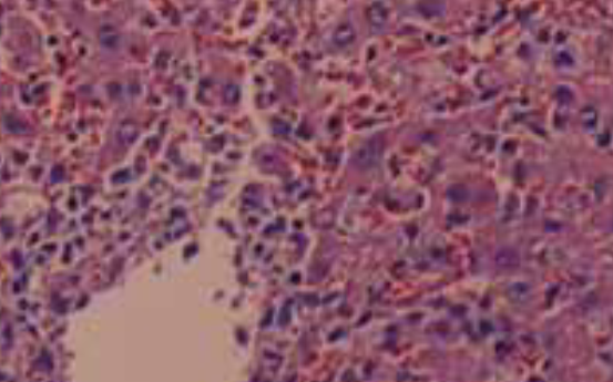
The objective of our study was to examine the hepatic protective mechanism of Ginkgo biloba extract (GBE) in rats with obstructive jaundice (OJ). Twenty rats underwent bile duct ligation and received daily intraperitoneal injections of either control saline or Ginkgo biloba extract for 14 days. Ten sham-operated rats had their bile duct exposed but not ligated or sectioned. Serum alanine transaminase (ALT) was analyzed for liver function tests and liver damage was further assessed by histologic examination. The levels of endothelin 1 (ET-1) and nitric oxide (NO) in blood and liver homogenate were measured. The serum alanine transaminase was elevated in the bile duct ligation rats (BDL rats); GBE could significantly lower serum transaminase level and ameliorate liver histological damage. ET-1 and NO levels in both plasma and liver tissue were also elevated in common bile duct (CBD)-ligated rats, but this increase was significantly decreased by GBE treatment. Moreover, the degree of liver damage severity positively correlates with high levels of ET-1 and NO. GBE mediated the liver protective effect at least in part by suppressing overproduction of ET-1and NO and restoring a proper balance between ET-1and NO to some extent.

Downloads

Download data is not yet available.
The hepatic protective mechanism of Ginkgo biloba extract in rats with obstructive jaundice

Downloads

Additional Files

Published

2011-11-20

How to Cite

1.
Weng M-Z, Zhou X-P, Jia J-G, Ding J, Fang C-F, Qin Y-Y, Tao S-F, Rao L-H, Li J-Y, Quan Z-W. The hepatic protective mechanism of Ginkgo biloba extract in rats with obstructive jaundice. Biomol Biomed [Internet]. 2011Nov.20 [cited 2023Dec.1];11(4):209-13. Available from: https://www.bjbms.org/ojs/index.php/bjbms/article/view/2547

Issue

Section

Pharmacology

INTRODUCTION

Obstructive jaundice (OJ) is characterized by an interruption to the drainage of bile in the biliary system, resulting in intracellular accumulation of bilirubin, bile acids and cholesterol. The biliary obstruction causes cholestatic liver injury, and the absence of bile in the intestine facilitates bacterial translocation across the intestinal mucosa, which, in turn, may cause sepsis and further liver injury [1]. The main treatment for obstructive jaundice includes surgical removal of obstruction and endoprosthesis introduced either percutaneously or endoscopically. This mechanism of liver injury during obstructive jaundice is involved in chemical mediators such as superoxide, nitric oxide (NO), endothelin 1 (ET-1) and cytokines [2, 3, 4, 5]. Among these cytokines, ET-1 and NO have been considered to be the main effectors in liver injury [3, 4, 5]. Obviously, it is important to reduce the release of ET-1and NO in the perioperative period in order to prevent or minimize the related complications.

Standardized Ginkgo biloba extract (GBE) derived from leaves and nuts of Ginkgo tree is a valuable therapeutic drug for a variety of disorders. A recent study showed that treatment significantly reduced pathological changes in liver and terminal ileum in obstructive jaundice [6]. However, currently the mechanism of action of GBE in obstructive jaundice is not clear. A number of studies provided evidence for a role of GBE in the regulation of production of NO and ET-1 [7, 8, 9, 10]. Since the ET-1and NO appears to be more important in obstructive jaundice, we postulate that GBE can exert a protective effect on liver injury induced by obstructive jaundice through the regulation of production of NO and ET-1. Thus, the present study was undertaken to verify the above hypothesis and elucidate GBE protective mechanisms related to liver injury induced by obstructive jaundice. To our knowledge, this is the first study to report GBE liver protective mechanism in obstructive jaundice.

MATERIAL AND METHODS

Drugs and animal models

GBE injection was obtained from Taiwan Chi Sheng Chemical Corporation with dosage of 17.5 mg/5 ml. All animal experiments were conducted following guidelines set forth in the National Institutes of Health Guide for the Care and Use of Laboratory Animals. Adult Wistar rats of either sex, weighing 330 to 370g were kept in a temperature-controlled environment, and were fed standard chow and water. Thirty Wistar rats were randomly divided into three groups, each involving 10 rats, and sex ratio in each group was the same (Male/Female 1:1). Sham-Operated group (SO group): The bile duct was mobilized but not ligated; Obstructive Jaundice group (OJ group): Ligation of the bile duct + injection of saline solution; Ginkgo biloba extract group (GBE group): Ligation of the bile duct + injection of Ginkgo biloba extract (50 mg//kg/day from the 1th to the 14th postoperative days). The rats were sacrificed by cervical dislocation 14 days later, and venous blood was collected and livers were harvested for hematoxylin/eosin staining.

Measurement of biochemical parameters

Plasma and tissue ET-1 concentrations were measured via radioimmunoassay (RIA; Phoenix Pharmaceuticals, Mountain View, CA). Plasma and tissue NO concentrations were measured via nitrate reductase assay (RIA; Phoenix Pharmaceuticals, Mountain View, CA). Serum ALT levels were assessed by L-glutamic acid colorimetric method using commercial kits (Nanjing Jiancheng Bio Company, C009, Nanjing, China). Serum total bilirubin level was assayed caffeine/sodium assay. All experimental procedures were carried out according to the manufacturer’s instructions.

Liver histological examination

Serially harvested liver specimens were fixed in 10% neutral-buffered formalin and embedded in paraffin. Sections were made at 5-mm and stained with hematoxylin and eosin (H&E).

Statistical analysis

All data were expressed as the mean ± SD. The differences among the groups were evaluated by using one-way analysis of variance followed by post-hoc test (Tukey’s HSD test). p values of less than 0.05 were considered as significant. The data were analyzed by statistical software (SPSS for Windows 10.0; SPSS, Chicago, Illinois, USA).

RESULTS

Sham group rats were agile, responsive to painful stimuli. There was no change in urine color in Sham group. All CBD-ligated rats were slightly jaundiced third days postoperative and profoundly jaundiced on the seventh day, and they had poor appetite, lost weight and listlessness. The rats in GBE group also demonstrated these symptoms above, such as poor appetite, lost weight and listlessness, etc. However, compared with the OJ group, they had less severe symptoms above. Although the Child-Pugh classification can be used clinically to assess patients’ general condition liver injury related, it has not been applied to animal studies. Therefore, the preliminary qualitative assessments of general condition of experimental animals were performed in our experiment. The levels of ALT and total bilirubin in the OJ groups and GBE group were significantly higher than that of the sham-operated group (Figure 1). But plasma ALT level in GBE group was found to be significantly reduced compared to OJ group (Figure 1 B), and there was no difference statistically significant in plasma total bilirubin level between OJ groups and GBE group (Figure 1 A). No histological damage was seen in the sham-operated group (Figure 2 A). Whereas marked destruction of the liver tissue with broad areas of necrosis was observed in OJ group (Figure 2 B). While liver injury was moderate in GBE group as compared to OJ group. Treatment with GBE clearly reduced this cholestatic liver damage (Figure 2 C). The plasma and tissue levels of ET-1 in OJ and GBE group increased compared with the Sham-Operated group, but its levels in the GBE group were significantly lower than in the OJ group (Figure 3 A). The plasma and tissue levels of NO were also measured. The levels of NO in blood and liver tissue in OJ group and GBE group increased significantly, compared with SO group, but the GBE group had significantly lower plasma and tissue NO levels than the OJ group (Figure 3 B). Together, as was shown in these data, ET-1 and NO concentrations in plasma and tissue were correlated to liver injury indicators (ALT, pathological index). The higher the concentration of ET -1and NO, the more severe liver damage, which suggested that liver damage severity correlated positively with high level of ET-1 and NO in plasma and tissue. In addition, a significant change was observed in ET-1/NO in Plasma and liver tissue. In sham group, ET-1/NO in Plasma and liver tissue was 0.78±0.02 and 0.49±0.09 respectively, and the counterpart in OJ group was 1.13±0.11 and 0.73±0.04 respectively, while the counterpart in GBE group was 0.80±0.06 and 0.56±0.03 respectively. It suggested that a proper balance between ET-1 and NO was restored to some extent in the GBE group.

FIGURE 1: The serum total bilirubin concentrations in 3 groups (A). The serum ALT concentrations in 3 groups (B). * p<0.01 vs SO group, Δ p<0.05 vs OJ group.
FIGURE 2: The haematoxylin and eosin staining of sections liver tissues. Normal hepatic architecture and lobular structure was observed in sham-operated control rats (A). Whereas liver section of OJ group rats showed severe destruction of the liver with diffuse severe bile duct hyperplasia, portal periportal necrotic lesions and loss of hepatic structure in periportal areas (B). In GBE group, moderate damage was seen with mild bile duct proliferation, minimal disorganization of the hepatocytes plates and rare hepatocytes necrosis(C). Original magnification: 100× (A, B, C).
FIGURE 3: The plasma and liver tissue concentrations of ET-1 in 3 groups (A). The plasma and liver tissue concentrations of NO in 3 groups (B). * p<0.01 vs SO group, Δ p<0.05 vs OJ group.

DISCUSSION

Hepatic injury is one of the main consequences of biliary obstruction, which markedly increases patient morbidity and mortality. Thus, novel treatment options are needed to reduce cholestasis-induced liver damage. The present study demonstrates that GBE treatment is effective in protecting rats from liver injury due to obstructive jaundice. GBE treatment markedly improved the CBD-ligated rats’ general condition, such as appetite, weight and mental state, etc. Moreover, BDL-induced increase in serum ALT, as assessed by liver function, was also reduced by GBE treatment. A primary consideration in the assessment of the efficacy of a potential therapeutic agent for hepatic injury is its effect on liver histology. Those livers from disease control (OJ group) had broad areas of necrosis; GBE group had remarkably improvement in pathological damage compared to OJ group. These results demonstrate that GBE effectively reduced the BDL-induced liver injury. In this preliminary study, we further examined the therapeutic mechanism of GBE. ET-1 is a 21 amino acid peptide with a powerful vasoconstrictor action that is involved in the pathogenesis of several hepato-biliary diseases, such as portal hypertension, liver failure and hepatopulmonary syndrome [11, 12]. High concentrations of ET-1 can decrease hepatic tissue oxygenation associated with sinusoidal vasoconstriction, and infusion of ET-1 into the portal vein markedly reduces sinusoidal blood flow, which subsequently results in hepatocellular damage [13]. Ruetten et al. [14] observed that the selective ETB receptor antagonist attenuated the degree of hepatocellular injury and dysfunction caused by endotoxin. Therefore, ET-1 has been considered to be one of the main effectors in the process of obstructive jaundice [3, 4, 11]. NO, a strong vasodilator is generated by all major cell type of liver, including hepatocytes, Kupffer, stellate, and sinusoidal endothelial cells. Biological effects of NO and its concentration are closely correlated [15]. Physiological concentration of NO dilates blood vessels and inhibits platelet adhesion and aggregation, but at higher concentrations it is cytotoxic. At high level, NO combines with other free radicals to form the powerful peroxynitrite, which is responsible for much of the liver damage. Under physiological conditions, ET-1and NO is mutually antagonistic in their effects on vascular tension. The equilibrium between NO and ET-1 is essential in maintaining liver’s blood supply. Biliary obstruction leads to excessive production of ET and NO [2, 3, 4, 5], which causes the destruction of this dynamic equilibrium and the disorders of hepatic hemodynamics, which further aggravates liver injury. Consistent with previous research, our data demonstrated that ET-1 and NO levels in both plasma and liver tissue in CBD-ligated rats were significantly elevated in comparison with the Sham-Operated group. Moreover, the degree of liver damage severity positively correlates with high levels of ET-1 and NO. Combined with other experimental results abovementioned, it was suggested that ET-1 and NO were important effectors in the process of obstructive jaundice. In the present study, treatment with GBE for 14 days decreased ET-1 and NO levels in both plasma and liver tissue in CBD-ligated rats (p< 0.01, p< 0.01), which suggested GBE mediated the protective effect of liver injury at least in part by suppressing overproduction of ET-1and NO. Standard Ginkgo biloba extract contains 22%-27% flavonoids that appear to possess strong free radical scavenging and anti-inflammatory properties [16], and 5%-7% terpenoids that inhibit platelet-activating factor (PAF) and decrease free radical release [17]. We demonstrated that GBE, as an anti-inflammatory factor, ameliorated inflammatory injury via inhibiting the overproduction of ET-1 in the obstructive jaundice. This is consistent with the findings of Sun et al. [9], who demonstrated that GBE partly reversed cerebral vasospasm and improves microcirculation by antagonizing the overproduction of ET-1, and relieved secondary ischemic brain injury. In a model of myocardial ischemia, Yuan et al. [8] also reported that GBE significantly reduced both myocardial ischemic size and infarction size by inhibiting the release of ET-1. GBE could act as free radical scavengers to capture oxygen-derived free radicals, such as NO, superoxide anion, hydroxyl and peroxyl radicals, and act as a donator of hydrogen atom to terminate pathological aggravation of free radical chain reaction and lipid peroxidation [9]. We have tested the scavenging ability of GBE toward NO in the experimental model of obstructive jaundice. Our data indicated that the levels of NO remarkably were reduced both in liver tissue and plasma, which provided evidence that the GBE may act as a good antioxidant in obstructive jaundice. Some data in the literature have indicated the extract and its ingredients could exhibit potent inhibitory effects on the expression of inducible nitric oxide synthase in hepatic fibrosis rats. Therefore the decrease in levels of NO in our experiment may in part due to the direct suppression of inducible nitric oxide synthase. In our experiment, GBE not only suppressed overproduction of ET-1and NO induced by obstructive jaundice, but also restored a proper balance between ET-1and NO to some extent, which sequently improved hepatic perfusion and attenuated the liver injury. A recent study showed that GBE treatment led to an increase of coronary artery blood flow through restoration of the equilibrium between NO and ET-1 [7]. Although the experimental model employed in the study is different from the present study, there is strong support for its role as restoration of the balance between ET-1 and NO in obstructive jaundice. In addition to antioxidation and antiinflammatory, GBE has other pharmacological effects including anti-apoptotic effects [18], modulation of immune response [19] and inhibition of platelet-activating factor [20]. Therefore, it was probable that additional hepatic protective mechanisms were also involved besides the abovementioned mechanism. This needs to be investigated in future studies.

CONCLUSION

This study found that treatment with GBE reduced liver injury in the obstructive jaundice model, and GBE mediated the liver protective effect at least in part by suppressing overproduction of ET-1and NO and restoring a proper balance between ET-1and NO to some extent. Although surgical and endoscopic decompression is the principal treatment of biliary obstruction in clinical practice, treatment with GBE may be helpful to reduce cholestasis-induced liver damage and its related complications in perioperative period.

Acknowledgements

ACKNOWLEDGEMENTS

This work was financially supported by Nano-specific Project of Science and Technology Commission of Shanghai Municipality (Project No.0952nm03800) and was sponsored by Research and Innovation Project of Shanghai Municipal Education Commission (Project No. 09YZ103). The authors have no conflict of interest to declare.

DECLARATION OF INTEREST

None to declare

REFERENCES

  1. , , , , (). Platelet-dependent accumulation of leukocytes in sinusoids mediates hepatocellular damage in bile duct ligation-induced cholestasis. Br J Pharmacol.
  2. , , , , , (). Hydrogen-rich saline protects against liver injury in rats with obstructive jaundice. Liver Int.
  3. , , , , , (). Multivariate analysis of factors associated with renal dysfunction in patients with obstructive jaundice. Br J Surg.
  4. , , , , , (). Effects of glycine on plasma and liver tissue changes of TNF-alpha, ET-1 and nitric oxide contents in rats with obstructive jaundice. world J Gastroenterol.
  5. , , , , , (). Effect of erythropoietin on oxidative stress and liver injury in experimental obstructive jaundice. Eur Surg Res.
  6. , , (). Pathophysiology of increased intestinal permeability in obstructive jaundice. World J Gastroenterol.
  7. , , , , (). Ginkgo biloba extract improves coronary artery circulation in patients with coronary artery disease: contribution of plasma nitric oxide and endothelin-1. Phytother Res.
  8. , , , , (). Protective effect of total flavones of rhododendra on ischemic myocardial injury in rabbits. Am J Chin Med.
  9. , , , , , (). Effects of extract of Ginkgo biloba on spasms of the basilar artery and cerebral microcirculatory perfusion in rats with subarachnoid hemorrhage. Clin Hemorheol Microcirc.
  10. , , , , , (). Ginkgo biloba extract improves oxidative organ damage in a rat model of thermal trauma. J Burn Care Rehabil.
  11. , , , , , (). Alteration of the renal regulatory hormonal pattern during experimental obstructive jaundice. Rev ESP Enferm Dig.
  12. , (). Endothelin-1 induced vascular smooth muscle cell proliferation is mediated by cytochrome p-450 arachidonic acid metabolites. Bosn J Basic Med Sci.
  13. , , , , , (). Altered expression of endothelin, vascular endothelial growth factor, and its receptor in hepatic tissue in endotoxemic rat. Exp Biol Med.
  14. , (). Effect of selective blockade of endothelin ETB receptors on the liver dysfunction and injury caused by endotoxemia in the rat. Br J Pharmacol.
  15. , (). Regulation of inducible nitric oxide synthase in hepatic sinusoidal endothelial cells. Am J Physiol.
  16. , , (). In-vivo and in-vitro assessment of the free-radical-scavenger activity of Ginkgo flavone glycosides at high concentration. J Pharm Pharmacol.
  17. , (). Characterization of 1-hydroxy-2-methyl-2-(E)-butenyl-4-diphosphate synthase (HDS) gene from Ginkgo biloba. Mol Biol Rep.
  18. , , , , , (). Effect of topical Ginkgo biloba extract on steroid-induced changes in the trabecular meshwork and intraocular pressure. Arch Ophthalmol.
  19. , , , , , (). Ginkgo biloba extract inhibits endotoxin-induced human aortic smooth muscle cell proliferation via suppression of toll-like receptor 4 ex-pression and NADPH oxidase activation. J Agric Food Chem.
  20. , , , , , (). Ginkgo biloba extract enhances antiplatelet and antithrombotic effects of cilostazol without prolongation of bleeding time. Thromb Res.