Epidemiology and mechanism of drug resistance of Mycoplasma pneumoniae in Beijing, China: A multicenter study

Authors

  • Dong-Xing Guo Beijing Key Laboratory for Research on Prevention and Treatment of Tropical Diseases, Beijing Tropical Medicine Research Institute, Beijing Friendship Hospital, Capital Medical University, Beijing, China
  • Wen-Juan Hu Department of Pediatrics, Civil Aviation General Hospital, Beijing, China
  • Ran Wei Beijing Key Laboratory for Research on Prevention and Treatment of Tropical Diseases, Beijing Tropical Medicine Research Institute, Beijing Friendship Hospital, Capital Medical University, Beijing, China
  • Hong Wang Department of Pediatrics, Civil Aviation General Hospital, Beijing, China
  • Bao-Ping Xu Department of Respiratory Diseases, Beijing Children’s Hospital, Capital Medical University, Beijing, China
  • Wei Zhou Department of Pediatrics, Peking University Third Hospital, Beijing, China
  • Shao-Jie Ma Department of Pediatrics, Civil Aviation General Hospital, Beijing, China
  • Hui Huang Department of Pediatrics, Civil Aviation General Hospital, Beijing, China
  • Xuan-Guang Qin Department of Pediatrics, Capital Medical University, Beijing Chaoyang Hospital, Beijing, China
  • Yue Jiang Department of Pediatrics, Capital Medical University, Beijing Chaoyang Hospital, Beijing, China
  • Xiao-Pei Dong Department of Pediatrics, Capital Medical University, Beijing Chaoyang Hospital, Beijing, China
  • Xiao-Yan Fu Department of Pediatrics, Capital Medical University, Beijing Chaoyang Hospital, Beijing, China
  • Da-Wei Shi Department of Pediatrics, Beijing Friendship Hospital, Capital Medical University, Beijing, China
  • Liang-Yu Wang Department of Pediatrics, Beijing Friendship Hospital, Capital Medical University, Beijing, China
  • A-Dong Shen Beijing Key Laboratory of Pediatric Respiratory Infection Diseases, Key Laboratory of Major Diseases in Children, Ministry of Education, National Clinical Research Center for Respiratory Diseases, National Key Discipline of Pediatrics (Capital Medical University), Beijing Pediatric Research Institute, Beijing Children’s Hospital, Capital Medical University, National Center for Children’s Health, Beijing, China
  • De-Li Xin Beijing Key Laboratory for Research on Prevention and Treatment of Tropical Diseases, Beijing Tropical Medicine Research Institute, Beijing Friendship Hospital, Capital Medical University, Beijing, China

DOI:

https://doi.org/10.17305/bjbms.2019.4053

Keywords:

Mycoplasma pneumoniae, epidemiology, drug resistance, infection

Abstract

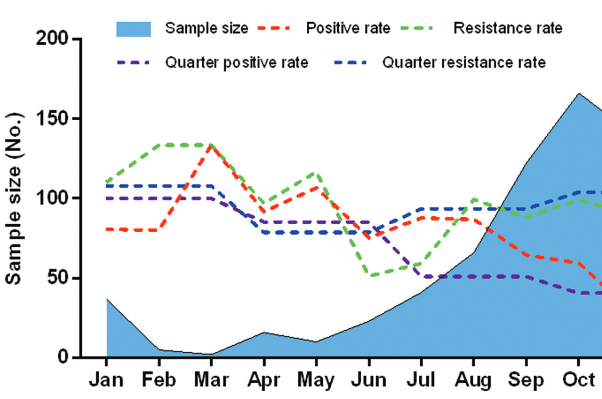
Mycoplasma pneumoniae (M. pneumoniae) is one of the most common causes of community-acquired respiratory tract infections (RTIs). We aimed to investigate the prevalence of M. pneumoniae infection, antibiotic resistance and genetic diversity of M. pneumoniae isolates across multiple centers in Beijing, China. P1 protein was detected by Nested PCR to analyze the occurrence of M. pneumoniae in pediatric patients with RTI. M. pneumoniae isolates were cultured and analyzed by Nested-PCR to determine their genotypes. Broth microdilution method was used to determine the minimum inhibitory concentration (MIC) of antibiotics. Out of 822 children with RTI admitted to 11 hospitals in Beijing, 341 (41.48%) were positive for M. pneumoniae by Nested PCR and 236 (69.21%) samples had mutations in 23S rRNA domain V. The highest proportion of M. pneumoniae positive samples was observed in school-age children (118/190; 62.11%) and in pediatric patients with pneumonia (220/389; 56.56%). Out of 341 M. pneumoniae positive samples, 99 (12.04%) isolates were successfully cultured and the MIC values were determined for 65 M. pneumoniae strains. Out of these, 57 (87.69%) strains were resistant to macrolides, and all 65 strains were sensitive to tetracyclines or quinolones. M. pneumoniae P1 type I and P1 type II strains were found in 57/65 (87.69%) and 8/65 (12.31%) of cultured isolates, respectively. Overall, we demonstrated a high prevalence of M. pneumoniae infection and high macrolide resistance of M. pneumoniae strains in Beijing. School-age children were more susceptible to M. pneumoniae, particularly the children with pneumonia. Thus, establishment of a systematic surveillance program to fully understand the epidemiology of M. pneumoniae is critical for the standardized use of antibiotics in China.

Downloads

Download data is not yet available.
Epidemiology and mechanism of drug resistance of Mycoplasma pneumoniae in Beijing, China: A multi-center study

Downloads

Additional Files

Published

2019-08-20

How to Cite

1.
Guo D-X, Hu W-J, Wei R, Wang H, Xu B-P, Zhou W, Ma S-J, Huang H, Qin X-G, Jiang Y, Dong X-P, Fu X-Y, Shi D-W, Wang L-Y, Shen A-D, Xin D-L. Epidemiology and mechanism of drug resistance of Mycoplasma pneumoniae in Beijing, China: A multicenter study. Biomol Biomed [Internet]. 2019Aug.20 [cited 2023Mar.26];19(3):288-96. Available from: https://www.bjbms.org/ojs/index.php/bjbms/article/view/4053

Issue

Section

Translational and Clinical Research

INTRODUCTION

Mycoplasma pneumoniae (M. pneumoniae) is a small, pliable, fastidious, and highly evolved pleomorphic bacteria lacking the cell wall, which was first isolated from a patient with primary atypical pneumonia [1,2]. M. pneumoniae is one of the most common pathogens causing community-acquired respiratory tract infections (RTIs) and has been recognized as a worldwide cause of primary atypical pneumonia [3-5]. It can affect people of all ages, especially the most vulnerable groups children and adolescents [6-8]. Nevertheless, as currently there are no reliable and rapid diagnostic tests for the detection of M. pneumoniae, the treatment of community-acquired pneumonia is empirical in most cases [9].

In addition to mild upper RTIs (URTIs; e.g., pharyngitis and sinusitis) and severe lower RTIs (LRTIs; e.g., bronchitis and pneumonia), M. pneumoniae may also cause damage to extrapulmonary systems [10,11]. M. pneumoniae infections mainly occur in preschool children, however, a recent report suggested that the incidence in infants is also high [12]. Although the symptoms are mild to moderate in most cases of M. pneumoniae infections, hospitalization is occasionally required in severe cases. In addition, multiple organ system damage may occur [13]. M. pneumoniae infection appears as a cyclic epidemic disease with intervals of four to seven years worldwide and persists for one to two years [10,14,15]. This periodicity in M. pneumoniae infection may be related to changes in the sequence of P1 adhesin, which is the main method of M. pneumoniae typing. According to the differences in the sequence of P1 adhesin gene MPN141, M. pneumoniae is divided into P1 type I and P1 type II with several subtypes [16,17]. The prevalence of M. pneumoniae subtypes differs between countries and years, and the dominance of one subtype is followed by the dominance of another M. pneumoniae subtype [18].

Although M. pneumoniae is sensitive to macrolide, tetracycline, and quinolone antibiotics, which affect the synthesis of proteins and nucleic acids, macrolides remain the first choice for the treatment of M. pneumoniae infection in children [19]. However, the phenomenon of M. pneumoniae resistance continues to increase worldwide, especially in China and Japan, where the resistance rate is reported to be over 90% [20,21]. Since 2004, tetracycline and quinolone antibiotics have been approved for use in children over eight years of age with macrolide resistance or refractory M. pneumoniae infection [22]. Therefore, with the increasing use of these two drugs in clinical practice, the drug resistance becomes an even more serious problem. Moreover, only a few studies have focused on the M. pneumoniae tetracycline or quinolone resistance in China.

Several serology-based studies were conducted in Beijing [23-25], however, they were all single-center studies and thus limited by their design. To the best of our knowledge, there are no multicenter studies investigating the changes in the prevalence of M. pneumoniae infections in Beijing. Therefore, in the present study, we collected samples from several major children’s hospitals in Beijing area to study the prevalence of M. pneumoniae infection and the antibiotic resistance of M. pneumoniae. In addition, we used molecular assays to identify the genotypes of isolated M. pneumoniae and to infer their potential antibiotic (macrolide, tetracycline, and quinolone) resistance mechanism.

MATERIALS AND METHODS

Patients’ samples and M. pneumoniae testing

This study was conducted at 11 centers in Beijing, China, from January 2014 to December 2014. Participating centers included Beijing Dongfang Hospital, The First Hospital of Tsinghua University, Beijing Children’s Hospital, China Meitan General Hospital, Beijing Chao-Yang Hospital, Civil Aviation General Hospital, Xiyuan Hospital, Beijing Changping Hospital of Integrated Chinese and Western Medicine, New Century Women’s and Children’s Hospital, New Century International Children’s Hospital, and Peking University Third Hospital. Ethics approvals were obtained from local or national institutional review boards, as appropriate. Tracheal swab specimens were collected from 822 pediatric patients with RTI symptoms. The pediatric patients were clinically diagnosed with pneumonia, URTI, or bronchitis. Patient data were collected, including sex, age, disease duration, and clinical diagnosis.

DNA was isolated from tracheal swab specimens using a Universal Genomic DNA Kit (Beijing Kangwei Century Biotech Co., Ltd., China), according to the manufacturer’s instructions. Nested polymerase chain reaction (PCR) was performed to test the presence of M. pneumoniae DNA in samples, according to the previous research [26]. Each M. pneumoniae-positive PCR sample was cultivated to obtain pure M. pneumoniae isolates.

Culture of M. pneumoniae-positive samples

Culture of M. pneumoniae was performed using PPLO (pleuropneumonia-like organisms) basic medium (BD-Difco, USA) containing 15% newborn calf serum, 10% fresh yeast extract (OXOID, UK), 0.4% phenol red indicator (Sigma, USA), 1% glucose, and 50000 U/100 mL penicillin, as described previously [21]. Tracheal swab specimens were inoculated on the M. pneumoniae liquid medium and maintained in an incubator at 37°C with a 5% CO2 atmosphere. The obtained isolates were stored at -80°C until further testing.

Measurement of minimum inhibitory concentration (MIC)

The measurement of MIC of antibiotics was performed using the standard broth microdilution method (standard MIC procedure) [27]. We used six antibiotics from three classes as follows: macrolides (erythromycin, azithromycin, and josamycin), tetracyclines (tetracycline and minocycline), and quinolones (levofloxacin). Antibiotic susceptibility test was conducted to distinguish between sensitive and clinically resistant strains according to the Clinical and Laboratory Standards Institute (CLSI) criteria [28]. The breakpoints of MIC were as follows: for macrolides, MIC ≥ 1.0 µg/ml was considered as resistant, 0.5 < MIC < 1.0 µg/ml as intermediate, and MIC ≤ 0.5 µg/ml as susceptible; for tetracyclines, MIC > 2.0 µg/ml was considered as resistant and MIC ≤ 2.0 µg/ml as susceptible; and for quinolones, MIC > 1.0 µg/ml was considered as resistant and MIC ≤1.0 µg/ml as susceptible. Two M. pneumoniae reference strains, M129 (ATCC 29342) and FH (ATCC 15531) were used as a drug-sensitive control.

M. pneumoniae genotyping and detection of resistance-related genes

P1 protein was detected by Nested PCR and classified according to the differences in the sequence of P1 adhesin gene MPN141 [29]. The reaction conditions were as follows: initial denaturation at 95°C for two minutes, followed by 30 cycles of 95°C for 30 seconds, 55°C for 30 seconds, and 72°C for 30 seconds, with a final extension step of 72°C for two minutes. PCR primer sequences are shown in Table 1.

TABLE 1: Primers used for Nested PCR analysis

The resistance-associated mutation in the 23S rRNA gene of M. pneumoniae was detected by Nested PCR. The reaction conditions were as follows: initial denaturation at 95°C for two minutes, followed by 35 cycles of 95°C for one minute, 55°C for one minute, and 72°C for 100 seconds, with a final extension step of 72°C for five minutes. The identification of nucleotide sequences was performed using BLAST against the NCBI GenBank database (http://blast.ncbi.nlm.nih.gov/Blast.cgi). The sequences of the resistance-associated genes and PCR primers are shown in Table 1.

Statistical analysis

Statistical analysis was performed using IBM SPSS Statistics for Windows, Version 19.0 (IBM Corp., Armonk, NY, USA). Quantitative data were presented as mean ± standard deviation (SD). Qualitative data were described as numbers or percentages. The normality of data distribution was tested by the Kolmogorov–Smirnov test. Qualitative variables were analyzed using Chi-squared test. Differences were considered statistically significant at p < 0.05.

RESULTS

Detection of M. pneumoniae in pediatric patients with RTI

The mean age of children with RTI was 4.68 ± 3.23 years (range, one month to 17 years). Out of 822 patients with RTI admitted to 11 hospitals in Beijing, 341 (41.48%) had a positive PCR for M. pneumoniae. Ninety-nine (12.04%, 99/822) M. pneumoniae-positive samples were successfully cultured. The sex of 779 patients was available. There were 417 males and 362 females. There was no statistically significant difference with regard to the M. pneumoniae positivity rates between male and female samples (38.85% [162/417] vs. 45.58% [165/362], p = 0.06; Figure 1A).

FIGURE 1: Distribution of Mycoplasma pneumoniae (M. pneumoniae) in children with RTIs. A) Distribution of M. pneumoniae-positive samples in children. There was no statistically significant difference in the M. pneumoniae positivity rates between boys and girls (p = 0.06). School-age children had the highest M. pneumoniae positivity rate, followed by preschool children and infants, with a significant difference (p < 0.001). Patients with pneumonia had the highest M. pneumoniae positivity rate, followed by those with URTI and bronchitis, with a significant difference (p < 0.001). B) Distribution of M. pneumoniae samples with mutations in domain V of 23S rRNA genes in children. No significant difference was found in the detection rate of mutant M. pneumoniae strains between boys and girls (p = 0.82). School-age children (7–17 years old) had the highest detection rate of M. pneumoniae mutant strains, followed by preschool children (3–6 years old) and infants (<2 years old), with a significant difference (p < 0.001). The majority of mutant strains were found in pneumonia samples, followed by URTI samples and bronchitis samples, with a significant difference (p < 0.001). *p < 0.05. RTI: Respiratory tract infection; URTI: Upper RTI.

The age of 776 children was collected. School-age children (7–17 years old) had the highest M. pneumoniae positivity rate (62.11%, 118/190), followed by preschool children [3–6 years old] (40.30%, 160/397) and infants [<2 years old] (24.87%, 47/189). There was a significant difference in the M. pneumoniae positivity rate between the three groups (p < 0.001).

The diagnosis of 775 children was available. The majority of M. pneumoniae-positive samples were found in patients with pneumonia (56.56%, 220/389), followed by those with URTI (28.71%, 60/209) and bronchitis (26.55%, 47/177). The M. pneumoniae detection rate in pneumonia samples was significantly higher than in URTI and bronchitis samples (p < 0.001), whereas there was no significant difference between URTI and bronchitis samples (p = 0.67).

Detection of mutations in domain V of the 23S rRNA gene

The 341 M. pneumoniae-positive PCR samples were analyzed by Nested PCR. Mutations in domain V of the 23S rRNA gene were found in 236 (69.21%) samples. The predominant mutation was A2063G (199/341, 58.36%), followed by A2064G (25/341, 7.33%). Twelve (3.52%) samples had the co-mutation of A2063G and A2064G. The mutation detection rate of 23S rRNA domain V in different groups is shown in Figure 1B. The detection rate of the mutant M. pneumoniae strains was 69.14% (112/162) in male patients and 70.30% (116/165) in female patients, with no significant difference between the sexes (p = 0.82). School-age children (7–17 years old) had the highest detection rate of M. pneumoniae mutant strains (77.78%, 26/91), followed by preschool children [3–6 years old] (71.07%, 113/159) and infants [<2 years old] (48.94%, 23/47). The detection rate of mutant M. pneumoniae strains was significantly higher in school-age and preschool children than in infants (p < 0.05). The majority of mutant strains were found in pneumonia samples (77.27%, 170/220), followed by URTI samples (59.57%, 28/47) and bronchitis samples (50.00%, 30/60). The percentage of mutant M. pneumoniae strains was significantly higher in pneumonia and bronchitis samples than in URTI samples (pneumonia vs. URTI, p < 0.001 and bronchitis vs. URTI, p = 0.012).

M. pneumoniae detection rate in different months

We analyzed the distribution of M. pneumoniae infection during all 12 months in 2014 (Figure 2) and found that the sample size varied from month to month, ranging from 2 to 181 positive samples. However, it should be noted that a smaller number of samples were collected during February and March, due to the Chinese Spring Festival. During the 12-month period, the M. pneumoniae positivity rate ranged from 22% (31/141) to 100% (2/2) and the resistance rate ranged from 38% (5/13) to 100% (3/3).

FIGURE 2: Distribution of Mycoplasma pneumoniae infections in children with respiratory tract infections (RTIs) over time, from January 2014 to December 2014. The sample size varied from month to month, ranging from 2 to 181 positive samples. During the 12-month period, the M. pneumoniae positivity rate ranged from 22% (31/141) to 100% (2/2) and the resistance rate ranged from 38% (5/13) to 100% (3/3). In terms of the quarterly distribution, the M. pneumoniae positivity rate was relatively higher in the first (63.64%) and second quarter (65.31%) of the year, followed by the third (56.33%) and fourth quarter (30.12%). Resistance rate was the highest in the first quarter (85.71%), followed by the fourth quarter (72.79%).

In terms of the quarterly distribution, the M. pneumoniae positivity rate was relatively higher in the first (63.64%) and second quarter (65.31%) of the year, followed by the third (56.33%) and fourth quarter (30.12%). Additionally, the resistance rate was the highest in the first quarter (85.71%), followed by the fourth quarter (72.79%).

Antibiotic resistance of M. pneumoniae isolates and related gene mutations

Out of 99 M. pneumoniae cultured isolates, MIC measurement was successfully performed in 65 M. pneumoniae isolates. As shown in Table 2, among these 65 M. pneumoniae isolates, 57 (87.69%) strains with A2063G mutation in domain V of the 23S rRNA gene were resistant to macrolides, and the MIC90 values were 1024 µg/ml (erythromycin), 128 µg/ml (azithromycin), and 8 µg/ml (josamycin). The other eight wild-type M. pneumoniae isolates were macrolide-sensitive strains with MIC ≤ 0.5 µg/ml. With regard to tetracyclines, all M. pneumoniae isolates were sensitive with MIC ≤ 2.0 µg/ml. Besides, all M. pneumoniae isolates were sensitive to levofloxacin with MIC 0.25 to 1.0 µg/ml.

TABLE 2: Antimicrobial susceptibility of Mycoplasma pneumoniae strains to antimicrobial agents (MIC range/MIC90, µg/ml)

One macrolide-resistant strain harbored a missense mutation (K27N) in L4 ribosomal protein. Eight macrolide-sensitive strains harbored the mutation M144V in L4 ribosomal protein and S170P in L22, which was consistent with the reference strain FH (Table 3). No mutation was found in the tetracycline-resistance genes (tet and 16S rRNA). The mutations in the quinolone-resistance genes are summarized in Table 4. For all 65 M. pneumoniae-cultured isolates, no mutation was found in the gyrB gene. Two strains had D149G, A533S, and C705 mutations in the gyrA gene. One strain had R454C and G652R mutations in the parC gene. Another six strains had a co-mutation in the gyrA and parC genes.

TABLE 3: Mutations in macrolide-resistance genes and susceptibility of M. pneumoniae isolates to macrolide antibiotics
TABLE 4: Mutations in quinolone-resistance genes and susceptibility of M. pneumoniae isolates to quinolone antibiotics

M. pneumoniae genotyping and susceptibility

M. pneumoniae P1 type I and P1 type II strains were found in 87.69% (57/65) and 12.31% (8/65) of cultured isolates, respectively. One of the P1 type I strains was sensitive to macrolides and the remaining 56 (98.25%) strains were resistant to macrolides. One P1 type II strain was resistant to macrolides and the remaining seven were sensitive to macrolides. The MIC values of various antibiotics for different M. pneumoniae P1 type strains are shown in Table 5. The results indicated that the MIC of macrolides for P1 type I strains was significantly higher than for P1 type II strains (p < 0.001) and no significant difference was detected in the MIC of tetracyclines (tetracycline, p = 0.06; minocycline, p = 0.43) and levofloxacin (p = 0.11) between the two subtypes.

TABLE 5: Mycoplasma pneumoniae P1 type strains and resistance phenotype analysis (MIC range/MIC90, µg/ml)

DISCUSSION

M. pneumoniae causes upper and lower RTIs in children and adults. Although the symptoms are mild and self-limited in the majority of patients, or the infections are asymptomatic, approximately 25% of patients will be hospitalized due to extrapulmonary complications or severe pneumonia [30]. It is generally known that the incidence and prevalence of M. pneumoniae vary in different periods and regions. Thus, the surveillance of M. pneumoniae infection is particularly important in the monitoring and prevention of acquired pneumonia. Here, we conducted for the first time a multicenter study of M. pneumoniae infection in Beijing, China. Overall, 822 tracheal swab specimens were collected from pediatric patients with RTI symptoms in 11 hospitals in Beijing. Out of 822 samples, 341 (41.48%) were positive for M. pneumoniae by PCR. Previous study showed that the positivity rate of M. pneumoniae ranged from 19.13% to 29.07% in Beijing [5,25,31]. Thus, we may conclude that M. pneumoniae infection in Beijing has a gradual upward trend. This may be due to the rise of drug-resistant strains caused by the overuse of antibiotics in recent years.

Excessive or inappropriate use of antibiotics may lead to a selective pressure for the development of antibiotic resistance [32]. This greatly increases the number of resistant strains and, consequently, the severity of clinical symptoms, making the treatment more difficult. The prevalence of M. pneumoniae infections has obvious regional differences. Ishiguro et al. reported that the macrolide resistance in different cities in Hokkaido significantly varied, from 0.0% to 100% [33]. Therefore, enhanced surveillance of drug-resistant bacteria across a country is essential for the prevention of M. pneumoniae infection. In M. pneumoniae infections, macrolide resistance has become a potential threat worldwide, especially in China. According to a national survey on antibiotic usage, the rate of macrolide use in China has been around 70% [34] and the rate of macrolide sales reached 7.9 billion in 2014. In such circumstances, the high prevalence of M. pneumoniae macrolide-resistant strains observed in our study is not unexpected. Similarly, other studies showed that more than 90% of M. pneumoniae infections in China were caused by resistant strains [35-37]. Based on these data, we assume that the macrolide-resistant strains have developed in the southern or northern China. On the other hand, in Western countries, the proportion of resistant bacteria has been relatively low, ranging from 1% to 10% [10,16,32,38]. This phenomenon is mainly owing to the careful use of macrolides in these areas. For that reason, a systematic and strict surveillance program is necessary to control the use of macrolides in our country and to manage the threat of emerging resistance of M. pneumoniae to the first-line antibiotic therapy.

Fortunately, tetracyclines and quinolones remained effective against clinical M. pneumoniae isolates. However, although we did not find tetracycline- or quinolone-resistant strains in the current study, an increasing number of Mycoplasma genitalium, Mycoplasma urealyticum, and Mycoplasma hominis strains resistant to these antibiotics has been isolated [39]. The drug resistance is primarily acquired and induced by the external environment. Therefore, with the increasing use of tetracycline and quinolone substitution therapy, the risk of development of clinically resistant strains should not be ignored. Additionally, it is worth noting that all cultured positive M. pneumoniae samples in our study had A2063G mutation in domain V of the 23S rRNA gene, which has been recognized as the most prevalent and highly associated with macrolide resistance. Although these M. pneumoniae positive strains harbored the same type of mutation, the correlation with resistance is not yet clear and further investigation is warranted to explain this phenomenon. In M. pneumoniae, resistance caused by 23S rRNA gene mutation is the most common resistance mechanism, because M. pneumoniae harbors only one copy of rRNA operon [40]. According to previous studies, mutations in the 23S rRNA gene, such as A-to-G transition or A-to-C transversion at position 2063 or 2064, respectively, predominantly cause the macrolide resistance in M. pneumoniae [41]. Furthermore, mutations at position 2617 in domain V of the 23S rRNA gene are associated with lower resistance to macrolides than the mutations at position 2063 or 2064 [41].

The positivity rate of M. pneumoniae infection and detection rate of 23S rRNA domain V gene mutations differed between the quarters of 2014 year and between different age groups of patients in our study. The highest M. pneumoniae infection rate was found in the second quarter and among school-age children. Additionally, we found no significant gender-related differences in the M. pneumoniae positivity rate. These results are consistent with previous epidemiological research on M. pneumoniae in China [5,42]. According to the age-based distribution, we speculate that the epidemiology of M. pneumonia in Beijing conforms to the traditional epidemiological model of infectious disease diffusion. In this model, the patients are linked by the characteristics of the bacterial strain, indicating a common exposure or person-to-person transmission [32]. Usually, school-age children live in a relatively closed environment and are in close contact with a large number of people, leading to a rapid transmission and outbreak of pathogens.

In our study, 57 (87.69%) clinical isolates were classified as P1 type I strains and 8 (12.31%) as P1 type II strains. The vast majority of P1 type I strains were macrolide-resistant, while most of the P1 type II isolates were macrolide-sensitive strains. The regular distribution of P1 subtypes among macrolide-resistant and macrolide-sensitive M. pneumoniae strains suggests an association between P1 subtype and the macrolide susceptibility. This potential correlation should be further investigated in studies with larger sample sizes. In addition, a more thorough analysis of the M. pneumoniae genome with whole-genome sequencing might provide more evidence for the resistance mechanism.

We found A2063G mutation in domain V of the 23S rRNA gene in 57 (87.69%) M. pneumoniae isolates, which was associated with high MIC90 of macrolides. Mutations in domain V of the 23S rRNA gene are the main cause of macrolides resistance, especially those at positions 2063 and 2064. Many studies have revealed that macrolides could inhibit protein synthesis by binding to large ribosomal subunits, so mutations in 23S rRNA may lead to decreased affinity of the ribosome for drug [21]. While many reports have investigated the role of the mutation in domain V of 23S rRNA gene, no study has analyzed mutations in the ribosomal proteins L4 and L22 [36]. Although we found several M. pneumoniae isolates with mutations in L4 and L22, the relationship between L4 and L22 mutations and macrolide resistance is yet unclear. Additionally, no mutation was detected in the tetracycline and quinolone resistance-associated genes, suggesting that tetracyclines and quinolones remain effective against clinical M. pneumoniae isolates.

There are several limitations in this study. First, this study only collected data for one year. Although we compared these data to the data from previous studies in Beijing, this may not be sufficient to observe changes in the prevalence of M. pneumoniae circulating types and epidemic trends over time. Second, due to the observational, non-randomized design of the study, we collected data in a retrospective manner, meaning that the information of some pediatric patients was unavailable and the quality of the data was dependent on the accuracy of the medical record.

CONCLUSION

We investigated M. pneumoniae infections in 11 hospitals in Beijing and found that school-age children were more susceptible to this disease, particularly pediatric patients with pneumonia. We also found that M. pneumoniae P1 type I may be the main cause of the epidemic in Beijing. The rates of macrolide resistance observed in this study were more than 85%. Fortunately, tetracyclines and quinolones remain effective against M. pneumoniae isolates in Beijing. In order to fully understand the biology and epidemiology of M. pneumoniae, the establishment of a systematic surveillance program is critical.

Acknowledgements

ACKNOWLEDGMENTS

The research was supported by the Natural Science Foundation of China (number 81271890) and Capital Medical Development Research Fund (number 2014-1-2094) and Capital Medical Development Research Fund (number 2016-1-2092).

DECLARATION OF INTERESTS

The authors declare no conflict of interests.

REFERENCES

  1. , , (). Epidemiology, clinical manifestations, pathogenesis and laboratory detection of Mycoplasma pneumoniae infections. FEMS Microbiol Rev. https://doi.org/10.1111/j.1574-6976.2008.00129.x
  2. (). Mycoplasma pneumoniae. Thorax.
  3. , , (). Mycoplasma pneumoniae:A potentially severe infection. J Clin Med Res. https://doi.org/10.14740/jocmr3421w
  4. (). Mycoplasma pneumoniae:A significant but underrated pathogen in paediatric community-acquired lower respiratory tract infections. Indian J Med Res. https://doi.org/10.4103/ijmr.IJMR_1582_16
  5. , , , , , (). Surveillance of Mycoplasma pneumoniae infection among children in Beijing from 2007 to 2012. Chin Med J (Engl). https://doi.org/10.3760/cma.j.issn.0366-6999.20131654
  6. (). Mycoplasma pneumoniae pneumonia requiring hospitalization, with emphasis on infection in the elderly. Arch Intern Med. https://doi.org/10.1001/archinte.1993.00410040054008
  7. (). Community-acquired pneumonia in children:Issues in optimizing antibacterial treatment. Paediatr Drugs. https://doi.org/10.2165/00148581-200305120-00005
  8. , , (). Mycoplasma pneumoniae in children:Carriage, pathogenesis, and antibiotic resistance. Curr Opin Infect Dis. https://doi.org/10.1097/QCO.0000000000000063
  9. (). Mycoplasma pneumoniae infections. Curr Opin Infect Dis. https://doi.org/10.1097/00001432-200104000-00012
  10. , , , , , (). Epidemic of Mycoplasma pneumoniae infection in Denmark, 2010 and 2011. Euro Surveill. https://doi.org/10.2807/ese.17.05.20073-en
  11. , , , (). Role of Mycoplasma pneumoniae and Chlamydia pneumoniae in children with community-acquired lower respiratory tract infections. Clin Infect Dis. https://doi.org/10.1086/319981
  12. , , , , , (). Epidemiological comparison of three Mycoplasma pneumoniae pneumonia epidemics in a single hospital over 10 years. Korean J Pediatr. https://doi.org/10.3345/kjp.2015.58.5.172
  13. , (). Mycoplasma pneumoniae and its role as a human pathogen. Clin Microbiol Rev. https://doi.org/10.1128/CMR.17.4.697-728.2004
  14. , , , , (). Early serologic diagnosis of Mycoplasma pneumoniae pneumonia:An observational study on changes in titers of specific-igM antibodies and cold agglutinins. Medicine (Baltimore). https://doi.org/10.1097/MD.0000000000003605
  15. , , , , (). Increased incidence of Mycoplasma pneumoniae infection in England and Wales in 2010:Multiocus variable number tandem repeat analysis typing and macrolide susceptibility. Euro Surveill. https://doi.org/10.2807/ese.16.19.19865-en
  16. , , , (). Prevalence, genotyping and macrolide resistance of Mycoplasma pneumoniae among isolates of patients with respiratory tract infections, Central Slovenia, 2006 to 2014. Euro Surveill. https://doi.org/10.2807/1560-7917.ES.2015.20.37.30018
  17. , , (). Genotyping of Mycoplasma pneumoniae clinical isolates reveals eight P1 subtypes within two genomic groups. J Clin Microbiol.
  18. , , , , (). First report of macrolide-resistant strains and description of a novel nucleotide sequence variation in the p1 adhesin gene in Mycoplasma pneumoniae clinical strains isolated in France over 12 years. J Clin Microbiol. https://doi.org/10.1128/JCM.01345-07
  19. , , (). Respiratory tract infections by Mycoplasma pneumoniae in children:A review of diagnostic and therapeutic measures. Eur J Pediatr. https://doi.org/10.1128/JCM.01345-07
  20. , , , , , (). Nationwide surveillance of macrolide-resistant Mycoplasma pneumoniae infection in pediatric patients. Antimicrob Agents Chemother. https://doi.org/10.1128/AAC.00663-13
  21. , , , , , (). Drug resistance mechanisms of Mycoplasma pneumoniae to macrolide antibiotics. Biomed Res Int. https://doi.org/10.1155/2014/320801
  22. , , (). Progresses of diagnosis and treatment of children with refractory Mycoplasma peumoniae. J Shanghai Jiao Tong Univ Med Sci.
  23. , , , , , (). Antibiotic sensitivity of 40Mycoplasma pneumoniae isolates and molecular analysis of macrolide-resistant isolates from Beijing, China. Antimicrob Agents Chemother. https://doi.org/10.1128/AAC.05627-11
  24. , , , , , (). Multiple-locus variable-number tandem-repeat analysis of 201Mycoplasma pneumoniae isolates from Beijing, China, from 2008 to 2011. J Clin Microbiol. https://doi.org/10.1128/JCM.02567-12
  25. , , , , , (). Surveillance of macrolide-resistant Mycoplasma pneumoniae in Beijing, China, from 2008 to 2012. Antimicrob Agents Chemother. https://doi.org/10.1128/AAC.02060-12
  26. , , , (). Amplification of P1 and 16S rRNA genes by nested PCR for detection of Mycoplasma pneumoniae in paediatric patients. Pathol Biol (Paris). https://doi.org/10.1016/j.patbio.2004.01.002
  27. , , , , , (). P1 gene of Mycoplasma pneumoniae in clinical isolates collected in Beijing in 2010 and relationship between genotyping and macrolide resistance. Chin Med J (Engl). https://doi.org/10.3760/cma.j.issn.0366-6999.20131643
  28. (). . Clinical and Laboratory Standards Institute:Performance Standards for Antimicrobial Susceptibility Testing:20thInformational Supplement.
  29. , , , , , (). Macrolide resistance determination and molecular typing of Mycoplasma pneumoniae by pyrosequencing. J Microbiol Methods. https://doi.org/10.1016/j.mimet.2010.06.004
  30. , (). Detection of Mycoplasma pneumoniae by real-time PCR. Methods Mol Biol. https://doi.org/10.1007/978-1-60327-353-4_10
  31. , , , , (). Mycoplasma pneumoniae infection and drug resistance in children:An analysis of 1026 cases. Zhongguo Dang Dai Er Ke Za Zhi.
  32. , , (). Investigations of Mycoplasma pneumoniae infections in the United States:Trends in molecular typing and macrolide resistance from 2006 to 2013. J Clin Microbiol. https://doi.org/10.1128/JCM.02597-14
  33. , , , , , (). Regional differences in prevalence of macrolide resistance among pediatric Mycoplasma pneumoniae infections in Hokkaido, Japan. Jpn J Infect Dis. https://doi.org/10.7883/yoken.JJID.2015.054
  34. (). Analysis and discussion of the high utilization rate of antibiotics. J Psychiatry.
  35. , , , , , (). Antimicrobial susceptibility of Mycoplasma pneumoniae isolates and molecular analysis of macrolide-resistant strains from Shanghai, China. Antimicrob Agents Chemother. https://doi.org/10.1128/AAC.01684-08
  36. , , , , , (). Molecular mechanisms of macrolide resistance in clinical isolates of Mycoplasma pneumoniae from China. Antimicrob Agents Chemother. https://doi.org/10.1128/AAC.01563-08
  37. , , , , , (). Macrolide-resistant Mycoplasma pneumoniae in adults in Zhejiang, China. Antimicrob Agents Chemother. https://doi.org/10.1128/AAC.04308-14
  38. , , , , , (). Increased macrolide resistance of Mycoplasma pneumoniae in france directly detected in clinical specimens by real-time PCR and melting curve analysis. J Antimicrob Chemother. https://doi.org/10.1093/jac/dkp160
  39. , (). Mutations in the 16S rRNA genes of Helicobacter pylori mediate resistance to tetracycline. J Bacteriol. https://doi.org/10.1128/JB.184.8.2131-2140.2002
  40. , , , , , (). In vitro selection and characterization of resistance to macrolides and related antibiotics in Mycoplasma pneumoniae . Antimicrob Agents Chemother. https://doi.org/10.1128/AAC.48.2.460-465.2004
  41. , , , , , (). Characterization and molecular analysis of macrolide-resistant Mycoplasma pneumoniae clinical isolates obtained in Japan. Antimicrob Agents Chemother. https://doi.org/10.1128/AAC.48.12.4624-4630.2004
  42. , , , , , (). Epidemiology and clinical profiles of Mycoplasma pneumoniae infection in hospitalized infants younger than one year. Respir Med. https://doi.org/10.1016/j.rmed.2015.04.006