Isoflurane exposure in infant rats acutely increases aquaporin 4 and does not cause neurocognitive impairment

Authors

  • Serdar Demirgan T.C. Health Ministry, Health Sciences University, Bagcilar Training and Research Hospital, Anesthesiology and Reanimation Clinic; Department of Molecular Biology and Genetics, Institute of Graduate Studies in Sciences, Istanbul University, Istanbul, Turkey https://orcid.org/0000-0001-8129-5004
  • Onat Akyol T.C. Health Ministry, Health Sciences University, Bagcilar Training and Research Hospital, Anesthesiology and Reanimation Clinic, Istanbul, Turkey https://orcid.org/0000-0003-1316-7160
  • Zeynep Temel Department of Neuroscience Institute of Health Sciences, Istanbul Medipol University, Istanbul, Turkey https://orcid.org/0000-0001-8563-5635
  • Aslıhan Şengelen Department of Molecular Biology and Genetics, Institute of Graduate Studies in Sciences, Istanbul University, Istanbul, Turkey https://orcid.org/0000-0003-2512-319X
  • Murat Pekmez Department of Molecular Biology and Genetics, Faculty of Science, Istanbul University, Istanbul, Turkey https://orcid.org/0000-0002-6150-8372
  • Recep Demirgan Department of Molecular Biology and Genetics, Institute of Graduate Studies in Sciences, Istanbul University, Istanbul, Turkey https://orcid.org/0000-0002-2653-4815
  • Mehmet Salih Sevdi T.C. Health Ministry, Health Sciences University, Bagcilar Training and Research Hospital, Anesthesiology and Reanimation Clinic, Istanbul, Turkey
  • Kerem Erkalp T.C. Health Ministry, Health Sciences University, Bagcilar Training and Research Hospital, Anesthesiology and Reanimation Clinic, Istanbul, Turkey
  • Ayşin Selcan T.C. Health Ministry, Health Sciences University, Bagcilar Training and Research Hospital, Anesthesiology and Reanimation Clinic, Istanbul, Turkey

DOI:

https://doi.org/10.17305/bjbms.2019.4116

Keywords:

Anesthesia, aquaporin 4 (AQP4), neurocognition, infant rat, isoflurane, pediatric anesthesia, 10-day-old rat

Abstract

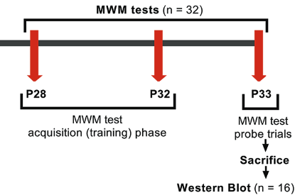
Isoflurane is commonly used in pediatric population, but its mechanism of action in cognition is unclear. Aquaporin 4 (AQP4) regulates water content in blood, brain, and cerebrospinal fluid. Various studies have provided evidence for the role of AQP4 in synaptic plasticity and neurocognition. In this study, we aimed to determine whether a prolonged exposure to isoflurane in infant rats is associated with cognition and what effect this exposure has on AQP4 expression. Ten-day-old [postnatal day (P) 10] Wistar albino rats were randomly allocated to isoflurane group (n = 32; 1.5% isoflurane in 50% oxygen for 6 hours) or control group (n = 32; only 50% oxygen for 6 hours). Acute (P11) and long-term (P33) effects of 6-hour anesthetic isoflurane exposure on AQP4 expression were analyzed in whole brains of P11 and P33 rats by RT-qPCR and Western blot. Spatial learning and memory were assessed on P28 to P33 days by Morris Water Maze (MWM) test. The analysis revealed that isoflurane increased acutely both mRNA (~4.5 fold) and protein (~90%) levels of AQP4 in P11 rats compared with control group. The increasing levels of AQP4 in P11 were not observed in P33 rats. Also, no statistically significant change between isoflurane and control groups was observed in the latency to find the platform during MWM training and probe trial. Our results indicate that a single exposure to isoflurane anesthesia does not influence cognition in infant rats. In this case, acutely increased AQP4 after isoflurane anesthesia may have a protective role in neurocognition.

Downloads

Download data is not yet available.
Isoflurane exposure in infant rats acutely increases aquaporin 4 and does not cause neurocognitive impairment

Downloads

Additional Files

Published

2019-08-20

How to Cite

1.
Demirgan S, Akyol O, Temel Z, Şengelen A, Pekmez M, Demirgan R, Sevdi MS, Erkalp K, Selcan A. Isoflurane exposure in infant rats acutely increases aquaporin 4 and does not cause neurocognitive impairment. Biomol Biomed [Internet]. 2019Aug.20 [cited 2023Sep.22];19(3):257-64. Available from: https://www.bjbms.org/ojs/index.php/bjbms/article/view/4116

Issue

Section

Molecular Biology

INTRODUCTION

Worldwide, millions of young children are exposed to anesthesia every year for various reasons. Risks that may result from anesthesia exposure are higher in neonates and infants than in adults. Inhaled anesthetics, such as isoflurane, are widely used in daily clinical practice in pediatric anesthesia. Numerous studies on newborn animals have demonstrated that the administration of volatile anesthetics, in various concentrations and durations, leads to neuronal degeneration via numerous pathways; however, neurocognitive impairment observed in these animals is not always attributed to the administration of inhaled anesthetics [1,2]. In addition, the effect of anesthesia depends on the postnatal day. There is more information in the literature on anesthesia effects in rats up to 7 days old (reflecting neonatal rats) compared with rats older than 7 days (e.g., 10-day-old rats representing an older neonatal or infant model). Thus, more research is needed for infant rat models [3,4].

Long-term neurological effects of single or multiple exposures to inhalational anesthetics are of great interest for brain cognitive development studies, especially the effects on synaptogenesis and neuronal network formation during learning and formation of memory. However, the molecular mechanisms of inhalational agents in neurocognitive impairment remain unclear [5]. In addition to the known role of astrocytes in providing structural and metabolic support to neurons, increasing evidence suggests that they also play a role in memory formation, signal transmission, and regulation of synaptic plasticity. Furthermore, astrocyte-specific proteins are of key importance in the maintenance of synaptic plasticity [6-8]. One of these proteins is aquaporin 4 (AQP4) [8,9]. AQP4 is a tetrameric membrane-bound protein of the water-channel protein family. Cerebral AQP4 is the predominant aquaporin found in the mammalian brain, including the cerebral cortex, corpus callosum, cerebellum, hypothalamus, and brain stem. The main function of cerebral AQP4, which is particularly expressed in perivascular astrocytes, is to control content in blood, brain, and cerebrospinal fluid [10]. Moreover, increasing evidence suggests that AQP4 does not only function as a water channel but also plays an important role in neurotransmission and neurocognition [8,11]. Few rodent studies have provided evidence regarding the role of AQP4 in synaptic plasticity and cognition [8,12]. In a post-mortem analysis of patients with Alzheimer’s disease exhibiting amyloid beta (Aβ) pathology, Zeppenfeld et al. reported diminished perivascular AQP4 levels. The authors also confirmed sustained higher levels of cortical AQP4 in elderly patients with normal cognition [11].

The aim of this experimental study was to investigate the effect of prolonged isoflurane anesthesia on cognition and cerebral AQP4 expression in 10-day-old infant rats. For the first time, in this study, we assessed the effect of isoflurane exposure on acute and long-term changes in AQP4 expression. We used Western blot and reverse transcription quantitative polymerase chain reaction (RT-qPCR) to analyze the levels of AQP4. Morris Water Maze (MWM) test was performed for spatial learning and memory assessment.

MATERIALS AND METHODS

Animals

This study was conducted in accordance with the regulations of the Animal Ethics Committee of Bagcilar Training and Research Hospital, Istanbul, Turkey, after obtaining the Institutional Review Board Approval (protocol number: 2016/109, 29th August 2016). All animal studies were conducted in Bagcilar Training and Research Hospital animal laboratory and molecular analyses were carried out at the Department of Molecular Biology and Genetics, Istanbul University. Ten-day-old [Postnatal day (P) 10] Wistar albino infant rats were used based on the reported age at which they are vulnerable to anesthesia-induced neurodegeneration [13]. All efforts were made to minimize the number of animals used and their suffering. Animals had free access to chow and tap water at all times before the experiments. Infant rats, weighing 14–18 g, were housed in cages at controlled room temperature (22–23°C) with a standard light/dark cycle (12-hour light/12-hour dark). Cognition was assessed in a blind manner.

Isoflurane exposure

The P10 rat infants were randomly assigned to two groups consisting of an equal number of males and females: isoflurane group (Group I, anesthesia, n = 32) and control group (Group C, no anesthesia, n = 32). In P10 rats from isoflurane group, anesthesia was induced with 1.5% isoflurane (Forane, Baxter International Inc., Deerfield, Illinois, USA) in 50% oxygen for 6 hours, while the rats in control group were exposed only to 50% oxygen for 6 hours. The procedures were performed in a temperature-controlled plexiglass chamber (dimensions: 13 × 13 × 20 cm) with eight rats at a time placed in the chamber. The isoflurane, oxygen, and carbon dioxide levels in the chamber were continuously monitored using infrared absorbance of the exhaled gas (Datex-Ohmeda S/5 Avance, General Electric, Boston, MA, USA). End-tidal CO2 levels were maintained at 1–2 mmHg pressure by adjusting the total gas flow rate between 5 and 7 l/minute. Normothermia (37°C ± 1°C) was preserved with heating pads. The respiratory frequency and depth and skin color of rats were observed during isoflurane exposure. Following isoflurane or oxygen exposure, all rats were returned to dams.

Experimental protocol

The P10 rats exposed to isoflurane (n = 16) and control rats (n = 16) were sacrificed after 24 hours (P11), and whole brain samples were collected for Western blot (n = 8) and RT-qPCR (n = 8). In a separate experiment, MWM test was performed for isoflurane-exposed (n = 16) and control rats (n = 16) on P28–P32 (acquisition phase). On P33 day (end of the probe trials) rats were sacrificed, and brain tissues were collected for Western blot (n = 8) and RT-qPCR (n = 8). The schematic representation of the experimental design is presented in Figure 1.

FIGURE 1: Schematic representation of the experimental design. Ten-day-old [postnatal day (P) 10] Wistar albino rats were randomly allocated to isoflurane group (1.5% isoflurane, 6 hours) or control group. Acute (P11) and long-term (P33) effects of isoflurane exposure on AQP4 expression were analyzed in whole brains by Western blot and reverse transcription quantitative polymerase chain reaction (RT-qPCR). Spatial learning and memory were assessed on P28 to P33 days by Morris Water Maze (MWM) test.

RT-qPCR analysis

Total RNA was isolated from brain tissue for RT-qPCR using the Magrev Total RNA Extraction Kit (Anatolia Geneworks, Turkey, Istanbul), according to the manufacturer’s instructions. Briefly, tissues were homogenized with PreLys buffer and vortexed. After homogenization, proteinase K was added to the homogenate (1:10 volume), incubated at 56°C for 30 minutes, and centrifuged at 10,000 × g for 5 minutes, at 4°C. Isolation of RNA from the supernatant was performed manually, using magnetic beads. The RNA was treated with DNase I (Quanta BioSciences, Gaithersburg, MD, USA). The AQP4 mRNA levels were measured using qScript One-Step SYBR Green RT-qPCR, on a Montania 4896 real-time PCR system (Anatolia Geneworks). The one-step RT-qPCR program used for cDNA synthesis was: 50°C for 30 minutes, Taq polymerase activation at 95°C for 14 minutes 30 seconds, followed by 45 cycles at 95°C for 30 seconds, 60°C for 45 seconds, and 72°C for 45 seconds, and melting curve analysis at 50–90°C. β-actin was used as the reference gene. The following primers were used: AQP4: 5¢-AGACAGAAGACTTGATCCT-3¢ (forward) and 5¢-GAAGATAATACCTCTCCAGAC-3¢ (reverse); β-actin: 5¢-GTGCGTGACATTAAAGAGA-3¢ (forward) and 5¢-CCGATAGTGATGACCTGA-3¢ (reverse). The expression of AQP4 was calculated using the 2-ΔΔCt method [14].

Western blot analysis

Western blot was performed using total brain lysates, to determine the changes in the expression of AQP4. Firstly, each brain tissue was frozen in liquid nitrogen. Tissues were pulverized to a powder with a mortar and pestle and homogenized in 2 ml of lysis buffer (Tris-buffered saline [TBS, 50 mM Tris, 150 mM NaCl, pH 7.6], 1% NP-40, 1% Triton X-100, 10% glycerol, 1 mM PMSF, and EDTA-free PIC [one tablet per 50 ml of buffer]). The homogenate was kept on ice for 30 minutes, homogenized again, and centrifuged at 10,000 × g for 10 minutes at 4°C. After centrifugation, the proteins were precipitated using the methanol-chloroform precipitation method [15] with minor modifications and solubilized in 1% sodium dodecyl sulfate (SDS). Total protein concentration was determined using the SMART BCA Protein Assay Kit (iNtRON Biotechnology, Seongnam, Gyeonggi, Korea). Samples for electrophoresis were prepared by mixing in a 1:1 ratio with sample buffer (25 mM Tris-HCl [pH 6.8], 2% SDS, 10% glycerol, 10% β-mercaptoethanol, and 0.002% bromophenol blue) and boiled for 4 minutes. Equal amounts of protein (30 µg/well) were loaded for 10% SDS polyacrylamide gel electrophoresis. Following electrophoresis, separated proteins were transferred to a PVDF membrane (Millipore, Darmstadt, Germany) using the Trans-Blot Turbo Transfer System (Bio-Rad, Hercules, CA, USA), and the membrane was blocked with 5% (w/v) non-fat dry milk (in TBST [TBS with 0.1% Tween20]) at room temperature for 1 hour. The membrane was incubated overnight with AQP4 rabbit polyclonal antibody (sc-20812 Santa Cruz Biotechnology [Dallas, TX, USA], working dilution 1:500) at 4°C and washed 5 times, for 5 minutes each, with TBST. The membrane was then incubated for 2 hours at room temperature with horseradish peroxidase (HRP)-conjugated goat anti-rabbit IgG (sc-2004 Santa Cruz Biotechnology [Dallas, TX, USA], working dilution, 1:5000). Chemiluminescence luminol reagent (ECL Plus Western Blotting Detection System [Amersham, MA, USA]) was used to visualize proteins, and protein expression was determined using the ImageLab 5.2.1 software (Bio-Rad). The data were normalized relative to glyceraldehyde 3-phosphate dehydrogenase (GAPDH, MA5-15738-HRP, ThermoFisher Scientific [Kwartsweg, Bleiswijk, Holland], working dilution, 1:2000).

MWM test

The MWM test with minor modifications was conducted by investigators blinded to the experimental groups, to evaluate the isoflurane-induced alterations in learning and memory [16]. The P28 rats exposed to isoflurane or oxygen were trained in the MWM for 6 consecutive days (5 days for training and 1 day for probe trial). A square escape platform of 10 × 10 cm was submerged 1 cm under the surface of the water (24°C ± 1°C) in a circular pool (diameter: 150 cm and depth: 60 cm). White tempera paint was used to make the water opaque. The escape latency (time needed to find the platform) and the target quadrant traveling time (time spent in the quadrant with the platform) were recorded by a digital chronometer and camera system. The rats were subjected to two sessions per day during the acquisition phase (training). All the rats were acclimated to the experimental environment for 10 minutes before testing. The quadrant with the platform and the point of entry for each rat were randomized and counterbalanced. Animals were allowed 60 seconds to locate the hidden platform, and if unsuccessful, they were gently guided to the platform and allowed to stay on it for 30 seconds. During the probe trials, the platform was removed, and the rat was placed in the opposite quadrant.

Acquisition phase (training)

During training, the ability of the rats to understand the spatial relationship between the cues and the escape platform (no cue rod) was evaluated. The platform was placed in one of the four quadrants and remained in the same position for all such trials. The entry point was random for each rat. The time taken to reach the platform was recorded by a digital chronometer and camera system.

Probe trial (memory test)

To assess the memory of the rats, probe trials were conducted on P33. The escape platform was removed, and the rats were placed in the opposite quadrant and allowed to swim for 60 seconds. The time spent in each quadrant, while attempting to locate the platform, was recorded. The data are expressed as the percentage of time spent by the animals in each of the quadrants.

Statistical analysis

The data are presented as mean ± standard deviation (SD) for each group. Statistical analysis and graph generation were performed using the GraphPad Prism version 7.00 for Windows, (GraphPad Software, La Jolla California USA). The statistical evaluation was performed with Student’s t-test or two-way analysis of variance, followed by Tukey’s post hoc test. The value of p < 0.05 was considered statistically significant.

RESULTS

Isoflurane exposure acutely increases mRNA and protein levels of AQP4 in infant rats

We used RT-qPCR and Western blot to analyze the effect of isoflurane anesthesia on mRNA and protein AQP4 expression, respectively. No statistically significant difference was observed in AQP4 expression depending on the sex (p > 0.05). Therefore, the subsequent results represent the data of all male and female rats in a group.

The RT-qPCR analysis (Figure 2) of all brain tissues collected from P11 rats exposed to oxygen (control group) or isoflurane on P10 revealed that 1.5% isoflurane increased the level of AQP4 mRNA by ~4.59 folds (p < 0.001) compared with control. However, the increase in AQP4 levels observed in P11 rats was not observed in P33 rats. We found that the levels of AQP4 in the isoflurane group on P33 decreased (~20.8 folds) compared with the isoflurane group on P11 (p < 0.001). In addition, there was no significant difference in AQP4 levels between oxygen-exposed P11 and P33 rats (p = 0.977). The melting temperature (Tm) for β-actin mRNA (82°C) and that for AQP4 mRNA was determined during the melting curve analysis after RT-qPCR.

FIGURE 2: mRNA levels of aquaporin 4 (AQP4) analyzed by reverse transcription quantitative polymerase chain reaction in rat brain on postnatal day (P) 11 and P33 in response to prolonged isoflurane anesthesia at P10. Group C: P10 rats were exposed to 50% oxygen for 6 hours; Group I: P10 rats were exposed to 1.5% isoflurane for 6 hours. Isoflurane increased the level of AQP4 mRNA by ~4.59 folds compared with control in P11 rats, however the AQP4 levels decreased over time. There was no significant difference in AQP4 levels between oxygen-exposed P11 and P33 rats (p = 0.977). The AQP4 mRNA levels were normalized to β-actin mRNA levels. Data are presented as mean ± standard deviation (n = 8/group). ***p < 0.001, Group C vs. Group I on P11; ###p < 0.001, Group I on P11 vs. P33. p < 0.001, determined by two-way analysis of variance followed by Tukey’s post hoc test.

With respect to AQP4 protein levels in brain samples collected from rats exposed to isoflurane or oxygen on P10, Western blot analysis showed (Figure 3) a ~90% increase for the isoflurane group compared with the group without anesthesia (p < 0.001). In contrast, no such statistically significant change was observed in the AQP4 levels between the isoflurane and control groups on P33 (p = 0.943). No statistically significant difference in AQP4 expression was observed between the P11 and P33 oxygen-exposed rats [control groups] (p = 0.110), but a significant reduction was observed between the isoflurane-treated groups (p < 0.001). The AQP4 expression in the isoflurane group on P33 was reduced by ~40% compared with the isoflurane group on P11 (p < 0.001).

FIGURE 3: Western blot results for aquaporin 4 (AQP4) levels in rat brain on postnatal day (P) 11 and P33 in response to prolonged isoflurane anesthesia at P10. Group C: P10 rats were exposed to 50% oxygen for 6 hours; Group I: P10 rats were exposed to 1.5% isoflurane for 6 hours. Isoflurane increased the AQP4 protein levels by ~90% compared with control in P11 rats. In contrast, AQP4 expression between the isoflurane and control groups on P33 did not differ significantly (p = 0.943). The AQP4 levels in the isoflurane group on P33 reduced by ~40% compared with the isoflurane group on P11. No statistically significant difference in AQP4 expression was observed between the P11 and P33 oxygen-exposed rats (p = 0.110). The AQP4 protein levels were normalized to glyceraldehyde 3-phosphate dehydrogenase (GAPDH). Data are presented as mean ± standard deviation (n = 8/group). ***p < 0.001, Group C vs. Group I on P11; ###p < 0.001, Group I on P11 vs. P33. p < 0.001, determined by two-way analysis of variance followed by Tukey’s post hoc test.

A single exposure to isoflurane does not cause long-term neurocognitive impairment in infant rats

In the MWM test acquisition (training) phase (5 days), all rats learned the task successfully, as evidenced by a reduction in the latency to locate the hidden platform. There was no difference between males and females (p > 0.05). Therefore, the results were presented regardless of sex. The exposure to 1.5% isoflurane for 6 hours had no significant effect on the latency to find the platform compared with the control group (Figure 4) [day 1 control vs. day 1 isoflurane, p = 0.984; day 2 control vs. day 2 isoflurane, p = 0.945; day 3 control vs. day 3 isoflurane, p = 0.258; day 4 control vs. day 4 isoflurane, p = 0.729; and day 5 control vs. day 5 isoflurane, p = 0.999].

FIGURE 4: The effect of isoflurane on performance in the Morris Water Maze (MWM) test. The latency of rats to find the hidden platform in the MWM on postnatal days (P) 28–32, Group I (1.5% isoflurane, 6 hours) compared with Group C (50% oxygen, 6 hours). The isoflurane exposure had no significant effect on the latency to find the platform compared with the control group. Data are presented as mean ± standard deviation of the two trials for each day (n = 16/group). Group C versus Group I on days 1, 2, 3, 4, and 5 were p = 0.984, p = 0.945, p = 0.258, p = 0.729, and p = 0.999, respectively. The p values were determined by two-way analysis of variance followed by Tukey’s post hoc test.

In the MWM test probe trials performed 24 hours after the last acquisition (training) phase, there was no statistical difference between the isoflurane group and control group (Figure 5, p = 0.275).

FIGURE 5: The effect of isoflurane on spatial memory in Morris Water Maze (MWM) test. Percentage of time spent in the target quadrant (t) relative to all quadrants of the MWM for postnatal day (P) 33 rats in Group I (1.5% isoflurane, 6 hours) compared with Group C (50% oxygen, 6 hours). There was no statistical difference between the isoflurane group and control group. Data are presented as mean ± standard deviation (n = 16/group). p = 0.275, determined by Student’s t-test.

DISCUSSION

Every year, many neonates and infants are exposed to inhaled anesthetics for various reasons. The safety of pediatric anesthesia is seriously questioned because a newborn’s brain continues to develop in the first few years of postnatal life and the effect of anesthesia on this development is not fully understood. It is known that exposure to anesthetic agents in the early stages of development can cause neurotoxicity. Nevertheless, due to the limited number of clinical trials, it is difficult to make a direct comparison between the clinical results of neonatal anesthesia-induced cognitive dysfunction and outcomes from studies on anesthesia-induced neurotoxicity in neonatal rats. In addition, the mechanism by which cognitive impairment occurs in such cases is still unclear [13]. Previous studies on action mechanisms of inhalational anesthetics frequently attribute the neuronal degeneration with cognitive dysfunction to cytotoxicity, oxidative stress, mitochondrial integrity, neuroinflammation, and neuroapoptosis [17-19].

Isoflurane is an inhalation anesthetic commonly used in the pediatric population. Animal studies have clearly demonstrated that the exposure of the neonatal rodent brain to isoflurane may deteriorate the neuronal development, mainly by inducing neuroapoptosis during synaptogenesis. In addition, it may lead to alterations in the blood pressure, arterial CO2 levels, and blood glucose concentration during the exposure and have negative effects on the stabilization of dendritic and synaptic architecture [13]. Most of the data obtained in previous studies is on 7-day-old mice or rats, where isoflurane exposure has been linked to apoptotic neurodegeneration [20]. Anesthesia exposure in rats up to 7 days old has been more standardized and studied than in infant rats [4]. It is accepted that 10-day-old rats can be used as infant models [3]. Spatial learning and memory are associated with synaptogenesis. A previous study showed that synaptogenesis in rodents started to accelerate on P7 and most of the synaptogenesis was completed on P14 [21]. It is also known that, in rats, the synaptogenesis begins in the cortex on P10 and ends on about P30 [22]. Due to these reasons, in the current study, we analyzed the effects of prolonged isoflurane exposure on cognition and expression of AQP4 in 10-day old infant rats, which is a protein that plays an important role in neurocognition.

Our MWM test results on P28 to P33 did not show any cognitive dysfunction in infant rats exposed to prolonged isoflurane (6 hours) on P10 compared with the control group. Factors that may influence the destructive effects of isoflurane on neonatal neuronal development are postnatal age, duration and number of exposures, concentration, and selective vulnerability of different brain regions. According to the literature, cognitive dysfunction following a single dose of neonatal isoflurane exposure depends primarily on the severity of neuroapoptosis on P4 to P10 and peaks on P7 [13]. However, some studies have demonstrated synaptic and neuronal dysfunction on P14 following isoflurane exposure [23]. In contrast, in a study with 7-day-old mice apoptotic cellular degeneration increased early after prolonged (6 hours) exposure to isoflurane (1.5%), but no significant decrease in adult neuronal density nor deficits in spatial learning or memory function were observed [24]. These controversial results from MWM tests emphasize the contribution of not only postnatal age and timing of synaptogenesis in determining the extent of cognitive dysfunction following neonatal isoflurane exposure but also the contribution of other factors such as severity of neuroinflammation and apoptosis [2,22].

Recently, astrocyte-specific proteins involved in the maintenance of synaptic plasticity have become the focal point of research on cognitive processes. AQP4, expressed by astrocytes, is an important protein that plays a role in synaptic plasticity and neurocognition, besides being the primary water channel in the mammalian brain [8,11]. As reported by electron microscopy studies, a tetrameric form of AQP4 has been identified on perivascular astrocytic membranes and in perisynaptic astrocytic processes in the brain [25], where its expression is remarkably restricted to astrocytic endfeet adjacent to blood vessels. Moreover, AQP4 has a heterogeneous distribution in the hippocampus, corpus callosum, cerebellum, hypothalamus, and brainstem [26]. The significance of AQP4 expression in synaptic plasticity has been established [9] and accumulating evidence in the literature supports the relationship between AQP4 and cognition. However, there has been no direct evidence to associate AQP4 expression and memory performance. Xu et al. demonstrated increased Aβ accumulation and memory impairment in MWM as a consequence of knockout of AQP4 in a mouse model of Alzheimer’s disease [27]. Consistent with this, Fan et al. showed reduced memory consolidation in AQP4 knockout mice [28]. On the other hand, Skucas et al. [12] demonstrated a lack of long-term plasticity and impairment of location-specific object memory in AQP4 knockout mice. Additionally, the authors did not detect any cognitive impairment in MWM test in AQP4 knockout mice. Similarly, Zhang et al. did not observe any differences in escape latency and quadrant preference between wild type and AQP4 null mice [9]. Our data did not reveal any significant difference in the latency to find the hidden platform between rats in which the AQP4 expression was acutely elevated 24 hours after isoflurane exposure compared with the rats with physiological AQP4 levels in the control group. The observed lack of impairment in cognition might be due to the neuroprotective effect of AQP4 expression against isoflurane-induced cognitive deficits in infant rats. Despite all these intriguing findings, more research is needed to clarify the exact mechanism of AQP4 in memory formation.

Some studies have shown that general inhalation anesthesia drugs, such as isoflurane, have neuroprotective effect. The neuroprotective effect of isoflurane exposure has been demonstrated in various hypoxia-ischemia-induced brain injury neonatal rat models [29,30]. Zhao et al. investigated the effect of isoflurane as a possible mechanism of anesthetic-induced neuroprotection of the rat hippocampus following hypoxia-ischemia. They found that isoflurane suppressed apoptosis induced by neonatal hypoxia-ischemia [30]. Sedlic et al. showed that isoflurane (2% isoflurane for 20 minutes) may delay reactive oxygen species (ROS)-induced cytoprotective opening of the mitochondrial permeability transition pore (mPTP) [31]. In addition to the neuroprotective effects of isoflurane, studies on neuroprotective effects of AQP4 are also available in the literature. The functions of AQP4 in different rat models of neonatal brain injury vary depending on the model. Badaut et al. [32] reported an inverse correlation between cytotoxic and vasogenic edema and AQP4 expression in P10 rats. The authors concluded that the edema formation might have been restricted by the induction of AQP4 following ischemic brain injury. In contrast, Liu et al showed that the inhibition of AQP4 expression alleviates brain edema and neurologic impairment and exerts anti-inflammatory effects following hypoxic-ischemic brain injury in P3 rats [33]. However, no in vivo study related to isoflurane exposure and brain AQP4 expression has been reported yet.

To explore the effect of isoflurane on AQP4 expression in the infant rat brain, we performed RT-qPCR and Western blot analysis 24 hours after a 6-hour exposure to 1.5% isoflurane. Our results indicated that the inhalation of 1.5% isoflurane for 6 hours markedly induced the mRNA and protein AQP4 levels 24 hours after the exposure, but that the AQP4 levels decreased over time. The increased AQP4 levels in P11 rats exposed to isoflurane were not observed in P33 rats, and the protein levels of AQP4 on P33 in exposed rats did not differ significantly from that of control rats. The acute increase of AQP4 levels after anesthesia may be explained by waste clearance in the brain, called the “glymphatic system”, as an emerging hypothesis. Because the brain lacks the lymphatic system, it requires an alternative mechanism for fluid and waste removal. In the glymphatic system, sweeping of the parenchymal fluid and solubilizing substances are facilitated by the glial AQP4 water channels [34,35]. It has been shown that the convective forces, generated by the hydrostatic pressure, drive water movement by AQP4 and mediate the cleaning function of AQP4 [35]. The AQP4-mediated water flow removes not only the interstitial brain fluid but also the interstitial waste and soluble proteins, supporting the hypothesis that the dysfunction or incorrect localization of AQP4 may contribute to the accumulation of proteins and consequently to neurodegenerative diseases [36]. A recent postmortem study by Zeppenfeld et al. showed a connection between perivascular AQP4 failure and Aβ accumulation [11]. Furthermore, isoflurane has previously been shown to exert a triggering effect on ROS generation, which leads to mPTP opening and increases caspase-3 levels [37]. Esposito et al. showed that AQP4 expression increases against ROS flow and that siRNA-mediated inhibition of AQP4 results in elevated levels of ROS. An increase in AQP4 expression has been considered to be a compensatory mechanism for increased ROS generation [38]. Whether the isoflurane-induced AQP4 expression augments neuronal or glial neuroprotective pathways against neonatal hypoxic-ischemic brain injury via the inhibition of apoptosis and inflammation or an increase of ROS remains unknown, and further studies are necessary to clarify the underlying mechanisms.

CONCLUSION

To the best of our knowledge, the present study is the first to examine the effect of anesthetic isoflurane, as a single agent, on both neurocognition and AQP4 expression in infant rats. Our results showed that 1.5% isoflurane anesthesia increases both mRNA and protein levels of AQP4 in P10 rats and that they decrease over time. No cognitive dysfunction in rats was observed due to isoflurane anesthesia. The increased expression of AQP4 resulting from isoflurane exposure may have prevented cognitive dysfunction in rats on P11 due to its neuroprotective effects including decreased apoptosis, neuroinflammation, and increased ROS clearance, in accordance with the novel role of the astrocyte-mediated AQP4 pathways. Future studies should focus on the effects of isoflurane exposure in AQP4 knockout rats and investigate its relationship with apoptosis, neuroinflammation, oxidative stress, and cognition in newborn and infant rats.

DECLARATION OF INTERESTS

The authors declare no conflict of interests.

REFERENCES

  1. , , , , , (). Isoflurane differentially affects neurogenesis and long-term neurocognitive function in 60-day-old and 7-day-old rats. Anesthesiology. https://doi.org/10.1097/ALN.0b013e31819c463d
  2. , , , , , (). Comparison of neurodegeneration and cognitive impairment in neonatal mice exposed to propofol or isoflurane. PLoS One. https://doi.org/10.1371/journal.pone.0099171
  3. , , , , , (). Prolonged exposure to isoflurane ameliorates infarction severity in the rat pup model of neonatal hypoxia-ischemia. Transl Stroke Res. https://doi.org/10.1007/s12975-011-0081-5
  4. , , , , , (). Pharmacological properties of various anesthetic protocols in 10-day-old neonatal rats. Exp Anim. https://doi.org/10.1538/expanim.17-0037
  5. , , , (). Neurodevelopmental implications of the general anesthesia in neonate and infants. Exp Neurol. https://doi.org/10.1016/j.expneurol.2015.03.028
  6. , , (). Mechanisms of actions of inhaled anesthetics. N Engl J Med. https://doi.org/10.1056/NEJMra021261
  7. , (). Astrocytes and synaptic plasticity. Neuroscientist. https://doi.org/10.1177/1073858409339215
  8. , , , , , (). Aquaporin-4 deficiency impairs synaptic plasticity and associative fear memory in the lateral amygdala:Involvement of downregulation of glutamate transporter-1 expression. Neuropsychopharmacology. https://doi.org/10.1038/npp.2012.34
  9. , , , , , (). Glia protein aquaporin-4 regulates aversive motivation of spatial memory in morris water maze. CNS Neurosci Ther. https://doi.org/10.1111/cns.12191
  10. , , (). Regulation and function of AQP4 in the central nervous system. Neurochem Res. https://doi.org/10.1007/s11064-015-1519-z
  11. , , , , , (). Association of perivascular localization of aquaporin-4 with cognition and Alzheimer disease in aging brains. JAMA Neurol. https://doi.org/10.1001/jamaneurol.2016.4370
  12. , , , , , (). Impairment of select forms of spatial memory and neurotrophin-dependent synaptic plasticity by deletion of glial aquaporin-4. J Neurosci. https://doi.org/10.1523/JNEUROSCI.6249-10.2011
  13. (). Anesthesia-related neurotoxicity and the developing animal brain is not a significant problem in children. Paediatr Anaesth. https://doi.org/10.1111/pan.12548
  14. , (). Analysis of relative gene expression data using real-time quantitative PCR and the 2(-delta delta C(T)) method. Methods. https://doi.org/10.1006/meth.2001.1262
  15. , (). A method for the quantitative recovery of protein in dilute solution in the presence of detergents and lipids. Anal Biochem. https://doi.org/10.1016/0003-2697(84)90782-6
  16. , (). Morris water maze:Procedures for assessing spatial and related forms of learning and memory. Nat Protoc. https://doi.org/10.1038/nprot.2006.116
  17. , , , , , (). Isoflurane-induced apoptosis of oligodendrocytes in the neonatal primate brain. Ann Neurol. https://doi.org/10.1002/ana.23652
  18. , , , , , (). The abolishment of anesthesia-induced cognitive impairment by timely protection of mitochondria in the developing rat brain:The importance of free oxygen radicals and mitochondrial integrity. Neurobiol Dis. https://doi.org/10.1016/j.nbd.2011.12.022
  19. , , , , , (). Selective anesthesia-induced neuroinflammation in developing mouse brain and cognitive impairment. Anesthesiology. https://doi.org/10.1097/ALN.0b013e3182834d77
  20. , , , (). Anesthesia induces neuronal cell death in the developing rat brain via the intrinsic and extrinsic apoptotic pathways. Neuroscience. https://doi.org/10.1016/j.neuroscience.2005.03.064
  21. , , , , , (). Synaptogenesis in the developing mouse visual cortex. Brain Res Bull. https://doi.org/10.1016/j.brainresbull.2009.08.028
  22. , (). Critical periods of vulnerability for the developing nervous system:Evidence from humans and animal models. Environ Health Perspect. https://doi.org/10.2307/3454543; https://doi.org/10.1289/ehp.00108s3511
  23. , , , , (). Isoflurane blocks synaptic plasticity in the mouse hippocampus. Anesthesiology. https://doi.org/10.1097/00000542-200106000-00021
  24. , , , , , (). The effects of neonatal isoflurane exposure in mice on brain cell viability, adult behavior, learning, and memory. Anesth Analg. https://doi.org/10.1213/ane.0b013e31818cdb29
  25. , , , , , (). Specialized membrane domains for water transport in glial cells:High-resolution immunogold cytochemistry of aquaporin-4 in rat brain. J Neurosci. https://doi.org/10.1523/JNEUROSCI.17-01-00171.1997
  26. , , , (). Aquaporin and brain diseases. Biochim Biophys Acta. https://doi.org/10.1016/j.bbagen.2013.10.032
  27. , , , , , (). Deletion of aquaporin-4 in APP/PS1 mice exacerbates brain aβaccumulation and memory deficits. Mol Neurodegener. https://doi.org/10.1186/s13024-015-0056-1
  28. , , , , , (). Aquaporin-4 promotes memory consolidation in morris water maze. Brain Struct Funct. https://doi.org/10.1007/s00429-011-0373-2
  29. , , , (). Postconditioning with isoflurane reduced ischemia-induced brain injury in rats. Anesthesiology. https://doi.org/10.1097/ALN.0b013e3181730257
  30. , , , , , (). Isoflurane postconditioning improved long-term neurological outcome possibly via inhibiting the mitochondrial permeability transition pore in neonatal rats after brain hypoxia-ischemia. Neuroscience. https://doi.org/10.1016/j.neuroscience.2014.09.006
  31. , , , , , (). Mitochondrial depolarization underlies delay in permeability transition by preconditioning with isoflurane:Roles of ROS and Ca2+. Am J Physiol Cell Physiol. https://doi.org/10.1152/ajpcell.00006.2010
  32. , , , , , (). Temporal and regional evolution of aquaporin-4 expression and magnetic resonance imaging in a rat pup model of neonatal stroke. Pediatr Res. https://doi.org/10.1203/PDR.0b013e3180db291b
  33. , , , (). Downregulation of aquaporin-4 protects brain against hypoxia ischemia via anti-inflammatory mechanism. Mol Neurobiol. https://doi.org/10.1007/s12035-016-0185-8
  34. , , , , , (). Impairment of glymphatic pathway function promotes tau pathology after traumatic brain injury. J Neurosci. https://doi.org/10.1523/JNEUROSCI.3020-14.2014
  35. , , , (). Filtering the muddied waters of brain edema. Trends Neurosci. https://doi.org/10.1016/j.tins.2015.04.009
  36. , (). Physiological roles of aquaporin-4 in brain. Physiol Rev. https://doi.org/10.1152/physrev.00011.2013
  37. , , , , , (). Anesthetics isoflurane and desflurane differently affect mitochondrial function, learning, and memory. Ann Neurol. https://doi.org/10.1002/ana.23536
  38. , , , , , (). Genomic and functional profiling of human Down syndrome neural progenitors implicates S100B and aquaporin 4 in cell injury. Hum Mol Genet. https://doi.org/10.1093/hmg/ddm322