Attributes of intestinal microbiota composition and their correlation with clinical primary non-response to anti-TNF-α agents in inflammatory bowel disease patients

Authors

  • Hanan Alatawi Department of Biological Sciences, Faculty of Science, King Abdulaziz University, Jeddah, Saudi Arabia; Department of Biological Sciences, University College of Haqel, University of Tabuk, Tabuk, Saudi Arabia https://orcid.org/0000-0003-3476-5821
  • Mahmoud Mosli Department of Internal Medicine, Faculty of Medicine, King Abdulaziz University, Jeddah , Saudi Arabia; Inflammatory Bowel Disease Research Group, King Abdulaziz University, Jeddah, Saudi Arabia https://orcid.org/0000-0002-6975-1297
  • Omar I. Saadah Department of Pediatrics, Faculty of Medicine, King Abdulaziz University, Jeddah, Saudi Arabia ; Inflammatory Bowel Disease Research Group, King Abdulaziz University, Jeddah, Saudi Arabia https://orcid.org/0000-0002-3744-9907
  • Vito Annese Department of Internal Medicine, Fakeeh University Hospital, Dubai, United Arab Emirates
  • Rashad Al-Hindi Department of Biological Sciences, Faculty of Science, King Abdulaziz University, Jeddah, Saudi Arabia https://orcid.org/0000-0002-4674-617X
  • Marfat Alatawy Department of Biological Sciences, Faculty of Science, King Abdulaziz University, Jeddah, Saudi Arabia; Department of Biological Sciences, University College of Haqel, University of Tabuk, Tabuk, Saudi Arabia
  • Hadba Al-Amrah Department of Biological Sciences, Faculty of Science, King Abdulaziz University, Jeddah, Saudi Arabia https://orcid.org/0000-0002-3783-1890
  • Dikhnah Alshehri Department of Biological Sciences, Faculty of Science, King Abdulaziz University, Jeddah, Saudi Arabia; Department of Biological Sciences, University College of Haqel, University of Tabuk, Tabuk, Saudi Arabia https://orcid.org/0000-0003-3261-8860
  • Ahmad Bahieldin Department of Biological Sciences, Faculty of Science, King Abdulaziz University, Jeddah, Saudi Arabia; Department of Genetics, Ain Shams University, Cairo, Egypt https://orcid.org/0000-0002-6496-7881
  • Sherif Edris Department of Biological Sciences, Faculty of Science, King Abdulaziz University, Jeddah, Saudi Arabia; Princess Al Jawhara Albrahim Centre of Excellence in Research of Hereditary Disorders (PACER-HD), King Abdulaziz University, Jeddah, Saudi Arabia https://orcid.org/0000-0002-2810-3736

DOI:

https://doi.org/10.17305/bjbms.2021.6436

Keywords:

Inflammatory bowel disease, non-responders, microbiota

Abstract

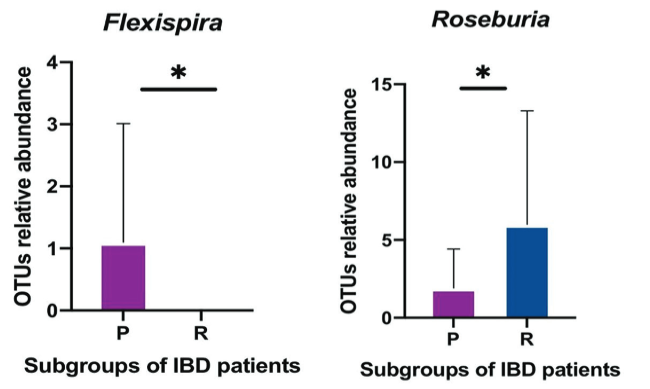
The largest microbial aggregation in the human body exists in the gastrointestinal tract. The microbiota in the host gastrointestinal tract comprises a diverse ecosystem, and the intestinal microbiota plays a vital role in maintaining gut homeostasis. This study aims to examine whether the gut microbiota influences unresponsiveness to anti-TNF-α treatments in primary nonresponder patients, and consequently identify the responsible microbes as biomarkers of unresponsiveness. Stool samples were collected from a cohort of patients with an established diagnosis of IBD, either ulcerative colitis (UC) or Crohn’s disease (CD), following completion of the induction phase of anti TNF therapy. 16S rRNA sequencing analysis was used to examine the pattern of microbiota communities in fecal samples. The quality and quantity of fecal microbiota were compared in responder and primary nonresponder IBD patients following anti-TNF-α therapy. As per our hypothesis, a difference in gut microbiome composition between the two patient subgroups was observed. A decreased abundance of short-chain fatty acid (SCFA)-producing bacteria, including Anaerostipes, Coprococcus, Lachnospira, Roseburia, and Ruminococcus, was detected in non-responsive patients, which was the hallmark of dysbiosis. Biomarkers of dysbiosis that were identified as predictors of clinical nonresponse, included Klebsiella, Eubacteriaceae, RF32, Bifidobacterium_animalis, and Muribaculaceae—previously known as S24-7. Signature biomarkers showed dramatic alteration in the composition of gut microbiota in patients who demonstrated primary nonresponse to anti-TNF-α agents. Dysbiosis, with features including a dropped biodiversity, augmentation in opportunistic pathogenic microbiota, and a lack of SCFA-producing bacteria, is a prominent feature of the microbiome of primary nonresponders to anti-TNF-α therapy.

Downloads

Download data is not yet available.

Downloads

Additional Files

Published

2022-06-01

How to Cite

1.
Alatawi H, Mosli M, Saadah OI, Annese V, Al-Hindi R, Alatawy M, Al-Amrah H, Alshehri D, Bahieldin A, Edris S. Attributes of intestinal microbiota composition and their correlation with clinical primary non-response to anti-TNF-α agents in inflammatory bowel disease patients. Biomol Biomed [Internet]. 2022Jun.1 [cited 2023Sep.23];22(3):412-26. Available from: https://www.bjbms.org/ojs/index.php/bjbms/article/view/6436

Issue

Section

Microbiology

INTRODUCTION

Ulcerative colitis (UC) and Crohn’s disease (CD) are the primary subtypes of inflammatory bowel disease (IBD), found in individuals with genetic susceptibility subject to particular environmental conditions [1]. IBD is a chronic and relapsing inflammatory condition of the gastrointestinal tract that occurs following immune system dysregulation [2]. Common clinical symptoms of IBD are abdominal pain, diarrhea, weight loss, and bloody stools [3,4].

One hundred trillion different microorganisms live in the human gut, including bacteria, viruses, fungi, and protozoa [5]. According to various molecular studies in a number of different cultures with various genetic profiles, more than 1000 species of microbiota colonize the gastrointestinal ecosystem, including Firmicutes, Bacteroidetes, Proteobacteria, and Actinobacteria [2,6].

The intestinal microbiota performs a vital role in the degradation of indigestible carbohydrates to produce short-chain fatty acids (SCFAs) and is important for vitamins synthesis (Vitamin K, Vitamin B12, and folic acid), amino acids synthesis, and regulation of fat metabolism. All these processes are necessary for the preservation of intestinal barrier functions [7]. The phyla Firmicutes and Bacteroidetes use the indigestible carbohydrates to produce SCFAs with the cooperation of bacteria specialized in the process of oligosaccharide fermentation, such as Bifidobacteria [8]. SFCAs – acetate, propionate, and butyrate – are the fundamental and essential end products of carbohydrate fermentation in the colon. Microbiota present in colonic epithelial cells consume butyrate to produce energy, while acetate and propionate remain in the intestinal cell [9].

Dysfunction in the composition and abundance of gut microbiota is associated with IBD severity. A significant decrease in the diversity of commensal bacteria, such as Bacteroides, Lactobacillus, Ruminococcus, Faecalibacterium, and Bifidobacterium, has also been observed in IBD patients. Low diversity of Roseburia spp. is associated with a high risk of IBD pathology [10].

A precise definition of primary nonresponse in IBD patients has not been established, nevertheless, an accepted definition with respect to the employment of anti-TNF therapy is a failure to fulfill clinical remission following the induction of a remedy period with documentation of adequate drug levels [11]. The role of the intestinal microbial composition in primary non-responders is not well understood. Several studies have shown no significant dissimilarity in the gut microbiome profile pre- and post-treatment with anti-TNF antagonists [12].

This study aims to determine whether the composition of gut microbiota has an effect on initial unresponsiveness to anti-TNF-α treatments in IBD patients. Accordingly, this study sets out to identify responsible microbiota and posits the utilization of the microbiota pattern as a biomarker to indicate unresponsiveness.

MATERIALS AND METHODS

Twenty Saudi Arabian IBD patients were recruited for this study from the outpatient Gastroenterology Department at King Abdulaziz University Hospital, in Jeddah, Kingdom of Saudi Arabia. The inclusion criteria were male or female patients that were 20–45 years old with a confirmed diagnosis of IBD, established through clinical, endoscopic, and histological criteria. Case record forms were obtained from patients, which included demographic and clinical data regarding gender, age, marital status, family history, diagnosis reports, environmental exposures (e.g., smoking), body mass index, and various laboratory parameters. The study was designed for patients treated with anti-TNF-α antagonists only (infliximab and adalimumab). The study mainly compared IBD patients that demonstrated no response (primary non-responders) to anti-TNF agents (n = 10) and patients that were judged to be responders to anti-TNF-α antagonists (n = 10). Primary non-response was defined as failure to demonstrate clinical remission following completion of the induction period anti-TNF therapy, that is, adalimumab or infliximab, without TDM. Similarly, response was defined as demonstration of clinical response or clinical remission at the end of induction.

The exclusion criteria were patients with contraindications to anti-TNF-α antagonists, including active tuberculosis, sepsis, or other severe opportunistic infections. Participants that were treated with medication, such as antibiotics, corticosteroids, mesalamine, and immunosuppressants, within 3 months before fecal collection were also excluded, as were pregnant or breastfeeding women and those that failed to submit stool samples as described at each phase of the study.

Sample collection and genomic DNA extraction

The fecal samples were collected, following completion of the induction phase of anti-TNF therapy, using the iSWAB-Microbiome Collection Kit and stored frozen at −20°C within 24 hours of collection. Genomic DNA of the gut microbes was extracted from the 200 μL fecal sample using PureLink Microbiome DNA Purification Kit (Invitrogen by Thermo Fisher Scientific, USA), following the instructions set out in the International Human Microbiome Standards project:

http://www.human-microbiome.org/

Polymerase chain reaction (PCR) DNA amplification and library preparation

Isolated genomic DNA of 20 stool samples was amplified for the target V3-V4 hypervariable regions of 16S rRNA, an intestinal bacteria DNA, using the universal primer set, Primer 341 Forward (5’-CCTACGGGNGGCWGCAG-3’) and Primer 806 Reverse (5-GACTACHVGGGTATCTAATCC-3’). The total PCR comprised 25 μL, as follows: 7.5 μL H2O, 12.5 μL DreamTaq PCR Master Mix, 3 μL DNA genome, and 1 μL forward/reverse primers. The thermal cycling condition of the PCR was carried out using an initial denaturation step at 95°C for 5 minutes followed by 40 cycles at 95°C for 30 seconds (denaturation), 56°C for 30 seconds (annealing), 72°C for 30 seconds (extension), and a final extension step at 72°C for 10 minutes, and a 4°C hold. A 5 μL of 5X DNA Loading Buffer Blue was added to the PCR product.

16S bioinformatics sequence analysis

Raw reads were refined using the Illumina MiSeq platform [13]. Paired end reads were generated using FLASH (v1.2.11) [14]. The reads sequenced with tags were clustered to operational taxonomic units (OTUs) at 97% sequence similarity using the USEARCH software (v7.0.1090) [15]. OTU representative sequences were classified according to the databases of 16S rDNA, with the Ribosomal Database Project Classifier (v.2.2) and Greengenes database [16], using 0.6 CI as a cutoff. A representative OTU phylogenetic tree was generated using the QIIME software (v1.80) (https://qiime2.org). A pre-filtering of selected OTUs was done according to the richness and evenness; at the initial analysis, richness and evenness were checked in all samples and OTUs that did not represent the richness and evenness in all samples were excluded from the study. The microbial alpha diversity indices were computed with the Mothur software (v1.31.2), and the microbiota beta diversity distance was generated by QIIME (v1.80). All unclassified bacteria features were discarded.

Ethical statement

This study was approved by the Research Committee, Unit of Biomedical Ethics, Faculty of Medicine at King Abdulaziz University, Jeddah, Saudi Arabia (Ref. No. 372-19). Informed consent was obtained from all patients.

Statistical analysis

An unpaired parametric t-test was incorporated using the Prism 9.0 software (GraphPad Software, Inc., San Diego, CA) and R (v3.1.1) to compare quantitative variables between groups and to identify taxa that had statistically significant differences. The resulting P value was modified using the Benjamini-Hochberg false discovery rate correction formula (function “p.adjust” in the stats package of R, v3.1.1). PERMANOVA was used to test significance among values. The linear discriminant analysis effect size (LEfSe) method was incorporated to reveal metagenomic biomarkers. The Pearson correlation coefficient was performed with a two-tailed test and a 95% CI in GraphPad Prism 9.0. Any differences between groups considered to be significant had p < 0.05.

Availability of data and material

The datasets (raw data), which supported the findings of the study, were deposited in the National Centre for Biotechnology Information (NCBI) with the unique BioProject ID: PRJNA673078 (https://dataview.ncbi.nlm.nih.gov/object/PRJNA673078).

RESULTS

Baseline characteristics

The study cohort included 20 patients with IBD (14 CD and 6 UC). The median age was 28 years (range, 20–45 years). Females constituted 65% (n = 13) of the cohort. The anti-TNF alpha treatment comprised infliximab in 11 patients (65%) and adalimumab in 9 patients (45%). Details of the study group are shown in Table 1.

TABLE 1: Clinical and demographic characteristics of IBD patient responders and non-responders to anti-TNF-α therapy.

Comparison of relative abundance of microbial community structure

The OTUs median number was statistically estimated to be 146 for primary non-responder patients (P) and 158 for responders (R). The expressed sequence tags were clustered into OTU with a 97% threshold Figure S1. The taxonomic composition distribution was used to identify the intestinal microbiota of IBD patients at phylum and genus levels [Figure 1A and B]. The largest number at the genera level was the Firmicutes phylum (57 genera), followed by Proteobacteria (18 genera), then Actinobacteria (18 genera), and then Bacteroidetes (11 genera) [Figure 1C].

FIGURE 1: The taxonomic composition distribution of relative abundance (A) at the phylum level, (B) the genus level among the samples of both groups; (P) refers to primary non-responders and (R) to responders. (C) is a phylogenetic tree showing the evolutionary distance in biological taxa.

At the phylum level, the fecal microbiota of primary non-responders were characterized by the increased relative abundance of Proteobacteria and Actinobacteria [Figure 2A] compared to responders [Figure 2B]. Interestingly, the populations of Firmicutes did not manifest obvious changes between the two primary subgroups of IBD patients (UC and CD) [Figure 2B]. The ratio of Bacteroidetes was lower in primary non-responders compared with responders. The Pearson correlation coefficient showed a strong positive correlation in gut microbiota composition between responder and non-responder IBD patients [Figure 2C]. Furthermore, an independent t-test showed no statistically significant difference between the relative abundance of predominant phyla of gut microbiota in response to anti-TNF-α treatment between the two IBD patient groups [Figures 3 and S1].

FIGURE 2: Predominant phyla: comparison of the relative abundance at the phyla level in faecal samples (A) primary non-responders, and (B) responders. (C) is a Pearson correlation coefficient graph showing r = 0.9900, 95% confidence interval = 0.9677 to 0.9969, and R squared = 0.9801.
FIGURE 3: Independent samples t-test statistically showing phyla of the gut microbiota. All P values were non-significant for responders (R=blue) and primary non-responders (P=purple) to anti-TNF-α therapies.

At the family level [Figure S2], the relative abundance of the Clostridiaceae family was stable in the two subgroups of patients. However, there was a remarkable increase in several families, including Corynebacteriaceae, Eubacteriaceae, Desulfovibrionaceae, Lachnospiraceae, Streptococcaceae, and Muribaculaceae – previously known as S24-7 – in non-responders. Furthermore, the diversity of Lactobacillaceae, Ruminococcaceae, Alcaligenaceae, Mogibacteriaceae, Peptostreptococcaceae, Veillonellaceae, Prevotellaceae, and Bacteroidaceae decreased in non-responders in comparison with responders.

At the genus level [Figure 1B] and species level [Figure S3], a rise and fall in the relative abundance of intestinal genera and species appeared to directly affect patient ability to respond to the drug. Several SCFA-producing bacteria were omitted in unresponsive patients (primary non-responders). These included Anaerostipes, Coprococcus, Lachnospira, Roseburia, and Ruminococcus. By contrast, some SCFA-producing bacteria showed an increase in relative abundance in primary non-responders, such as Blautia, Faecalibacterium, Lachnobacterium, Odoribacter, Oscillospira, and Pseudoramibacter eubacterium. An increased diversity of several pathogenetic gut microbiota was observed in primary non-responders, including Enterococcus, Fusobacterium (adherent-invasive bacteria), Clostridium, Streptococcus, Staphylococcus, Actinomyces, Corynebacterium, Rothia, Rhodococcus, Atopobium, Listeria, Peptostreptococcus, Clostridium, Campylobacter, Flexispira, Morganella, Actinobacillus, Selenomonas, Haemophilus, Acinetobacter, and Klebsiella. Furthermore, a great abundance of opportunistic pathogens such as Muribaculaceae, Proteus, Odoribacter, Alistipes, Veillonella, and Pseudomonas was recorded in fecal samples of primary non-responders. An independent t-test showed a statistically significant association (p < 0.05) for six genera and seven species [Figure 4]. These results suggest that primary non-responders to anti-TNF-α treatment possess a type of unstable gut microbiota structure.

FIGURE 4: Independent samples t-test showing responders (R=blue) and primary non-responders (P=purple) with statistically significant P values in Pediococcus (*P< 0.034983), Proteus (*P< 0.040331), Shigella (*P< 0.010478), Roseburia (*P< 0.04166), Flexispira (*P< 0.034983), and Klebsiella (*P< 0.016427)

Alpha and beta diversity

The alpha diversity was measured in observed species (Sobs) (p = 0.79118), using the Chao1 estimator (p = 0.63053), ACE (p = 0.57874), Shannon diversity (p = 0.79594), and Simpson diversity (p = 0.63053) indices [Figure S4 and Table S1]. In general, in terms of alpha diversity, there was no significant difference in relative abundance between responders and primary non-responders [Figure 5 and Table S2].

FIGURE 5: Plot of microbial alpha diversity in patients responding to anti-TNF-α treatment, calculated using observed species (A) [Sobs], (B) Chao, (C) ACE, (D) Shannon, and (E) Simpson indices in different faecal samples. Alpha diversity was non-significant. Primary non-responders (P) = purple, and responders (R) = blue.

The beta diversity analysis was estimated using the Bray–Curtis dissimilarity, for unweighted and weighted UniFrac distances Figure S5. These tools showed that the composition of intestinal microbiota of primary non-responders overall shifted toward dysbiosis compared with responders. Principal coordinate analysis and 2D-weighted UniFrac showed a significant overlap between the gut microbiota structure of responders and primary non-responders [Figure 6A and B]. The weighted UniFrac heat map was used to determine the intestinal microbiota profile distances in IBD patients, between responders and non-responders. The microbial community composition of primary non-responders showed the proportion of change (disorder) compared with responders [Figure 6C]. The weighted UniFrac phylogenetic tree indicated that the community shared a lot of evolutionary history at the phylum level. However, at the genus and species level, the microbial community tends to diverge. Overall, half of the phylogenetic tree of unresponsive individuals were unique to responders, with a distance of 0.5 (fraction) [Figure 6D].

FIGURE 6: (A) Principal coordinate analysis (PCoA). (B) 2D-weighted UniFrac profile of β-diversity for microbial diversity among stool samples. (C) Weighted-UniFrac heat map. (D) Phylogenetic tree indicating presence of clear clustering in microbial structure.

LEfSe analysis

The LEfSe analysis suggests that Klebsiella sp. and Bifidobacterium animalis robustly correlated with primary non-response [Figure 7A]. Furthermore, Eubacteriaceae, Streptococcaceae (RF32), and Muribaculaceae (S24-7) families are characterized by non-responsiveness to anti-TNF-α agents [Figure7A and B]. Response to anti-TNF-α agents was distinguished by the presence of Bacteroides caccae, hence, the Bacteroidetes phylum can be considered as a biomarker for response.

FIGURE 7: LEfSE shows (A) Negative LDA scores were enriched in primary non-responders with five taxa. Positive LDA were enriched in responders with one taxon; red (P) indicates primary non-responders and green (R) indicates responders. (B) Cladogram tree of biomarkers.

DISCUSSION

This study examined the microbiota pattern of IBD patients that did not respond to anti-TNF-α therapies compared to those of demonstrated response [4,17]. The gut microbiota were compared to assess the nature of the dysbiosis and identify the microbes that could potentially be responsible for dysbiosis, and consequently to a lack of response. The present study depended on next-generation sequencing technologies, especially for the 16S rRNA gene sequencing, which permits describing the gut microbiota architecture. The 16S rRNA gene sequencing is a taxonomic genomic marker that is specialized for examination of a particular part of the microbiota, the bacteria, and archaea, regardless of other microorganisms, such as virobiota, mycobiota, and eukaryota [18]. The overall vision of the study is to identify microbiota structures that could be used as a prognostic indicator for clinical non-response to anti-TNF-α therapy in Saudi Arabian patients with IBD. According to the previous literature, it has been shown that high concentrations of Klebsiella, S24-7, Eubacteriaceae, and Bifidobacterium animalis are associated with clinical nonresponse to anti-TNF-α treatment, while low concentrations of B. caccae can be used as a biomarker for response. Patients that demonstrated nonresponse to treatment had a characteristic reduction of SCFA-producing bacteria or a shift toward an inflammation-producing bacterium [19]. Furthermore, a greater number of opportunistic pathogens such as Muribaculaceae, Proteus, Odoribacter, Alistipes, Veillonella, and Pseudomonas were recorded in fecal samples of primary non-responders.

In this study, we did not observe a great dissimilarity in gut microbiota composition between responders and non-responders. Only moderate differences were found, with limited statistical significance. Of particular note, the majority of the findings were consistent with some previous studies on the gut microbiome in the context of different diseases, such as spondyloarthritis [12,20]. An increased abundance of Proteobacteria and Actinobacteria in non-responders was observed in this cohort. An outgrowth of these phyla has been associated with gastric bypass, metabolic disruption, inflammation, and cancer [21]. By contrast, the proportion of Bacteroidetes phylum decreased in non-responders compared with responders. An unexpected result was the detection of an elevation of Firmicutes phylum biodiversity in non-responders compared with responders. This finding conflicts with several studies that demonstrated a reduced concentration of the Firmicutes phylum in non-responders [18].

At the family level, the non-responders showed a remarkable decline in the Bacteroidaceae and Lactobacillaceae populations. Members of the Lactobacillaceae family have an essential role in the fermentation of kefir, which has been suggested to be a probiotic that may play a role in the treatment of IBD [22]. However, an increased abundance of Bifidobacteriaceae, Lachnospiraceae, Enterobacteriaceae, and Muribaculaceae was observed in non-responders compared to responders. A logical interpretation of the presence of increased populations of SCFA-producing bacteria including Bifidobacteriaceae, Lachnospiraceae, and Muribaculaceae in primary non-responders is a shift toward harboring pro-inflammatory bacteria, thereby promoting inflammation and ultimately leading to worsening conditions in primary non-responders [19].

A variation in the diversity of genera within one family – the Lachnospiraceae family – was detected where intestinal genera had a varied presence in non-responders. The abundance of Roseburia and Coprococcus genera decreased while the concentration of Blautia and Clostridium increased. Furthermore, a variation in abundance was observed in the Ruminococcaceae family: Where the diversity of Feaclbacterium genus increased while the diversity of the Ruminococcus genus decreased. This is consistent with some previous studies [23].

The Enterococcus genus is considered an aggressive genus that is characteristically highly abundant in the fecal samples of non-responders. The virulence and bacteriocinogenic features of the Enterococcus genus have been detected in the human gut. The Enterococcus genus has been associated with intestinal inflammation and a number of other infections, such as bloodstream and urinary tract infections, endocarditis, and peritonitis [24]. The decreased abundance of Escherichia was observed in primary non-responders compared with responders in the present study, which is inconsistent with several previous IBD studies [25]. Aside from these findings, the Fusobacterium genus increased in non-responsive patients, which coheres with some previous studies [26].

The concentration of Klebsiella and Proteus increased in primary non-responders. However, the Bacteroides fragilis (spp.) population, a human commensal bacterial, decreased in non-responders. This result corresponds with the previous studies [27]. A deficiency in several SCFA-producing bacteria in the fecal samples of non-responders was found, including the genera Anaerostipes, Coprococcus, Lachnospira, Roseburia, and Sutterella. This outcome is in line with a number of the previous studies’ [28]. SCFA-producing bacteria contribute to shaping the architecture of the gut community, organize transepithelial transport, are important for intestinal motility, and regulate the microbial homeostasis within the intestine. Moreover, SCFA has important immunomodulatory and anti-inflammatory characteristics [29]. Despite the fact that B. animalis and Faecalibacterium prausnitzii are used as probiotics, an increase in the concentration of these species was observed in primary non-responders and therefore may be used as a biomarker (B. animalis) for similar patients. This increase may be interpreted in rare cases as a result of high consumption of probiotics, associated with a high risk of adverse reactions between microbiota and their hosts. Consequently, this phenomenon may be linked with a number of diseases, such as bacterial infections and sepsis [30].

The Desulfovibrio genus, which belongs to the Proteobacteria phylum, is heterogeneous sulfate-reducing bacteria [31]. An increase in the Desulfovibrio genus has been associated with an elevated glutathione and riboflavin metabolism [32], rising toxins production, and increased bacterial genes attached with virulence agents. In the present study, the abundance of the Desulfovibrio genus was increased in non-responders compared with responders. The high diversity of Desulfovibrio in such patients is consistent with a number of other studies [33].

The present study is limited by its small sample size and lack of TDM measurements, which is typically used to document adequate trough levels and absence of anti-drug antibodies that are necessary to prove primary nonresponse. Nevertheless, this is the first study to examine the microbiota of Saudi IBD patients as potential biomarkers for response to anti-TNF therapy and as such can help pave the way for further larger studies.

CONCLUSION

The fecal microbiota of primary non-responders to anti-TNF-α therapy demonstrated dysbiosis, in addition to other features, such as decreased biodiversity, augmentation in opportunistic pathogenic microbiota, and a lack of SCFA-producing bacteria. A decreased abundance of SCFA-producing bacteria, including Anaerostipes, Coprococcus, Lachnospira, Roseburia, and Ruminococcus, was detected in unresponsive patients. Particular fecal microbiota may be used as biomarkers to predict clinical response to anti-TNF-α medication. The potential biomarkers for primary non-response patients identified were Klebsiella, Eubacteriaceae, RF32, B. animalis, and Muribaculaceae.

REFERENCES

  1. , , , , , (). Nrf2 suppresses macrophage inflammatory response by blocking proinflammatory cytokine transcription. Nat Commun. https://doi.org/10.1038/ncomms11624
  2. , , , , , (). Gut microbial colonisation in premature neonates predicts neonatal sepsis. Arch Dis Child Fetal Neonatal Ed. https://doi.org/10.1136/fetalneonatal-2011-301373
  3. , , , , , (). Interactions between intestinal microbiota and host immune response in inflammatory bowel disease. Front Immunol. https://doi.org/10.3389/fimmu.2017.00942
  4. , , , (). Epidemiology and natural history of inflammatory bowel diseases. Gastroenterology. https://doi.org/10.1053/j.gastro.2011.01.055
  5. , , , , , (). Evolution of mammals and their gut microbes. Science. https://doi.org/10.1126/science.1155725
  6. , , , , , (). Dysbiosis of gut microbiota in inflammatory bowel disease:Current therapies and potential for microbiota-modulating therapeutic approaches. Bosn J Basic Med Sci. https://doi.org/10.17305/bjbms.2020.5016
  7. , (). Learning tolerance while fighting ignorance. Cell. https://doi.org/10.1016/j.cell.2009.07.024
  8. (). Physiological role of gut microbiota for maintaining human health. Digestion. https://doi.org/10.1159/000444066
  9. , , (). Carbohydrate fermentation in the human colon and its relation to acetate concentrations in venous blood. J Clin Invest. https://doi.org/10.1172/jci111847
  10. , , , , , (). Mucosal flora in inflammatory bowel disease. Gastroenterology. https://doi.org/10.1053/gast.2002.30294
  11. , , , , (). Efficacy, tolerability, and predictors of response to infliximab therapy for Crohn's disease:A large single centre experience. J Crohns Colitis. https://doi.org/10.1016/j.crohns.2011.07.011
  12. , , , , , (). Primary non-response in inflammatory bowel disease, definition, potential causes, therapeutic drug monitoring and microbiota-a review. Appl Ecol environ Res. https://doi.org/10.15666/aeer/1804_55055525
  13. , , , , , (). An improved dual-indexing approach for multiplexed 16S rRNA gene sequencing on the Illumina MiSeq platform. Microbiome. https://doi.org/10.1186/2049-2618-2-6
  14. , (). FLASH:Fast length adjustment of short reads to improve genome assemblies. Bioinformatics. https://doi.org/10.1093/bioinformatics/btr507
  15. , (). Error filtering, pair assembly and error correction for next-generation sequencing reads. Bioinformatics. https://doi.org/10.1093/bioinformatics/btv401
  16. , , , , , (). Greengenes, a chimera-checked 16S rRNA gene database and workbench compatible with ARB. Appl Environ Microbiol. https://doi.org/10.1128/aem.03006-05
  17. , , , , , (). Metabonomics and the gut microbiome associated with primary response to anti-TNF therapy in Crohn's disease. J Crohns Colitis. https://doi.org/10.1093/ecco-jcc/jjaa039
  18. , , , , , (). Microbiota composition may predict anti-TNF alpha response in spondyloarthritis patients:An exploratory study. Sci Rep. https://doi.org/10.1038/s41598-018-23571-4
  19. , , , , , (). Characteristics of faecal microbiota in paediatric Crohn's disease and their dynamic changes during infliximab therapy. J Crohns Colitis. https://doi.org/10.1093/ecco-jcc/jjx153
  20. , , , , , (). The oral and gut microbiomes are perturbed in rheumatoid arthritis and partly normalized after treatment. Nat Med.
  21. , , (). Proteobacteria:Microbial signature of dysbiosis in gut microbiota. Trends Biotechnol. https://doi.org/10.1016/j.tibtech.2015.06.011
  22. , , , , , (). Local and systemic immune mechanisms underlying the anti-colitis effects of the dairy bacterium lactobacillus delbrueckii. PLoS One. https://doi.org/10.1371/journal.pone.00←3
  23. , , , , , (). Altered intestinal microbiota-host mitochondria crosstalk in new onset Crohn's disease. Nat Commun. https://doi.org/10.1038/ncomms13419
  24. , , , , , (). Virulence factors of Enterococcus strains isolated from patients with inflammatory bowel disease. World J Gastroenterol. https://doi.org/10.3748/wjg.v19.i23.3562
  25. , , , , , (). Host-mediated inflammation disrupts the intestinal microbiota and promotes the overgrowth of enterobacteriaceae. Cell Host Microbe. https://doi.org/10.1016/j.chom.2007.06.010
  26. , , , , , (). Fusobacterium varium localized in the colonic mucosa of patients with ulcerative colitis stimulates species-specific antibody. J Gastroenterol Hepatol. https://doi.org/10.1046/j.1440-1746.2002.02834.x
  27. , , , , , (). Induction of persistent colitis by a human commensal, enterotoxigenic Bacteroides fragilis, in wild-type C57BL/6 mice. Infect Immun. https://doi.org/10.1128/iai.00814-08
  28. , , , , , (). Reduced abundance of butyrate-producing bacteria species in the fecal microbial community in Crohn's disease. Digestion. https://doi.org/10.1159/000441768
  29. , , , , , (). Metabolites produced by commensal bacteria promote peripheral regulatory T-cell generation. Nature. https://doi.org/10.1038/nature12726
  30. , (). Risk and safety of probiotics. Clin Infect Dis.
  31. , , , (). Desulfovibrio desulfuricans bacteremia and review of human Desulfovibrio infections. J Clin Microbiol. https://doi.org/10.1128/jcm.41.6.2752-2754.2003
  32. (). Inventory of a reservoir:Friends And foes. Indian J Med Res.
  33. , , , , , (). Integrated metagenomics/metaproteomics reveals human host-microbiota signatures of Crohn's disease. PLoS One. https://doi.org/10.1371/journal.pone.0049138

Conflicts of interest: The authors declare no conflicts of interest.

Funding: The authors received no specific funding for this work.