

Review of roles of RNA-binding proteins on NAFLD and the related pharmaceutical measures
Abstract
Nonalcoholic fatty liver disease (NAFLD) is the most common chronic liver disease worldwide and poses a serious threat to public health. NAFLD is considered a risk factor for metabolic syndrome (MS) and is closely associated with type 2 diabetes mellitus (T2DM), obesity, dyslipidemia, and cardiovascular disease. Recently, increasing attention has been paid to the role of RNA-binding proteins (RBPs) in the pathogenesis of NAFLD. A growing body of research has linked RBPs—such as human antigen R (HuR), sequestosome 1 (p62), polypyrimidine tract-binding protein 1 (PTBP1), and heterogeneous nuclear ribonucleoproteins (hnRNPs)—to lipogenesis and inflammation, both of which contribute to NAFLD through mechanisms involving transcriptional regulation, alternative splicing, RNA stability, polyadenylation, and subcellular localization. However, these findings are often fragmented and lack a comprehensive synthesis. The interactions and mechanisms between RBPs and NAFLD have not yet been thoroughly reviewed. This article provides an overview of the roles and mechanisms of various RBPs in NAFLD, summarizing current knowledge with the aid of figures and tables. In particular, it highlights the influence of HuR on NAFLD through multiple pathways, categorizing its effects based on increased or decreased expression. Furthermore, it reviews drugs that alleviate NAFLD by modulating RBPs, aiming to offer valuable insights for drug-targeted therapies based on RBP regulatory networks.
Keywords
RNA binding proteins RBP nonalcoholic fatty liver disease NAFLD therapy mechanismFull text
Introduction
Nonalcoholic fatty liver disease (NAFLD) is one of the most prevalent liver disorders worldwide, affecting over 25% of the adult population and imposing significant social and economic burdens [1]. It is characterized by the accumulation of lipids in hepatocytes and elevated levels of liver-related enzymes. Steatosis is diagnosed when at least 5% of liver cells exhibit excessive fat deposition, particularly in individuals who either abstain from alcohol or limit their intake to no more than 20 g/day for women and 30 g/day for men [2, 3]. NAFLD encompasses a spectrum ranging from simple intrahepatic fat accumulation (steatosis) to nonalcoholic steatohepatitis (NASH), which involves hepatocyte inflammation and cell death and may progress to fibrosis, cirrhosis, and hepatocellular carcinoma (HCC) [4, 5]. NASH plays a central role in the progression of NAFLD, with hepatic fibrosis identified as the primary predictor of liver-related mortality [6]. A recent model predicts a 178% increase in liver-related deaths associated with NASH by 2030 [7]. More recently, researchers in the field have proposed the term “MAFLD” (metabolic associated fatty liver disease) as a more appropriate overarching label, reflecting the growing recognition of the condition’s metabolic underpinnings. Like NAFLD, MAFLD represents the hepatic manifestation of a multisystem disorder [8]. Nevertheless, this review will continue to use the term NAFLD to align with its widespread usage in the literature reviewed.
The leading explanation for the etiology and pathophysiology of NAFLD is the “multiple factors” hypothesis. This model encompasses fat accumulation, insulin resistance (IR), oxidative stress, mitochondrial dysfunction, endoplasmic reticulum stress (ERS), disruptions in hepatic lipid and bile acid (BA) metabolism, alterations in the gut microbiota, and genetic predispositions [9]. NAFLD is a multifactorial disease strongly associated with metabolic syndrome (MS), type 2 diabetes mellitus (T2DM), and obesity, with IR recognized as a central component in its pathogenesis [10]. As the disease progresses, IR worsens and activates lipogenic enzymes via sterol regulatory element-binding protein 1c (SREBP1c), thereby promoting lipogenesis [11, 12]. Furthermore, IR diminishes insulin’s ability to suppress lipolysis in adipose tissue, leading to elevated levels of free fatty acids (FFAs) delivered to the liver [13, 14].
RNA-binding proteins (RBPs) play a central role in coordinating RNA processing and post-transcriptional gene regulation (PTGR), including the maturation, localization, stabilization, and translation of both coding and non-coding RNAs [15]. RBPs interact with RNA through RNA-binding domains (RBDs), which are primarily found in the 5′ and 3′ untranslated regions (UTRs) of target RNAs. Most RBPs contain multiple RBDs, enhancing their affinity and specificity for target mRNAs. A single RBP can regulate the expression of numerous mRNAs, while multiple RBPs may bind to the same mRNA, either cooperating or competing in their regulatory functions [16, 17]. Loss of RBP function or functional mutations can disrupt cellular homeostasis and contribute to metabolic disorders such as NAFLD [18, 19].
Although existing studies have explored the role of RBPs in NAFLD, their scope is often limited by the vast diversity of RBPs. The primary aim of this work is to provide a comprehensive review of RBPs that have a relevant impact on NAFLD, with a detailed summary of their roles and underlying mechanisms. Additionally, growing interest in drug development targeting RBP-regulated pathways has led to the emergence of several RBP-based therapeutic strategies. This review also discusses such drug candidates, reflecting the critical role of RBPs and their RNA interactions in the progression of NAFLD.
RBPs in NAFLD
Hu proteins
The embryonic lethal abnormal vision (ELAV)/Hu proteins represent an important family of RBPs, comprising four mammalian members encoded by distinct genes: HuB, HuC, HuD, and human antigen R (HuR) (also known as human antigens B, C, D, and R). These proteins play key roles in enhancing gene expression at the post-transcriptional level [20]. HuR, which is broadly expressed across tissues, stabilizes and/or promotes the translation of mRNAs encoding pro-inflammatory mediators in the cytoplasm, serving as a critical regulator of inflammatory and immune responses [21]. In contrast, HuB, HuC, and HuD are primarily expressed in neurons, where they contribute to neuronal differentiation, axonal growth, and the maintenance of neuronal integrity [22].
HuR
HuR, encoded by the ELAV-like 1 (Elavl1) gene, is located on human chromosome 19p13.2 [23]. Its mRNA is approximately 6 kb in length and encodes a protein with a molecular weight of 36 kDa. Functionally, HuR regulates post-transcriptional gene expression by modulating the stability and activity of its target RNAs. Structurally, HuR contains three RNA recognition motifs (RRMs): a tandem RRM1 and RRM2, connected by a flexible linker, and a C-terminal RRM3 [24]. Although primarily localized in the nucleus, HuR can translocate to the cytoplasm upon specific stimuli, a process mediated by a shuttling sequence located between RRM2 and RRM3 [20]. In the cytoplasm, HuR interacts with its target mRNAs to exert diverse regulatory effects—most commonly promoting mRNA stability and enhancing translation, although in certain tissues it may also suppress these functions [25, 26]. Recently, HuR has gained attention for its critical roles in cell signaling, inflammation, fibrosis, and the development of HCC [27]. Several studies have also identified hepatic HuR as a key player in NAFLD progression through its regulation of lipid and glucose metabolism, modulation of lipid transport, and suppression of adipogenesis [28–30]. A recent study reported a significant reduction in HuR expression in the livers of mice fed a high-fat diet (HFD). However, in contrast to this observation, a separate study by Zhang et al. [31] found that hepatic HuR deficiency actually worsened HFD-induced NAFLD. These conflicting results may be due to differences in the HFD conditions used across studies involving HuR-deficient mice. In the latter study, HuR deficiency led to increased levels of triglyceride (TG) and cholesterol ester (CE) species, along with decreased expression of genes involved in cholesterol biosynthesis and the BA-activated farnesoid X receptor (FXR)/retinoid X receptor (RXR) pathway. As a result, these mice exhibited heightened inflammation and fibrosis, ultimately progressing to HCC-like tumor development [28]. In HuR-deficient livers, serum taurocholic acid (TCA), macrophage markers, innate immune response genes, multiple chemokines, and the long non-coding RNA (lncRNA) LINC01018 were significantly upregulated. In contrast, levels of tauroursodeoxycholic acid (TUDCA)—a BA known to inhibit ERS—were reduced [28, 32]. Additionally, genes involved in fatty acid biosynthesis, including acetyl-CoA carboxylase (Acc1), fatty acid synthase (Fas), elongation of very-long-chain fatty acids member 6 (Elovl6), and fatty acid desaturases 1 and 2 (Fads1 and Fads2), were markedly upregulated [32]. As summarized in Figure 1, HuR plays a regulatory role in NAFLD progression by modulating ERS and fatty acid metabolism.
The lncRNA H19, a 2.3 kb RNA molecule, has emerged as a key regulator of hepatic lipid metabolism and ERS [33, 34]. H19 promotes steatosis and lipid accumulation by modulating several pathways, including the miR-130a/peroxisome proliferator-activated receptor gamma (PPARγ) axis, MLX-interacting protein-like (MLXIPL) expression, and mechanistic target of rapamycin complex 1 (mTORC1) signaling [35, 36]. Mechanistically, HuR deficiency leads to upregulation of H19 and sphingosine-1-phosphate receptor 2 (S1PR2), while suppressing sphingosine kinase 2 (SphK2) expression, thereby promoting inflammation and hepatic lipid accumulation (as illustrated in Figure 1). In summary, HuR functions as a critical regulator of hepatic lipid metabolism, enterohepatic BA homeostasis, inflammation, and fibrosis. Hepatocyte-specific deletion of HuR exacerbates these pathological changes, underscoring its potential as a therapeutic target for NAFLD [32].
Phosphatase and tensin homolog deleted on chromosome 10 (PTEN) functions as a lipid phosphatase that dephosphorylates phosphatidylinositol-3,4,5-trisphosphate (PIP3), thereby inhibiting downstream signaling of phosphatidylinositol 3-kinase (PI3K) and reducing AKT activity [37]. In the liver, PTEN plays a critical role in regulating lipogenesis, glucose metabolism, and hepatocyte homeostasis. Liver-specific PTEN deficiency promotes NAFLD and hepatocarcinogenesis, while also improving glucose tolerance [38]. In contrast, mice overexpressing PTEN are protected against hepatic steatosis [39] (see Figure 1). HuR binds to the 3′UTR of PTEN mRNA, enhancing its stability and translation. In HuR-deficient mice, overexpression of PTEN alleviates diet-induced hepatic fat accumulation, highlighting a protective axis involving HuR and PTEN in NAFLD pathogenesis [30].
HuR interacts with intron 24 of Apob pre-mRNA, the 3′UTR of UQCRB mRNA, and the 5′UTR of NDUFB6 mRNA, thereby regulating Apob mRNA splicing and the translation of UQCRB and NDUFB6. Hepatocyte-specific deletion of HuR reduces the expression of Apob, UQCRB, and NDUFB6 in mice, impairing ATP synthesis and hepatic lipid transport, which in turn exacerbates HFD-induced NAFLD [31]. As illustrated in Figure 1, HuR deficiency worsens NAFLD under HFD conditions. Given HuR’s complex role in insulin sensitivity, a therapeutic strategy combining HuR modulation with insulin sensitizers—such as thiazolidinediones, glucagon-like peptide-1 (GLP-1) receptor agonists, biguanides (e.g., metformin), and dipeptidyl peptidase IV (DPP-4) inhibitors—may provide more effective treatment for NAFLD [40]. Interestingly, increases in the expression of HuR, NDUFB6, CYCS, Apob-100, UQCRB, and Apob-48 following oral metformin administration may represent an off-target effect of metformin.
LINC01018 is a non-conserved intergenic lncRNA located on chromosome 5 and is highly expressed in the liver. In patients with NAFLD, hepatic expression of LINC01018 is significantly reduced compared to healthy controls. Interestingly, dietary intervention with low carbohydrate intake in NAFLD patients restores LINC01018 expression, along with the upregulation of genes associated with its activity. LINC01018 modulates fatty acid metabolism through its interaction with HuR [41]. In humanized mouse models, reducing hepatic HuR expression by more than 60% results in a marked decrease in the expression of three key genes regulated by LINC01018: ADH1C, CYP4A11, and ALDH5A1. This downregulation contributes to the development and progression of NAFLD (Figure 1).
Apolipoprotein A-IV (APOA4) is a plasma lipoprotein primarily synthesized in the liver and small intestine [1]. In mice, APOA4 promotes hepatic TG secretion, thereby increasing plasma TG levels [42]. APOA4-AS, an antisense lncRNA, acts as a coordinated regulator of APOA4 expression, exhibiting a parallel expression pattern. Both APOA4-AS and APOA4 are elevated in the livers of individuals with NAFLD. Knockdown of APOA4-AS in vitro and in vivo reduces APOA4 expression, leading to decreased plasma TG and total cholesterol (TC) levels in mice. Mechanistically, APOA4-AS exerts its regulatory function by directly binding to HuR, which stabilizes APOA4 mRNA. Deletion of HuR leads to downregulation of both APOA4-AS and APOA4 transcripts [43]. As illustrated in Figure 1, HuR contributes to increased TG and TC levels through its stabilizing effect on APOA4-AS and APOA4.
CCAAT/enhancer-binding protein beta (C/EBPβ), a member of the C/EBP transcription factor family, plays a pivotal role in initiating adipogenesis and contributes to diabetes pathogenesis by regulating key metabolic enzymes such as phosphoenolpyruvate carboxykinase (PEPCK) in both adipose tissue and the liver. In models of NASH induced by a methionine–choline-deficient (MCD) diet, C/EBPβ expression is markedly upregulated. Hepatic deletion of C/EBPβ protects against excessive TG accumulation, inflammation, ERS, and oxidative stress. In contrast, overexpression of C/EBPβ exacerbates activation of PPARγ and nuclear factor kappa B (NF-κB) signaling pathways [44]. Previous studies have shown that HuR facilitates the nucleocytoplasmic transport and stability of C/EBPβ mRNA during the early stages of adipogenesis [45, 46] (see Figure 1). While direct evidence in the context of liver disease is still lacking, these findings suggest that HuR may regulate C/EBPβ expression and thereby influence the development and progression of NAFLD.
HAMP, the gene encoding human hepcidin, is primarily expressed in the liver and plays a central role in regulating iron absorption from the duodenum and iron release from macrophages [47]. In the context of NAFLD, studies have shown that elevated levels of saturated fatty acids in the liver enhance HuR translocation from the nucleus to the cytoplasm. This translocation promotes HuR binding to the 3′UTR of HAMP mRNA, resulting in increased HAMP expression in hepatocytes [48]. Modulating this HuR-mediated regulation of HAMP may offer therapeutic potential in slowing or preventing the progression of NAFLD (Figure 1).
Treatment with obeticholic acid (OCA) increases the expression of low-density lipoprotein receptor (LDLR) in the liver, leading to reduced plasma levels of low-density lipoprotein cholesterol (LDL-C) in mice. This effect is mediated through activation of the FXR, which induces the expression of HuR—a post-transcriptional regulator that stabilizes LDLR mRNA [49]. Deletion of HuR blocks OCA-induced upregulation of LDLR and impairs plasma cholesterol clearance via the LDLR pathway (Figure 1).
ABCA1 plays a key role in reverse cholesterol transport by facilitating cholesterol efflux and the biogenesis of high-density lipoprotein (HDL). HuR enhances ABCA1 expression by binding to its 3′UTR and stabilizing its mRNA. Silencing HuR reduces ABCA1 expression and impairs cholesterol efflux to apolipoprotein A1 (APOA1) in both hepatocytes and macrophages [50]. As illustrated in Figure 1, HuR supports reverse cholesterol transport and HDL formation by promoting ABCA1 expression.
A recent study found that mice lacking HuR specifically in adipose tissue were more susceptible to HFD-induced metabolic dysfunction, exhibiting increased IR and inflammation. HuR reduces lipid accumulation and lowers serum TC and HDL cholesterol (HDL-C) by stabilizing and promoting the translation of adipose TG lipase (ATGL) mRNA [29] (Figure 1).
HuR protects liver cells from oxidative damage caused by excessive fat accumulation by regulating the stability of manganese-dependent superoxide dismutase (MnSOD) and heme oxygenase-1 (HO-1) mRNAs. Researchers have identified ten HuR-binding sites within the 3′UTR of MnSOD mRNA, and HuR knockdown leads to reduced MnSOD mRNA and protein levels [51]. In mice, decreased MnSOD levels intensify oxidative stress, worsening the pathological features of NASH [52]. Similarly, lower MnSOD levels have been observed in NASH patients, with certain genetic variants affecting MnSOD function and correlating with increased NASH risk [53, 54]. Recent studies have also shown that administration of the HO-1 inducer hemin significantly alleviates steatosis, inflammation, and fibrosis in mice fed an MCD diet, while also reducing serum ALT and AST levels by inhibiting both canonical and non-canonical Wnt signaling pathways [55]. In MCD-induced models of NASH, HuR has been shown to regulate the stability of both MnSOD and HO-1 mRNAs. Reduced HuR expression correlates with decreased MnSOD and HO-1 levels, suggesting that impaired HuR-mediated regulation of antioxidant enzymes may contribute to the progression from simple steatosis to NASH [56] (Figure 1).
Angiotensin II (Ang II) and its type 1 receptor (AT1R) have been identified as key contributors to the progression of NAFLD. AT1R blockers have shown promise in ameliorating fatty liver and are being explored as potential therapeutic agents for NAFLD [57, 58]. AT1R-associated protein (ATRAP, also known as AGTRAP) functions as a negative regulator of AT1R by directly binding to it, thereby attenuating the effects of Ang II [59]. S-adenosylmethionine (SAM), a methyl donor involved in epigenetic and protein modifications, has been shown to influence the expression of ATRAP by modulating HuR activity [60]. Specifically, ATRAP mRNA interacts with HuR, and SAM preserves HuR methylation, which is essential for the nucleocytoplasmic shuttling of ATRAP mRNA. This, in turn, enhances ATRAP protein production and may help mitigate NAFLD progression (Figure 1). These findings underscore HuR’s critical role in mRNA transport, a regulatory mechanism with implications for NAFLD pathogenesis [61]. Additionally, several bioactive compounds—including berberine, quercetin, and apigenin—have been found to inhibit HuR, thereby reducing the expression of HuR-regulated genes, particularly those involved in inflammation. This anti-inflammatory effect may be beneficial in the treatment of NAFLD (Figure 1) [62].
Circular RNA poly(A) binding protein nuclear 1 (circPABPN1) has been shown to recruit HuR, thereby preventing its interaction with PABPN1 mRNA and ultimately reducing PABPN1 translation [63]. However, the potential role of PABPN1 in the onset and progression of NAFLD remains largely unexplored [64, 65]. Based on the above analysis, we summarize the key interactions between HuR and various molecular pathways implicated in NAFLD pathogenesis (Figure 1).
Human antigen D
HuD plays a critical role in regulating TG levels in pancreatic β-cells. Research has shown that reduced HuD expression leads to increased intracellular TG accumulation in βTC6 cells by downregulating the post-transcriptional expression of insulin-induced gene 1 (INSIG1), a key inhibitor of lipid synthesis. This downregulation also enhances the nuclear localization of SREBP1c, thereby activating genes involved in lipogenesis [66]. However, similar regulatory effects of HuD have not yet been observed in the liver, highlighting the need for further investigation into its potential role in hepatic lipid metabolism.
lncRNAs
lncRNAs are single-stranded RNA molecules ranging in length from approximately 200 base pairs to 10 kilobases and represent the most abundant class of non-coding RNAs in the human genome—outnumbering microRNAs (miRNAs) [67, 68]. LncRNAs regulate nearly every aspect of post-transcriptional RNA processing, including pre-mRNA splicing, cleavage and polyadenylation, translational control, nuclear export, RNA stability, localization, and editing [17]. RBPs can recognize and bind specific RNA sequences to form heterogeneous nuclear ribonucleoprotein (hnRNP) complexes. In mammalian cells, the hnRNP family comprises at least 20 nuclear RBPs [69, 70]. Several hnRNPs associated with lncRNAs—such as hnRNPI, hnRNPU, and hnRNPA1—have been implicated in NAFLD. However, there remains a substantial gap in our understanding of their precise roles, molecular mechanisms, and expression profiles in the context of NAFLD.
hnRNPI/Polypyrimidine tract-binding protein 1(PTBP1)
PTBP1, also known as PTB or hnRNP I, belongs to the PTBP family, which also includes PTBP2 and PTBP3. All three proteins share a common structure comprising four RRMs [71]. PTBP1 is widely expressed in non-neural tissues and in neurogenic cells. PTBP2 (also known as nPTB) is predominantly expressed in neurons, spermatocytes, and myoblasts, while PTBP3 (also referred to as ROD1) is primarily found in hematopoietic and hepatic tissues. PTBP3 functions as a splicing factor and has been implicated in HCC development [72]. Its downregulation inhibits activation of the PI3K/AKT signaling pathway, thereby suppressing HCC growth, migration, and invasion [73]. However, to date, no studies have directly linked PTBP3 to NAFLD. PTBP1, originally characterized as a histone-associated hnRNP in HeLa cells, binds to hnRNA and is also referred to as hnRNP I [74, 75]. It plays essential roles in mRNA stabilization, alternative splicing, and nucleocytoplasmic transport by binding to polypyrimidine-rich tracts in pre-mRNAs [75]. The lncRNA H19 shares multiple binding sites with PTBP1 [76]. Through its interaction with PTBP1, H19 modulates hepatic metabolism and contributes to the progression of fatty liver disease [77]. Interestingly, overexpression of H19 has also been reported to protect against obesity and improve insulin sensitivity, according to research by Schmidt et al. [78]. Conversely, inhibition of H19 enhances the differentiation of human adipose-derived stem cells and promotes lipid accumulation by targeting PTBP1 [76]. Disruption of hepatic lipid homeostasis induces the expression of both H19 and PTBP1, which enhances their interaction. This promotes PTBP1 binding to Srebp1c mRNA and protein, resulting in greater mRNA stability, increased proteolytic cleavage, enhanced nuclear translocation, and elevated transcriptional activity of SREBP1c—further stimulating lipogenesis. These findings reveal an H19/PTBP1/SREBP1 feedforward loop that amplifies signaling and contributes to NAFLD progression [77]. In addition, human lncRNA metabolic regulator 1 (hLMR1) enhances PTBP1 binding to the promoters of Sc5d, Lss, Fdps, and hydroxymethylglutaryl-CoA synthase 1 (Hmgcs1), thereby promoting the transcription of genes involved in cholesterol metabolism [79]. Circular RNA (circRNA) circMBOAT2, located on chromosome 2p25, promotes lipid metabolic reprogramming in intrahepatic cholangiocarcinoma (ICC) through the circMBOAT2/PTBP1/FASN axis. It binds to PTBP1 and protects it from ubiquitin-dependent degradation. This altered lipid profile influences cell membrane composition, energy metabolism, and redox balance, potentially affecting NAFLD progression [80]. Furthermore, hyperglycemia and elevated FFA levels stimulate pancreatic and duodenal homeobox 1 (PDX1) expression, which in turn enhances the transcription of metastasis-associated lung adenocarcinoma transcript 1 (MALAT1). This may contribute to β-cell dysfunction via the PDX1/MALAT1/PTBP1 axis, potentially linking this pathway to NAFLD [81]. Finally, PTBP1 also affects the invasiveness and metastatic potential of hepatocarcinoma by regulating the alternative splicing of Axl exon 10 [82].
hnRNPU
A recent study identified a novel lncRNA, regulator of hyperlipidemia (lncRHL), which suppresses hepatic very-low-density lipoprotein (VLDL) secretion. lncRHL exerts its effect by binding to and stabilizing hnRNP U (hnRNPU). In turn, hnRNPU transcriptionally activates Bmal1, leading to reduced VLDL secretion. When lncRHL is deficient, hnRNPU becomes destabilized and degraded more rapidly, resulting in suppressed Bmal1 transcription and increased VLDL secretion in liver cells. The lncRHL/hnRNPU/Bmal1/microsomal TG transfer protein (MTTP) axis presents a promising regulatory pathway for maintaining hepatic and plasma lipid homeostasis [83]. Inactivation of hnRNPU in liver cells exacerbates HFD-induced NASH by inducing a truncated form of the tyrosine kinase receptor B (TrkB), which promotes liver inflammation, hepatocyte death, and fibrosis [84]. Additionally, the family with sequence similarity 3D (FAM3D)–formyl peptide receptor 1 (FPR1) signaling axis has been shown to upregulate hnRNPU expression. This increase enhances lipid oxidation and reduces fat accumulation in obese mice by recruiting the glucocorticoid receptor (GR) to the promoter region of the short-chain acyl-CoA dehydrogenase (SCAD) gene [85].
Brown fat lncRNA 1 (Blnc1) and lnc-BATE1 are both key regulators of brown adipocyte differentiation [86]. lnc-BATE1 binds to hnRNPU in trans, directing lncRNAs to specific subnuclear domains to facilitate their functional activity [87]. This interaction forms a ribonucleoprotein complex that is essential for brown adipogenesis [86]. The transcription factor zinc finger and BTB domain-containing 7b (Zbtb7b) plays a crucial role in the development of brown and beige adipose tissue by promoting the assembly of the Blnc1/hnRNPU complex [88].
Lnc-RAP-1, also known as Firre, is another lncRNA that binds to hnRNPU [87]. Inhibition of lnc-RAP-1 has been shown to impair lipid accumulation and reduce the expression of adipocyte markers during white preadipocyte differentiation [89]. Additionally, hnRNPU recruits Blnc1 to the transcription factor EBF2, forming the Blnc1/hnRNPU/EBF2 ribonucleoprotein complex, which promotes the expression of thermogenic genes [90]. While these mechanisms are clearly involved in fat metabolism, their roles in liver metabolism and NAFLD remain largely unexplored.
hnRNPA1
hnRNPA1, a highly abundant member of the hnRNP family, is known to stabilize mRNAs and regulate their expression [70]. Recent findings by Zhao et al. [91] revealed significantly elevated levels of TGs and TC in both liver tissues and serum of hnRNPA1-knockout mice. Loss of hnRNPA1 in murine skeletal muscle was shown to exacerbate IR and hepatic steatosis under HFD conditions. This effect is attributed to hnRNPA1’s interaction with glycogen synthase 1 (gys1) mRNA, which promotes glycogen synthesis and helps maintain insulin sensitivity. Additionally, Gui et al. [92] demonstrated that hnRNPA1 regulates lipid metabolism by binding to the lncRNA H19 and enhancing the translation of key fatty acid oxidation genes, such as carnitine palmitoyltransferase 1B (CPT1b) and PPARγ coactivator 1-alpha (PGC1α), thereby improving IR. In a steatosis model using HepG2 cells supplemented with FFAs, hnRNPA1 binding to the 5′UTR of SREBP-1a was shown to increase via activation of the p38 MAPK signaling pathway [93]. This interaction promotes cap-independent translation of SREBP-1a, activating SREBP-responsive genes involved in lipid metabolism. Furthermore, the lncRNA SHGL suppresses both lipogenesis and gluconeogenesis in the liver by recruiting hnRNPA1 to enhance translation of calmodulin (CALM1–3) mRNAs. This results in increased cellular calmodulin (CaM) protein levels, which activate the PI3K/Akt signaling pathway and inhibit the mTOR/SREBP-1c axis [94]. These combined effects independently suppress gluconeogenesis and lipogenesis in hepatocytes, offering a promising therapeutic strategy for treating hepatic steatosis via the lncSHGL/hnRNPA1 regulatory axis (Figure 2) [95].
hnRNPA2B1
Lnc-HC forms a ribonucleoprotein complex with hnRNPA2B1, targeting transcripts of CYP7A1 and ABCA1, two key genes involved in lipid and cholesterol metabolism [96]. In vitro studies have shown that this interaction leads to the nuclear retention and subsequent degradation of CYP7A1 and ABCA1 mRNAs, resulting in cholesterol accumulation within hepatocytes [97].
hnRNPC
Umbilical cord-derived mesenchymal stem cell extracellular vesicles (UCMSC-EVs) deliver circ-Tulp4 into hepatocytes, where circ-Tulp4 inhibits the HNRNPC/ABHD6 axis. This inhibition reduces apoptosis and alleviates DM-NAFLD, offering a novel therapeutic strategy for targeting DM-NAFLD through modulation of cell death pathways [98].
p62/insulin like growth factor 2 (IGF2) mRNA binding protein 2
The human IGF2 mRBPs (IMP1–3 or IGF2BP1–3) were first identified in 1999 due to their interaction with IGF2 leader 3 mRNA [99]. These proteins play a key role in modulating RNA dynamics in a transcript-specific manner across the genome [100]. While the expression of Imp1 and Imp3 markedly declines after birth, Imp2 remains widely expressed postnatally [101]. p62, a splice variant of IGF2BP2 lacking exon 10, retains all six characteristic RNA-binding motifs despite this deletion [102].
Studies have shown that liver-specific overexpression of p62 induces histological steatosis in approximately 60% of animals, without causing overt liver damage [103]. Transgenic animals with elevated p62 levels on a normal diet develop fatty liver [103] and are also prone to developing NASH [104]. When these p62-overexpressing animals are fed an MCD diet, they exhibit increased expression of Ccl2 [104]. In contrast, IMP2-deficient mice are highly resistant to HFD-induced fatty liver [105]. These mice display reduced fat mass—especially during HFD feeding—along with lower circulating lipid levels, decreased hepatic TG accumulation, and improved glucose tolerance and insulin sensitivity [105]. Notably, most lipid species are elevated in p62-induced steatosis, with TGs showing the most significant increase [106]. When p62 liver-specific transgenic mice are fed an MCD diet, they develop earlier and more severe fibrosis [104, 107], suggesting that IMP2 accelerates NAFLD progression. Furthermore, following treatment with diethylnitrosamine (DEN), livers of p62-transgenic mice exhibit increased inflammation and an enhanced ductular reaction (DR), characterized by dedifferentiated cells that drive the progression toward steatohepatitis-associated cirrhosis [108].
Overexpression of IMP2 in hepatocytes may disrupt miRNA regulation, impairing the translation efficiency of its target RNAs and leading to aberrant fatty acid metabolism, thereby contributing to steatosis [109]. Animal studies suggest that p62 promotes NASH progression by driving hepatic iron deposition and free cholesterol production, which in turn lead to lipid peroxidation and inflammation via NF-κB activation [110]. Research by Stephan Laggai on p62 transgenic mice revealed an elevated C18:C16 fatty acid ratio and increased expression of fatty acid elongase 6 (ELOVL6), accompanied by liver inflammation [111]. ELOVL6 expression is specifically associated with steatotic processes and has been linked to hepatic inflammation [112]. Mechanistically, p62 promotes hepatic C18 fatty acid production through SREBP1-dependent induction of ELOVL6, contributing to NASH in both mice and humans [113]. ELOVL6 overexpression has been associated with NASH development. In mice fed a HFD, miR-130b expression increases [114], while activation of the AKT pathway suppresses hepatic adipogenesis and gluconeogenesis in NAFLD models [115]. Recent findings indicate that in NAFLD mice, downregulation of miR-130b-5p reduces lipid accumulation by upregulating IGFBP2, whereas miR-130b-5p overexpression enhances lipid accumulation by inhibiting IGFBP2. In HFD-fed mice, downregulation of miR-130b-5p or overexpression of IGFBP2 boosts IGFBP2 levels and increases AKT phosphorylation, resulting in the suppression of lipid synthesis genes (SREBP-1, SCD1, LXRα, ChREBP, Acc1, and Fas). This ultimately inhibits lipid accumulation and improves IR in NAFLD. In summary, miR-130b-5p exacerbates lipid accumulation and IR in NAFLD by inhibiting the AKT pathway via suppression of IGFBP2 [116]. Hepatocyte-specific deletion of IMP2 modestly promotes diet-induced fatty liver by impairing fatty acid oxidation, due to increased degradation of IMP2 client mRNAs, such as Cpt1a and Pparα, without significantly affecting lipogenic gene expression [117]. Repression of IGFBP2 is common in both NAFLD and NASH patients and is often attributed to DNA methylation, with expression levels varying across NAFLD cases [118]. Notably, hypermethylation of IGFBP2 precedes the onset of hepatic steatosis in dietary NAFLD models—even when mice are metabolically stable—highlighting its potential as an early biomarker for liver disease risk. Additionally, IGFBP2 expression is age-dependent, showing decreased levels in young mice prone to HFD-induced obesity [119].
Tristetraprolin (TTP)
TTP contains two zinc finger motifs that enable RNA binding. Although its role in liver physiology remains incompletely understood and somewhat inconsistent across studies, evidence suggests that hepatic TTP contributes to the progression of steatosis, inflammation, and fibrosis. Deletion of TTP prevents steatosis in mice fed an MCD diet, possibly by enhancing VLDL secretion [120]. Conversely, bone marrow-specific deletion of TTP reduces serum levels of TGs, TC, and VLDL/LDL, but promotes hepatic steatosis and alters the expression of genes involved in lipid metabolism and inflammation, such as Srebp1, Saa1, and Ccr2 [121]. TTP shares common mRNA targets with HuR, another RBP implicated in hepatic steatosis. Therefore, downregulation of TTP may indirectly promote steatosis by reducing competition with HuR for these shared targets [122].
Carbon monoxide (CO) promotes the sequestration of plasminogen activator inhibitor-1 (PAI-1) into stress granules (SGs), and CO-induced activation of TTP enhances PAI-1 degradation during SG assembly. This CO-dependent TTP activation reduces PAI-1 levels in SGs, potentially alleviating age-related NAFLD and highlighting TTP as a novel therapeutic target in age-associated liver disease [123]. Inhibition of linc-SCRG1 reduces the expression of fibrosis-related genes by suppressing TTP expression [124]. Linc-SCRG1 inhibits TTP, leading to the inactivation of hepatic stellate cell (HSC) phenotypes [124]. Studies have shown that metformin-induced activation of TTP decreases TNF-α production in Kupffer cells (KCs), thereby preventing hepatocyte necroptosis. Additionally, TTP-mediated destabilization of Ras homolog enriched in brain (Rheb) enhances lipophagy in primary hepatocytes and mouse liver, positioning TTP as a promising target for reducing hepatosteatosis [125]. TTP regulates TNF-α levels by binding to AU-rich elements (AREs) in the TNF-α mRNA transcript [126]. Interestingly, TNF-α itself can upregulate TTP expression, suggesting a reciprocal regulatory loop between TTP and TNF-α that may influence NAFLD progression [127]. Recent findings propose a novel role for TTP in metabolic regulation, particularly in hepatic glucose and lipid metabolism [128]. Sawicki et al. demonstrated that TTP post-transcriptionally represses fibroblast growth factor 21 (FGF21), a liver-derived hormone involved in insulin sensitivity. Loss of TTP results in elevated FGF21 levels, suggesting that hepatic TTP influences both liver and systemic insulin responsiveness. Thus, targeting hepatic TTP could represent a promising approach for treating NAFLD (Figure 3) [129].
Cytoplasmic polyadenylation element-binding protein 1 (CPEB1)
CPEB1 is an mRNA-binding protein that regulates translation through cytoplasmic polyadenylation. It binds to the cytoplasmic polyadenylation element (CPE) in the 3′UTR of target mRNAs and interacts with three regulatory proteins: Gld2, PARN, and Maskin [130]. Elevated levels of CPEB1 have been implicated in pathological angiogenesis in chronic liver disease [131] and in promoting HCC stemness and chemoresistance [132]. In contrast, its homolog CPEB4 has been found to counteract hepatic steatosis under ERS conditions [133]. A previous study indirectly linked circRNA-002581 to CPEB1 regulation via sequestration of miRNA-122 (miR-122) [134]. More recently, the circRNA-002581–miR-122–CPEB1 axis has been shown to play an active role in NASH pathogenesis through modulation of the PTEN–AMPK–mTOR pathway and suppression of autophagy. In NASH models, knockdown of circRNA-002581 reduced lipid accumulation, ALT and AST levels, hydrogen peroxide (H2O2), pro-inflammatory cytokines, and apoptosis, while increasing ATP levels—suggesting circRNA-002581 as a potential therapeutic target for NASH (Figure 4) [135]. Furthermore, microarray analysis of mRNAs regulated by CPEB1 revealed that CPEB1 deficiency results in widespread impairment of insulin signaling. CPEB1 knockout mice exhibit IR, as CPEB1 normally represses the translation of Stat3 and Pten mRNAs. These findings suggest that CPEB1 may contribute to NAFLD by disrupting glucose homeostasis [136].
Tat-activating regulatory DNA-binding protein-43 (TDP-43)
TDP-43 contains two RRM domains flanked by an N-terminal domain and a glycine-rich C-terminal region. Structurally, it resembles members of the hnRNP family and is involved in RNA processing [137]. Mutations in TDP-43 can cause its mislocalization from the nucleus to the cytoplasm, where it may aggregate abnormally [138]. Overexpression of TDP-43 has been shown to increase interleukin-6 (IL-6) levels in pre-adipocytes, macrophages, and adipocytes [139]. Steatosis activates NF-κB signaling via upstream activation of IKKβ, leading to increased production of TNF-α, IL-6, and IL-1β. These cytokines recruit and activate KCs, mediating inflammation and contributing to the progression of NASH [139]. A positive correlation has been observed between IL-6 levels and body mass index (BMI), and increasing soluble IL-6 receptor alpha (sIL-6Rα) and gp130/sIL-6Rβ levels has been shown to alleviate NAFLD in obesity [140]. Additionally, inhibiting IL-6/signal transducer and activator of transcription 3 (STAT3) signaling has been found to reduce I148M variant-mediated susceptibility to NAFLD [141]. TDP-43 also plays a role in lipid metabolism regulation through its interaction with the liver-specific lncRNA lncLSTR. This complex regulates Cyp8b1, a key enzyme in BA synthesis, enhancing the BA pool. This, in turn, promotes ApoC2 expression via FXR activation, leading to the stimulation of lipoprotein lipase and increased plasma TG clearance [142].
Yes-associated protein 1 (YAP)
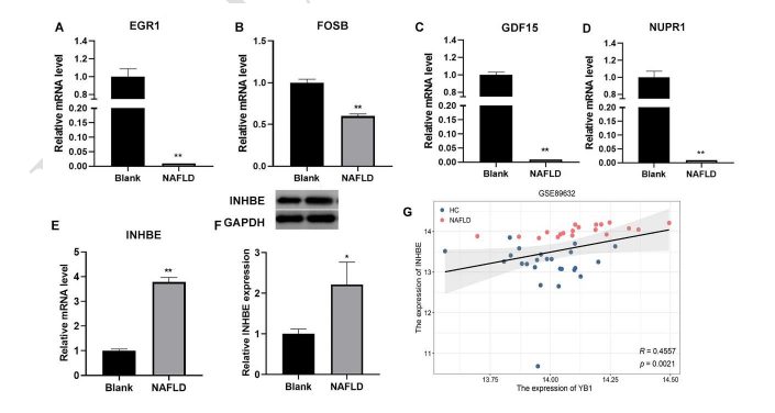
YAP1 or YAP plays a key role in liver repair, cell fate determination, and tumorigenesis. The lncRNA lncARSR prevents YAP phosphorylation, leading to activation of insulin receptor substrate 2 (IRS2) and increased expression of the SREBP-1c gene. IRS2 subsequently activates the PI3K/AKT pathway, inducing lipogenic gene expression and accelerating NAFLD progression [143]. Another study has shown that large tumor suppressor kinase 2 (LATS2) regulates YAP activity in NAFLD by modulating its phosphorylation status [144]. In macrophages, the STING–YAP axis regulates steatosis by reprogramming lipid metabolism through a pathway involving transmembrane protein 205 (TMEM205), mitofusin 2 (MFN2), and protein disulfide isomerase (PDI). TMEM205, a YAP target gene, activates AMPKα, which interacts with hepatocyte MFN2, promoting PDI–hypoxia-inducible factor-1α (HIF-1α) signaling and degradation of perilipin 2 (PLIN2) on lipid droplets (LDs). Macrophage-specific STING deficiency enhances nuclear YAP activity, reducing lipid accumulation and PLIN2 expression under HFD-induced oxidative stress [145]. Studies have also highlighted the involvement of the Wnt/β-catenin signaling pathway in regulating hepatic lipid metabolism [146, 147]. Ma et al. observed elevated levels of Y-box binding protein 1 (YB-1) and β-catenin in the livers of NAFLD mice. They further demonstrated that YB-1 influences lipid synthesis and β-oxidation via the Wnt/β-catenin pathway [148]. In addition, the lncRNA MAYA (MST1/2-Antagonizing for YAP Activation) promotes hepatocyte senescence by downregulating YAP expression [149], and hepatocyte senescence is known to contribute to the pathogenesis of NAFLD [150].
In monkeys with hepatic steatosis, increased nuclear localization of YAP has been observed in hepatocytes [151]. Studies have shown that Si-Ni-San, a traditional Chinese medicine formula, reduces YAP expression and mitigates lipid droplet accumulation in liver cells affected by NAFLD [152]. Rosmarinic acid (RA) also downregulates YAP protein levels and ameliorates NAFLD by modulating the YAP/TAZ–PPARγ/proliferator-activated receptor γ coactivator-1α (PGC-1α) signaling pathway [153]. Curcumol, a natural compound, inhibits hepatocyte senescence through YAP/nuclear receptor coactivator 4 (NCOA4)-mediated regulation of ferritinophagy in NAFLD. Supplementation with curcumol improves liver damage and reduces hepatic steatosis in HFD-fed golden hamsters [154]. The lncRNA SRD5A3-AS1 inhibits miR-1205, resulting in upregulation of NF2 expression. In turn, NF2 suppresses YAP activity, reducing cell proliferation and lowering the levels of inflammatory and fibrotic markers, such as IL-6, TGF-β1, and α-SMA in NAFLD [155]. Lian-Mei-Yin (LMY), a traditional Chinese medicine long used for treating liver disorders, has been found to reduce hepatic steatosis in both zebrafish and mouse models of NAFLD in a time- and dose-dependent manner. Its mechanism involves inhibition of Yap1-mediated activation of Foxm1, a key factor in NAFLD progression [156].
YAP expression in hepatic HSCs and KCs is critical for the development of fibrosis and the progression of NASH [157]. In mice with diet-induced NAFLD, hepatic expression of CYR61 increases in a YAP-dependent manner and is associated with fibrosis development [158]. As a key effector of the Hippo pathway, YAP plays an early and essential role in HSC activation [159], thereby promoting liver scarring during NASH progression [160]. Inhibition of the Hippo/YAP signaling pathway is necessary for magnesium isoglycyrrhizinate to suppress HSC inflammation and activation [161]. Recent research has identified a novel link between the gene CSN6, which stabilizes HMGCS1, and YAP activation through mevalonate metabolism. Targeting the CSN6–HMGCS1–YAP1 axis may reveal a potential therapeutic vulnerability in NAFLD-associated HCC [162]. Additionally, YAP activation in NASH may be linked to the DR. During NASH development, YAP activation occurs prior to DR and may contribute to it by promoting hepatocyte dedifferentiation [163].
Yb-1
YB-1 or YBX1 is a multifunctional DNA- and RBP characterized by a conserved cold shock domain (CSD) [164]. Its broad range of functions stems from its ability to interact with nucleic acids, form homomultimers, and assemble into complexes with other proteins. YBX1 regulates numerous DNA- and RNA-dependent processes, including transcription, splicing, translation, DNA repair, and mRNA stability [157, 165].
Adipocyte autophagy plays a significant role in the pathogenesis of NAFLD. Autophagy is elevated in the white adipose tissue (WAT) of mice fed a HFD, and suppression of autophagy in WAT has been shown to alleviate hepatic steatosis, inflammation, and fibrosis [166]. Studies have demonstrated that YB-1 facilitates adipogenesis by enhancing autophagy mediated by Unc-51–like kinases 1 and 2 (ULK1 and ULK2). Further investigation revealed that YB1 specifically binds to m5C-modified Ulk1 transcripts, stabilizing their mRNA. In addition to acting as an RBP, YB1 also functions as a DNA-binding protein that promotes Ulk2 transcription. Together, these actions increase ULK1 and ULK2 levels, thereby enhancing autophagy and promoting adipogenesis. Elevated YB1 expression in WAT increases autophagy and stimulates adipose tissue expansion in mice. Due to its regulatory roles in autophagy and adipogenesis, YB1 may represent a promising therapeutic target for combating obesity and related metabolic disorders [167]. The well-characterized lncRNA HOX Transcript Antisense RNA (HOTAIR) has also been shown to interact with YBX1, promoting cell proliferation through regulation of YBX1 target genes [168]. However, its direct impact on NAFLD remains to be fully elucidated and warrants further investigation.
Eukaryotic initiation factor 4E (EIF4E)
EIF4E is an mRNA cap-binding protein essential for the interaction between mRNA and ribosomes, facilitating cap-dependent translation through its interaction with eukaryotic initiation factor 4G (eIF4G) [169]. Yan et al. [170] found that elevated plasma levels of EIF4E may causally contribute to the development of NAFLD in the European population. Wang et al. [171] showed that inflammatory stress enhances the phosphorylation of both mammalian target of rapamycin (mTOR) and EIF4E, which in turn promotes the translation of recombinant Cluster of Differentiation 36 (CD36). CD36 facilitates the uptake of long-chain fatty acids, leading to lipid accumulation and the onset of NAFLD [172]. Rapamycin has been shown to reduce CD36 expression by inhibiting the mTOR pathway and downstream phosphorylation events, thereby alleviating NAFLD [173]. Furthermore, a clinical trial conducted by Kubrusly et al. [174] demonstrated that EIF4E levels are elevated in patients with NASH-related cirrhosis.
Astrocyte elevated gene-1 (AEG-1)
AEG-1, also known as metadherin (MTDH), is a 582-amino acid protein anchored to the endoplasmic reticulum membrane [175]. Elevated AEG-1 expression has been observed in individuals with NASH who developed steatosis following a HFD. Molecular analyses reveal that AEG-1 regulates fatty acid β-oxidation (FAO) by inhibiting the activation of PPAR alpha (PPARα), while simultaneously promoting de novo lipogenesis (DNL) and TG accumulation through enhanced translation of mRNAs encoding enzymes involved in fatty acid synthesis. AEG-1 also activates the NF-κB signaling pathway, contributing to hepatic inflammation and fibrosis. In hepatocyte-specific AEG-1 transgenic mice (Alb/AEG-1), inhibition of PPARα and FAO leads to spontaneous NASH development—a condition that is reversed in hepatocyte-specific AEG-1 knockout (AEG-1ΔHEP) mice, suggesting a protective role against diet-induced NASH in the absence of AEG-1 [176]. AEG-1 contains an LXXLL motif at amino acids 21–25 [177], which contributes to its steatotic activity and also regulates its inflammatory and tumorigenic functions, helping maintain a balance in AEG-1’s activity [175]. AEG-1 is also regulated post-translationally via S-palmitoylation at cysteine 75, mediated by the palmitoyltransferase ZDHHC6. This modification negatively regulates AEG-1, restraining its inflammatory and oncogenic functions. Inhibiting depalmitoylases increases AEG-1 palmitoylation, which may suppress both NASH and HCC [178]. In Alb/AEG-1 mice, PPARα inhibition leads to decreased FAO and increased Fas translation, driving DNL and activating NF-κB-mediated inflammation—together contributing to NASH pathology. Therapeutically, hepatocyte-targeted delivery of AEG-1 siRNA via nanoparticles significantly protects wild-type mice from HFD-induced NASH. Thus, AEG-1 inhibition represents a promising therapeutic strategy for NASH patients [176].
Quaking (QKI) 5
Sirtuins (SIRT1–7) regulate diverse cellular functions through post-translational modification of proteins. Among them, SIRT1 is closely associated with metabolic regulation and has been implicated in the development of NAFLD due to its role in deacetylating various cellular proteins [179]. In mice, downregulation of SIRT1 via small hairpin RNA induces hepatic steatosis, underscoring its protective role in liver metabolism. PPARγ, predominantly expressed in adipose tissue, is upregulated in the livers of NAFLD patients. Increased hepatic PPARγ activity promotes lipid storage and contributes to steatosis [180]. QKI, a member of the signal transduction and activators of RNA (STAR) family of RBPs, is expressed in the liver, with QKI5 being the predominant isoform. Research by Weiyan demonstrated that SIRT1 deacetylates QKI5, thereby influencing TG synthesis in NAFLD mouse models. This regulatory interaction activates the transcription factor Forkhead box protein O1 (FoxO1) through post-transcriptional regulation of PPARα, leading to the inhibition of TG synthesis and slowing NAFLD progression [181].
Endothelial differentiation-related factor 1 (EDF1)
EDF1, also known as hMBF-1, is a highly conserved intracellular protein composed of 148 amino acids. EDF1 functions as a coactivator for several nuclear receptors involved in lipid metabolism, including liver X receptor alpha (LXRα), steroidogenic factor 1, liver receptor homolog 1, and PPARγ [182, 183]. It facilitates the recruitment of the lncRNA Blnc1, leading to the formation of the LXR ribonucleoprotein transcriptional complex [184]. In cooperation with the LXRα/RXR beta (RXRβ) complex, EDF1 enhances the activity of the SREBP1c promoter, with Blnc1 further amplifying the transcriptional activity of this complex. Overexpression of EDF1 in hepatocytes stimulates the expression of lipogenic genes, and co-expression of Blnc1 intensifies this effect. Additionally, EDF1 is essential for PPARγ transcriptional activation during 3T3-L1 adipocyte differentiation [183], indicating its potential significance in the pathogenesis of NAFLD.
DEAD-box family
DEAD-box protein 1 (DDX1)
DDX1 is a member of the DEAD-box RNA helicase family and is involved in various RNA and DNA processing events, including mRNA translation, miRNA maturation, rRNA processing, tRNA splicing, and the repair of DNA double-strand breaks [185–187]. Studies have shown that DDX1 directly binds to insulin mRNA, and upon stimulation with FFA, DDX1 becomes phosphorylated and dissociates from insulin mRNA, leading to reduced insulin translation [188]. Additionally, DDX1 regulates insulin translation by interacting with eukaryotic initiation factors EIF3a and EIF4B [188]. DDX1 deficiency has been shown to impair calcium influx and insulin secretion in pancreatic β cells [189]. Given its role in insulin metabolism, we hypothesize that DDX1 may influence the development of NAFLD. However, there are currently no direct studies linking DDX1 to NAFLD.
p68 and p72
p68 and p72, members of the DEAD-box RNA helicase family, are RBPs involved in RNA helicase activity and RNA–protein complex remodeling. They interact with the noncoding RNA Steroid Receptor RNA Activator (SRA), which functions as a transcriptional coactivator for PPARγ and promotes adipocyte differentiation in vitro. SRA-deficient mice are resistant to HFD-induced obesity, exhibiting reduced fat mass and increased lean body content. Their livers display fewer lipid droplets, lower expression of lipogenic genes, and reduced hepatic steatosis [190]. In cell models, knockdown of SRA inhibits adipocyte differentiation [191], and SRA has also been shown to promote hepatic steatosis by repressing ATGL expression [192].
Lin28
LIN28 was originally identified as a regulator of developmental timing in Caenorhabditis elegans [193]. In humans, LIN28 modulates the degradation of let-7 miRNAs and plays roles in various cancers [194, 195]. There are two isoforms of LIN28: LIN28A, which is localized primarily in the cytoplasm, and LIN28B, which is found in both the cytoplasm and nucleus. Unlike LIN28A, LIN28B functions predominantly in the nucleus, where it sequesters primary let-7 transcripts and inhibits their processing [196]. LIN28B was first identified as being overexpressed in HCC [197].
C1632 inhibits LIN28, promoting lipid catabolism and ketogenesis while reducing SREBP1-mediated lipogenesis. These effects collectively limit intracellular lipid accumulation in HepG2 and AML12 cells. In both genetic and dietary mouse models of NAFLD, C1632 activates an anti-steatotic response, suggesting that LIN28 inhibition may offer therapeutic potential for the prevention or treatment of NAFLD [198].
Zhu et al. found that transgenic mice overexpressing LIN28A or LIN28B are resistant to obesity and exhibit improved glucose tolerance. In contrast, muscle-specific Lin28a knockout and let-7 overexpression in mice resulted in glucose intolerance. The regulatory effects of LIN28A/B and let-7 on glucose metabolism are mediated through the insulin–PI3K–mTOR signaling pathway [199]. Additionally, in osteosarcoma cells with high LIN28B expression, aerobic glycolysis was enhanced, while mitochondrial function was impaired [200]. These findings underscore the critical role of LIN28 isoforms in regulating glucose homeostasis, which may influence NAFLD development through their effects on glucose metabolism.
Rpl 8
RCRIN, a conserved read-through circRNA that inhibits metabolic dysfunction-associated steatotic liver disease (MASLD), is downregulated in patients with MASLD. In normal hepatocytes, RCRIN binds to the RPL8 protein and recruits the E3 ubiquitin ligase RNF2 to mediate its degradation. This reduces the number of RPL8-containing ribosomes, thereby inhibiting lipid accumulation and ERS. In MASLD hepatocytes, decreased RCRIN levels lead to the accumulation of RPL8, which contributes to the formation of RPL8-containing ribosomes. This, in turn, enhances lipid accumulation and ERS in the liver. Notably, overexpression of RCRIN or silencing of Rpl8 significantly inhibits the development and progression of MASLD. These findings suggest that RCRIN and RPL8 may serve as valuable biomarkers for MASLD and related metabolic liver disorders. However, whether circulating RCRIN levels in serum could function as a diagnostic marker for MASLD warrants further investigation [201].
Sarcopenia-related RBPs
Rosenberg first coined the term sarcopenia in 1989 to describe the age-related decline in skeletal muscle mass and volume [202]. Sarcopenia is now recognized as an extrahepatic manifestation of NAFLD [203]. NAFLD and sarcopenia may share common underlying mechanisms, including IR, vitamin D deficiency, chronic inflammation, and reduced physical activity. Sarcopenia has emerged as a novel risk factor for the development of NAFLD [204]. In a prospective observational cohort study, individuals with low muscle mass were found to have a higher risk of developing NAFLD [205]. Those with sarcopenia face a 2.3- to 3.34-fold increased risk of NAFLD [206], and a striking 24-fold increased risk of liver fibrosis [204]. In patients with biopsy-proven NAFLD, sarcopenia has been associated with NASH and significant fibrosis, independent of obesity, inflammation, or IR [207]. Among young and middle-aged populations, both the prevalence and severity of MAFLD are strongly associated with sarcopenia [208]. Evaluating sarcopenia has proven valuable for risk assessment in MAFLD patients [209]. In a prospective study of 225 Caucasian individuals, the prevalence of sarcopenia increased linearly with liver fibrosis severity. Even after adjusting for confounding factors, sarcopenia remained significantly correlated with hepatic steatosis severity [210]. Given the overlapping pathophysiological mechanisms between NAFLD and sarcopenia, it remains unclear whether sarcopenia precedes or follows NAFLD progression [211].
In response to lipopolysaccharide (LPS), TNFα mRNA can be stabilized through phosphorylation of TTP, which promotes TTP binding to the 3′UTR of TNFα mRNA [212]. IL-6 mRNA interacts with multiple RBPs, including HuR, TTP, and AU-rich element-binding protein 1 (AUF1, also known as hnRNP D), suggesting their involvement in the regulation of IL-6 and their potential role in muscle wasting in the elderly [213–215]. QKI protein levels increase during myogenesis and regulate alternative splicing by binding to ACUAA motifs [216]. Depletion of RNA-binding Fox homolog proteins 1 and 2 (RBFOX1 and RBFOX2) in mice leads to severe muscle mass loss and altered splicing of numerous transcripts, highlighting their essential role in muscle maintenance [217]. Masuda et al. [218] reported that in aging skeletal muscle, AUF1 expression increases, TIA-1 and TTP levels decrease, while HuR expression remains unchanged. Polyadenylate-binding nuclear protein 1 (PABPN1) has also emerged as a candidate RBP implicated in muscle aging. Its levels decline in aged muscle, and genetic reduction of PABPN1in mouse models results in muscle atrophy [219, 220]. hnRNP H1 (hnRNPH1) regulates the alternative splicing of RBFOX2 [221], and PABPN1levels have been shown to decrease with age in skeletal muscle [222]. In summary, these RBPs likely contribute to RNA processing and splicing in skeletal muscle and may influence NAFLD development through their role in sarcopenia.
Interplay across multiple RBPs in NAFLD
Thousands of overlapping binding sites for TTP and HuR have been identified across more than 1300 genes. While TTP promotes mRNA decay, HuR stabilizes and enhances the translation of its target mRNAs. RNA immunoprecipitation (RNA-IP) experiments have shown that TTP can directly bind to and destabilize HuR mRNA. High expression or aberrant nuclear/cytoplasmic distribution of HuR, along with decreased TTP levels, has been observed in various types of cancers [223, 224]. The lncRNA NEAT1, which exists in two major isoforms (NEAT1_v1 and NEAT1_v2), has been implicated in NAFLD. A study by Ahne demonstrated that NEAT1 is oppositely regulated by TTP and HuR in the context of NAFLD [225]. Some RBPs are capable of binding to numerous mRNA targets, potentially producing competitive or synergistic regulatory effects. Such interactions have been explored in other diseases and pathophysiological processes [226–228]. However, studies investigating the combined regulation of specific RNAs by multiple RBPs in the progression or treatment of NAFLD remain limited. Further research is needed to clarify these complex regulatory networks and their therapeutic potential.
Based on the above content, several common and relatively well-studied RBPs implicated in NAFLD are summarized in Table 1. Additionally, Table 2 lists several drugs that can modulate the expression or activity of these RBPs, offering potential therapeutic strategies for the treatment or alleviation of fatty liver disease.
| RBPs | Expression | Related RNA | Expression | Mechanism | Results | NAFLD | Reference |
|---|---|---|---|---|---|---|---|
| HuR | ↓ | Acc1, Fas, Elovl6, Fads1 & 2-related genes | ↑ | Fatty acid biosynthesis | Fatty acid biosynthesis | ↑ | [32] |
| ↓ | H19 | ↑ | miR-130a/PPARγ axis, MLXIPL expression, mTORC1 signaling | Steatosis and lipid accumulation | ↑ | [35, 36] | |
| ↑ | PTEN mRNA | ↑ | increases stability and translation of PTEN | Lipid depositionin hepatocytes↓ | ↓ | [30] | |
| ↓ | Apob pre-mRNA, UQCRB, NDUFB6 mRNA | ↓ | Regulate splicing of Apob mRNA and translation of UQCRB and NDUFB6 | Liver lipid transport and ATP synthesis↓ | ↑ | [31, 40] | |
| ↓ | ADH1C, CYP4A1 1, ALDH5A 1 | ↓ | LINC01 018 binds to HuR and regulate its activity | Regulates hepatic fatty acid metabolism | ↑ | [41] | |
| ↑ | APOA4 mRNA | ↑ | HuR–A POA4-AS complex stabilizes APOA4 mRNA | Increases TGs and TC | ↑ | [43, 43] | |
| ↑ | C/EBPβ mRNA | ↑ | Binds to 3’UTR of C/EBP βmRNA and increases its stability and translation | Aggravates hepatic PPARγ activation, NF-κB activation | ↑ | [44–46] | |
| ↑ | HAMP mRNA | ↑ | Binds to the 3’UTR of HAMP mRNA, up-regulate HAMP mRNA in hepatocytes | Lipid content↑ | ↑ | [48] | |
| ↑ | LDLR mRNA | ↑ | Stabilizes LDLR mRNA, increase LDLR in the liver | LDLC in the plasma ↓ | ↓ | [49] | |
| ↑ | ABCA1 mRNA | ↑ | Promotes ABCA1 through 3’UTR binding-mediated mRNA stabilization | Cholesterol efflux, HDLC biogenesis | ↓ | [50] | |
| ↑ | ATGL mRNA | ↑ | Increases its stability and protein level of ATGL mRNA | TC and HDLC↓ | ↓ | [29] | |
| ↓ | MnSOD and HO-1 mRNA | ↓ | Protective role of HuR against oxidative stress ↓ | Oxidative stress ↑ | ↑ | [56] | |
| HnRNPI (PTBP1) | ↑ | H19, SREBP1 | ↑ | H19/PTBP1/SREBP1 forward amplifying pathway | Lipogenic program | ↑ | [77] |
| Sc5d, Lss, Fdps, and Hmgcs1 | ↑ | hLMR1 enhances the binding of PTBP1 on the promoters of Sc5d, Lss, Fdps, and Hmgcs1 | Cholesterol synthesis, adipogenic differentiation | ↑ | [79] | ||
| hnRNPU | ↑ | lncRHL | ↑ | lncRHL/hnRNPU/BMAL1/MTTP axis | VLDL secretion in hepatocytes | ↓ | [83] |
| hnRNPA1 | ↓ | gys1 mRNA | ↓ | gys1 mRNA stability↓ | Glycogen storage ↓ IR↑ | ↑ | [91] |
| ↑ | lncRNA SHGL | ↑ | Activates PI3K/Akt pathway activate CaM/Akt pathwa y repress mTOR/SREBP-1C pathway | Hyperglycemia and steatosis↓ | ↓ | [92, 95] | |
| H19 | ↑ | Increases translation of PGC1a and CPT1b | Lipidectopic deposition and IR↓ | ↓ | [93] | ||
| hnRNPC | ↓ | circ-Tulp4 | ↑ | Inhibits the HNRNPC/ABHD6 axis | Reduces apoptosis | ↓ | [98] |
| p62 | ↑ | miR-130b-5p | ↓ | AKT pathway | Expression of lipid synthesis genes↓ | ↓ | [116] |
| TTP | ↓ | FGF21 mRNA | ↑ | FGF21↑ | Improves glucose tolerance and insulin sensitivity | ↓ | [129] |
| CPEB1 | ↓ | miR-122 | ↓ | CircRNA-002581-miR-122-CPEB1 axis PTEN-AMPK-mTOR pathway | Autophagy ↑ | ↓ | [134–136] |
| TDP-43 | ↑ | lncLSTR | ↑ | TDP-43/FXR/apoC2 pathway | TG↓ | ↓ | [142] |
| YAP | ↑ | lncARSR | ↑ | LncARSR activates IRS2/AKT pathway by reducing YAP1 phosphorylation | Lipid accumulation | ↑ | [143] |
| MAYA | ↓ | Regulates iron overload in hepatocytes | Cellular senescence↓ | ↓ | [149, 150] | ||
| ↓ | SRD5A3AS1 | ↑ | SRD5A3-AS1 inhibite miR-1205, upregulating NF2, upregulate NF2 negatively regulate YAP1 | NAFLD cell proliferation↓, IL-6, TGF-β1, α-SMA ↓ | ↓ | [155] | |
| YBX1 | ↑ | ULK1 mRNA | ↑ | YBX1 enhances ULK1- and ULK2-mediated autophagy | Adipogenesis | ↑ | [167] |
| AEG-1 | ↑ | Fatty acid synthesis related mRNA | ↑ | Fatty acid synthesis↑ | DNL and TG↑ | ↑ | [176] |
| EDF1 | ↑ | Blnc1 RNA | ↑ | Facilitates formation of LXR ribonucleoprotein transcriptional complex | Stimulates lipogenic gene expression | ↑ | [183, 184] |
| p68 and p72 | ↑ | SRA | ↑ | Adipocyte differentiation repress adipose triglyceride lipase | Lipogenesis | ↑ | [190–192] |
| RPL 8 | ↑ | RCRIN | ↓ | Form of RPL8-containing ribosomes | Lipid accumulation and ERS | ↑ | [201] |
Expression: The increase or decrease in the expression levels of RBPs and related RNAs. Results: The impact of RBPs on NAFLD and its metabolism through relevant mechanisms. HuR: Human antigen R; NAFLD: Nonalcoholic fatty liver disease; TG: Triglyceride; TC: Total cholesterol; ATGL: Adipose triglyceride TG lipase; C/EBPβ: CCAAT/enhancer-binding protein beta; PPARγ: Proliferator-activated receptor gamma; NF-κB: Nuclear factor kappa B; FXR: Farnesoid X receptor; APOA4: Apolipoprotein A-IV; MnSOD: Manganese-dependent superoxide dismutase; HO-1: Heme oxygenase-1; mTORC1: Mechanistic target of rapamycin complex 1; Acc1: Acetyl-CoA carboxylase; Fas: Fatty acid synthase; Elovl6: Elongation of very-long-chain fatty acids member 6; Fads1&2: Fatty acid desaturases 1 and 2; SphK2: Sphingosine kinase 2; ERS: Endoplasmic reticulum stress; TTP: Tristetraprolin; RBP: RNA-binding protein; YAP: Yes-associated protein 1; AEG-1: Astrocyte elevated gene-1; HMGCS1: Hydroxymethylglutaryl-CoA synthase 1; MLXIPL: MLX-interacting protein-like; 3’ UTR: 3′ untranslated region; SREBP1c: Sterol regulatory element-binding protein 1c; hLMR1: Human lncRNA metabolic regulator 1; PTBP1: Polypyrimidine tract-binding protein 1; MTTP: Microsomal triglyceride TG transfer protein; VLDL: Very-low-density lipoprotein; gys1: Glycogen synthase 1; PI3K: Phosphatidylinositol 3-kinase; mTOR: Mammalian target of rapamycin; hnRNPA1: Heterogeneous nuclear ribonucleoprotein A1; CaM: Calmodulin; FGF21: Fibroblast growth factor 21; CPEB1: Cytoplasmic polyadenylation element-binding protein 1; TDP-43: Tat-activating regulatory DNA-binding protein 43; IRS2: Insulin receptor substrate 2; YXB1: Y-box binding protein-1; ULK1: Unc-51–like kinases 1; ULK2: Unc-51–like kinases 2; EDF1: Endothelial differentiation-related factor 1; Blnc1: Brown fat lncRNA 1; SRA: Steroid receptor RNA activator; GDNL: De novo lipogenesis; CPT1b: Carnitine palmitoyltransferase 1B; PGC1α: PPARγ coactivator 1-alpha.
| RBPs | Drugs | Diseases | Function | Stage | Reference |
|---|---|---|---|---|---|
| HuR | TUDCA | NAFLD | Inhibites ERS | Animal experiment | [28] |
| HuR | Insulin sensitizers | NAFLD | Ameliorates insulin resistance | Speculation | [40] |
| HuR | OCA | NAFLD | Inducts hepatic HuR expression | Animal experiment | [49] |
| HuR | SAM | NAFLD | Maintains HuR methylation | Animal experiment | [61] |
| HuR | Flavonoids | NAFLD | Inhibites HuR and reduce expression of HuR target genes | Speculation | [62] |
| TTP | Metformin | NASH | Decreases TNF-α production in KCs | Animal experiment | [125] |
| YAP | Si-Ni-San | NAFLD | Reduces YAP expression | Cell and animal experiment | [152] |
| YAP | RA | NAFLD | Down-regulates the expression of YAP | Cell and animal experiment | [153] |
| YAP | Curcumol | NAFLD | Inhibites hepatocyte senescence | Cell and animal experiment | [154] |
| YAP | SRD5A3-AS1 | NAFLD | Negatively regulates YAP | Animal experiment | [155] |
| YAP | LMY | NAFLD | Suppresses YAP1-mediated Foxm1 activation | Animal experiment | [156] |
| YAP | Magnesium isoglycyrrhizinate | HSC | Inhibites Hippo/YAP signaling pathway | Cell and animal experiment | [161] |
| YAP | HMGCS1 | NAFLD related cancer | Activates YAP | Cell and animal experiment | [162] |
| EIF4E | Rapamycin | NAFLD | Inhibites phosphorylation of EIF4E | Cell and animal experiment | [171–173] |
| AEG-1 | ZDHHC6 | NASH | Increases AEG-1 palmitoylation | Animal experiment | [178] |
| Lin28 | C1632 | NAFLD | Inhibites Lin28 | Cell and animal experiment | [198] |
TUDCA: Tauroursodeoxycholic acid; NAFLD: Nonalcoholic fatty liver disease; SAM: S-adenosylmethionine; NASH: Nonalcoholic steatohepatitis; TTP: Tristetraprolin; ERS: Endoplasmic reticulum stress; HuR: Human antigen R; RBP: RNA-binding protein; OCA: Obeticholic acid; YAP: Yes-associated protein 1; AEG-1: Astrocyte elevated gene-1; EIF4E: Eukaryotic initiation factor 4E; LMY: Lian-Mei-Yin; HMGCS1: Hydroxymethylglutaryl-CoA synthase 1; RA: Rosmarinic acid; HSC: Hepatic stellate cell; KC: Kupffer cell.
Conclusion
Despite the growing body of literature on RBPs and their roles in NAFLD, existing research remains fragmented and lacks comprehensive, systematic reviews. Given the high prevalence and clinical significance of NAFLD, a thorough understanding of the post-transcriptional regulatory functions of RBPs is essential for the development of innovative RNA-based therapies. This review aims to clarify the pathogenesis of NAFLD by highlighting recent advances in our understanding of the involvement of various RBPs in disease development. Ultimately, we anticipate that a more complete picture of the dynamic, RBP-mediated regulatory network in NAFLD will emerge. Correcting gene expression abnormalities through targeted modulation of RBPs holds promise as an effective therapeutic strategy. RNA-based therapies that mimic protective RBPs or inhibit pathogenic ones could provide new avenues for intervention. However, improving target specificity remains a major challenge that must be addressed. Moreover, while direct links between specific RBPs and NAFLD are still being established, most existing data derive from in vivo and in vitro models. Clinical studies in patients are urgently needed to validate these findings and assess the utility of diagnostic or therapeutic panels based on the RBPs discussed in this review. Despite the many unresolved questions surrounding RBPs in NAFLD, the current body of knowledge and accumulating evidence suggest that RBPs may open a new era in the treatment of fatty liver disease.
Future research in this field could evolve along several technological fronts, including genomics, high-throughput screening (HTS), and gene editing, to deepen our understanding and expand applications related to RBPs in NAFLD. The widespread adoption of HTS methods, such as RNA sequencing (RNA-seq), will facilitate the identification of novel RBPs and potential therapeutic targets, accelerating the discovery of candidate molecules for NAFLD treatment and shortening the drug development timeline. Gene editing technologies, particularly CRISPR-Cas9, will aid in the creation of precise NAFLD animal models, offering new avenues for advancing precision medicine. As these technologies continue to develop, drug development costs are expected to decrease significantly. Moreover, RBP-targeted therapies may exhibit more precise mechanisms of action, potentially reducing side effects and adverse reactions. This could enhance therapeutic outcomes while lowering the economic burden on patients.
References
- Lazarus JV, Mark HE, Anstee QM, Arab JP, Batterham RL, Castera L, et al. Advancing the global public health agenda for NAFLD: a consensus statement. Nat Rev Gastroenterol Hepatol 2022;19(1):60–78. https://doi.org/10.1038/s41575-021-00523-4
- Walker RG, Barrandon O, Poggioli T, Dagdeviren S, Carroll SH, Mills MJ, et al. Exogenous GDF11, but not GDF8, reduces body weight and improves glucose homeostasis in mice. Sci Rep 2020;10(1):4561. https://doi.org/10.1038/s41598-020-61443-y
- Gariani K, Philippe J, Jornayvaz FR. Non-alcoholic fatty liver disease and insulin resistance: from bench to bedside. Diabetes Metab 2013;39(1):16–26. https://doi.org/10.1016/j.diabet.2012.11.002
- Musso G, Cassader M, Gambino R. Non-alcoholic steatohepatitis: emerging molecular targets and therapeutic strategies. Nat Rev Drug Discov 2016;15(4):249–74. https://doi.org/10.1038/nrd.2015.3
- Michelotti GA, Machado MV, Diehl AM. NAFLD, NASH and liver cancer. Nat Rev Gastroenterol Hepatol 2013;10(11):656–65. https://doi.org/10.1038/nrgastro.2013.183
- Schwabe RF, Tabas I, Pajvani UB. Mechanisms of fibrosis development in nonalcoholic steatohepatitis. Gastroenterology 2020;158(7):1913–28. https://doi.org/10.1053/j.gastro.2019.11.311
- Estes C, Razavi H, Loomba R, Younossi Z, Sanyal AJ. Modeling the epidemic of nonalcoholic fatty liver disease demonstrates an exponential increase in burden of disease. Hepatology 2018;67(1):123–33. https://doi.org/10.1002/hep.29466
- Wu YL, Li HF, Chen HH, Lin H. Emergent roles of circular RNAs in metabolism and metabolic disorders. Int J Mol Sci 2022;23(3):1032. https://doi.org/10.3390/ijms23031032
- Salvoza N, Giraudi PJ, Tiribelli C, Rosso N. Natural compounds for counteracting nonalcoholic fatty liver disease (NAFLD): advantages and limitations of the suggested candidates. Int J Mol Sci 2022;23(5):2764. https://doi.org/10.3390/ijms23052764
- Choudhury J, Sanyal AJ. Insulin resistance and the pathogenesis of nonalcoholic fatty liver disease. Clin Liver Dis 2004;8(3):575–94. https://doi.org/10.1016/j.cld.2004.04.006
- Shimomura I, Bashmakov Y, Horton JD. Increased levels of nuclear SREBP-1c associated with fatty livers in two mouse models of diabetes mellitus. J Biol Chem 1999;274(42):30028–32. https://doi.org/10.1074/jbc.274.42.30028
- Donnelly KL, Smith CI, Schwarzenberg SJ, Jessurun J, Boldt MD, Parks EJ. Sources of fatty acids stored in liver and secreted via lipoproteins in patients with nonalcoholic fatty liver disease. J Clin Invest 2005;115(5):1343–51. https://doi.org/10.1172/JCI23621
- Seppälä-Lindroos A, Vehkavaara S, Häkkinen AM, Goto T, Westerbacka J, Sovijärvi A, et al. Fat accumulation in the liver is associated with defects in insulin suppression of glucose production and serum free fatty acids independent of obesity in normal men. J Clin Endocrinol Metab 2002;87(7):3023–8. https://doi.org/10.1210/jcem.87.7.8638
- Bugianesi E, Gastaldelli A, Vanni E, Gambino R, Cassader M, Baldi S, et al. Insulin resistance in non-diabetic patients with non-alcoholic fatty liver disease: sites and mechanisms. Diabetologia 2005;48(4):634–42. https://doi.org/10.1007/s00125-005-1682-x
- Gerstberger S, Hafner M, Tuschl T. A census of human RNA-binding proteins. Nat Rev Genet 2014;15(12):829–45. https://doi.org/10.1038/nrg3813
- Pope SD, Medzhitov R. Emerging principles of gene expression programs and their regulation. Mol Cell 2018;71(3):389–97. https://doi.org/10.1016/j.molcel.2018.07.017
- Van Nostrand EL, Freese P, Pratt GA, Wang X, Wei X, Xiao R, et al. A large-scale binding and functional map of human RNA-binding proteins. Nature 2020;583(7818):711–9. https://doi.org/10.1038/s41586-020-2077-3
- Salem ESB, Vonberg AD, Borra VJ, Gill RK, Nakamura T. RNAs and RNA-binding proteins in immuno-metabolic homeostasis and diseases. Front Cardiovasc Med 2019;6:106. https://doi.org/10.3389/fcvm.2019.00106
- Schieweck R, Ninkovic J, Kiebler MA. RNA-binding proteins balance brain function in health and disease. Physiol Rev 2021;101(3):1309–70. https://doi.org/10.1152/physrev.00047.2019
- Hinman MN, Lou H. Diverse molecular functions of Hu proteins. Cell Mol Life Sci 2008;65(20):3168–81. https://doi.org/10.1007/s00018-008-8252-6
- Srikantan S, Gorospe M. HuR function in disease. Front Biosci (Landmark Ed) 2012;17(1):189–205. https://doi.org/10.2741/3921
- Perrone-Bizzozero N, Bird CW. Role of HuD in nervous system function and pathology. Front Biosci (Schol Ed) 2013;5(2):554–63. https://doi.org/10.2741/S389
- Ma WJ, Furneaux H. Localization of the human HuR gene to chromosome 19p13.2. Hum Genet 1997;99(1):32–3. https://doi.org/10.1007/s004390050305
- Díaz-Quintana A, García-Mauriño SM, Díaz-Moreno I. Dimerization model of the C-terminal RNA recognition motif of HuR. FEBS Lett 2015;589(10):1059–66. https://doi.org/10.1016/j.febslet.2015.03.013
- Lourou N, Gavriilidis M, Kontoyiannis DL. Lessons from studying the AU-rich elements in chronic inflammation and autoimmunity. J Autoimmun 2019;104:102334. https://doi.org/10.1016/j.jaut.2019.102334
- Grammatikakis I, Abdelmohsen K, Gorospe M. Posttranslational control of HuR function. Wiley Interdiscip Rev RNA 2017;8(1):10.1002/wrna.1372. https://doi.org/10.1002/wrna.1372
- Liu R, Wu K, Li Y, Sun R, Li X. Human antigen R: a potential therapeutic target for liver diseases. Pharmacol Res 2020;155:104684. https://doi.org/10.1016/j.phrs.2020.104684
- Subramanian P, Gargani S, Palladini A, Chatzimike M, Grzybek M, Peitzsch M, et al. The RNA binding protein human antigen R is a gatekeeper of liver homeostasis. Hepatology 2022;75(4):881–97. https://doi.org/10.1002/hep.32153
- Li J, Gong L, Liu S, Zhang Y, Zhang C, Tian M, et al. Adipose HuR protects against diet-induced obesity and insulin resistance. Nat Commun 2019;10(1):2375. https://doi.org/10.1038/s41467-019-10348-0
- Tian M, Wang J, Liu S, Li X, Li J, Yang J, et al. Hepatic HuR protects against the pathogenesis of non-alcoholic fatty liver disease by targeting PTEN. Cell Death Dis 2021;12(3):236. https://doi.org/10.1038/s41419-021-03514-0
- Zhang Z, Zong C, Jiang M, Hu H, Cheng X, Ni J, et al. Hepatic HuR modulates lipid homeostasis in response to high-fat diet. Nat Commun 2020;11(1):3067. https://doi.org/10.1038/s41467-020-16918-x
- Wang Y, Tai YL, Way G, Zeng J, Zhao D, Su L, et al. RNA binding protein HuR protects against NAFLD by suppressing long noncoding RNA H19 expression. Cell Biosci 2022;12(1):172. https://doi.org/10.1186/s13578-022-00910-7
- Lee SY, Hong IK, Kim BR, Shim SM, Sung Lee J, Lee HY, et al. Activation of sphingosine kinase 2 by endoplasmic reticulum stress ameliorates hepatic steatosis and insulin resistance in mice. Hepatology 2015;62(1):135–46. https://doi.org/10.1002/hep.27804
- Nagahashi M, Takabe K, Liu R, Peng K, Wang X, Wang Y, et al. Conjugated bile acid-activated S1P receptor 2 is a key regulator of sphingosine kinase 2 and hepatic gene expression. Hepatology 2015;61(4):1216–26. https://doi.org/10.1002/hep.27592
- Wang H, Cao Y, Shu L, Zhu Y, Peng Q, Ran L, et al. Long non-coding RNA (lncRNA) H19 induces hepatic steatosis through activating MLXIPL and mTORC1 networks in hepatocytes. J Cell Mol Med 2020;24(2):1399–412. https://doi.org/10.1111/jcmm.14818
- Liu J, Tang T, Wang GD, Liu B. LncRNA-H19 promotes hepatic lipogenesis by directly regulating miR-130a/PPARγ axis in non-alcoholic fatty liver disease. Biosci Rep 2019;39(7):BSR20181722. https://doi.org/10.1042/BSR20181722
- Maehama T, Dixon JE. The tumor suppressor, PTEN/MMAC1, dephosphorylates the lipid second messenger, phosphatidylinositol 3,4,5-trisphosphate. J Biol Chem 1998;273(22):13375–8. https://doi.org/10.1074/jbc.273.22.13375
- Horie Y, Suzuki A, Kataoka E, Sasaki T, Hamada K, Sasaki J, et al. Hepatocyte-specific Pten deficiency results in steatohepatitis and HCCs. J Clin Invest 2004;113(12):1774–83. https://doi.org/10.1172/JCI20513
- Garcia-Cao I, Song MS, Hobbs RM, Laurent G, Giorgi C, de Boer VC, et al. Systemic elevation of PTEN induces a tumor-suppressive metabolic state. Cell 2012;149(1):49–62. https://doi.org/10.1016/j.cell.2012.02.030
- Van Wagner LB, Rinella ME. The role of insulin-sensitizing agents in the treatment of nonalcoholic steatohepatitis. Therap Adv Gastroenterol 2011;4(4):249–63. https://doi.org/10.1177/1756283X11403809
- Ruan X, Li P, Chen Y, Shi Y, Pirooznia M, Seifuddin F, et al. In vivo functional analysis of non-conserved human lncRNAs associated with cardiometabolic traits. Nat Commun 2020;11(1):45. https://doi.org/10.1038/s41467-019-13688-z
- VerHague MA, Cheng D, Weinberg RB, Shelness GS. Apolipoprotein A-IV expression in mouse liver enhances triglyceride secretion and reduces hepatic lipid content by promoting very lowdensity lipoprotein particle expansion. Arterioscler Thromb Vasc Biol 2013;33(11):2501–8. https://doi.org/10.1161/ATVBAHA.113.301948
- Qin W, Li X, Xie L, Li S, Liu J, Jia L, et al. A long non-coding RNA, APOA4-AS, regulates APOA4 expression depending on HuR in mice. Nucl Acids Res 2016;44(13):6423–33. https://doi.org/10.1093/nar/gkw341
- Rahman SM, Schroeder-Gloeckler JM, Janssen RC, Jiang H, Qadri I, Maclean KN, et al. CCAAT/enhancing binding protein beta deletion in mice attenuates inflammation, endoplasmic reticulum stress, and lipid accumulation in diet-induced nonalcoholic steatohepatitis. Hepatology 2007;45(5):1108–17. https://doi.org/10.1002/hep.21614
- Gantt K, Cherry J, Tenney R, Karschner V, Pekala PH. An early event in adipogenesis, the nuclear selection of the CCAAT enhancer-binding protein (C/EBP) mRNA by HuR and its translocation to the cytosol. J Biol Chem 2005;280(26):24768–74. https://doi.org/10.1074/jbc.M502011200
- Cherry J, Jones H, Karschner VA, Pekala PH. Post-transcriptional control of CCAAT/enhancer-binding protein beta (C/EBPbeta) expression: formation of a nuclear HuR-C/EBPbeta mRNA complex determines the amount of message reaching the cytosol. J Biol Chem 2008;283(45):30812–20. https://doi.org/10.1074/jbc.M805659200
- Parajes S, González-Quintela A, Campos J, Quinteiro C, Domínguez F, Loidi L. Genetic study of the hepcidin gene (HAMP) promoter and functional analysis of the c.-582A > G variant. BMC Genet 2010;11:110. https://doi.org/10.1186/1471-2156-11-110
- Lu S, Mott JL, Harrison-Findik DD. Saturated fatty acids induce post-transcriptional regulation of HAMP mRNA via AU-rich element-binding protein, human antigen R (HuR). J Biol Chem 2015;290(40):24178–89. https://doi.org/10.1074/jbc.M115.648212
- Singh AB, Dong B, Kraemer FB, Xu Y, Zhang Y, Liu J. Farnesoid X receptor activation by obeticholic acid elevates liver low-density lipoprotein receptor expression by mRNA stabilization and reduces plasma low-density lipoprotein cholesterol in mice. Arterioscler Thromb Vasc Biol 2018;38(10):2448–59. https://doi.org/10.1161/ATVBAHA.118.311122
- Ramírez CM, Lin CS, Abdelmohsen K, Goedeke L, Yoon JH, Madrigal-Matute J, et al. RNA binding protein HuR regulates the expression of ABCA1. J Lipid Res 2014;55(6):1066–76. https://doi.org/10.1194/jlr.M044925
- Lebedeva S, Jens M, Theil K, Schwanhäusser B, Selbach M, Landthaler M, et al. Transcriptome-wide analysis of regulatory interactions of the RNA-binding protein HuR. Mol Cell 2011;43(3):340–52. https://doi.org/10.1016/j.molcel.2011.06.008
- He J, Hu B, Shi X, Weidert ER, Lu P, Xu M, et al. Activation of the aryl hydrocarbon receptor sensitizes mice to nonalcoholic steatohepatitis by deactivating mitochondrial sirtuin deacetylase Sirt3. Mol Cell Biol 2013;33(10):2047–55. https://doi.org/10.1128/MCB.01658-12
- Delli Bovi AP, Marciano F, Mandato C, Siano MA, Savoia M, Vajro P. Oxidative stress in non-alcoholic fatty liver disease. an updated mini review. Front Med (Lausanne) 2021;8:595371. https://doi.org/10.3389/fmed.2021.595371
- Huang YS, Chang TE, Perng CL, Huang YH. Genetic variations of three important antioxidative enzymes SOD2, CAT, and GPX1 in nonalcoholic steatohepatitis. J Chin Med Assoc 2021;84(1):14–8. https://doi.org/10.1097/JCMA.0000000000000437
- Du J, Ren W, Zhang Q, Fu N, Han F, Cui P, et al. Heme oxygenase-1 suppresses Wnt signaling pathway in nonalcoholic steatohepatitis-related liver fibrosis. Biomed Res Int 2020;2020:4910601. https://doi.org/10.1155/2020/4910601
- Ferrigno A, Campagnoli LIM, Barbieri A, Marchesi N, Pascale A, Croce AC, et al. MCD diet modulates HuR and oxidative stress-related HuR targets in rats. Int J Mol Sci 2023;24(12):9808. https://doi.org/10.3390/ijms24129808
- Borém LMA, Neto JFR, Brandi IV, Lelis DF, Santos SHS. The role of the angiotensin II type I receptor blocker telmisartan in the treatment of non-alcoholic fatty liver disease: a brief review. Hypertens Res 2018;41(6):394–405. https://doi.org/10.1038/s41440-018-0040-6
- Toblli JE, Muñoz MC, Cao G, Mella J, Pereyra L, Mastai R. ACE inhibition and AT1 receptor blockade prevent fatty liver and fibrosis in obese Zucker rats. Obesity (Silver Spring) 2008;16(4):770–6. https://doi.org/10.1038/oby.2007.114
- Li N, Wang HX, Han QY, Li WJ, Zhang YL, Du J, et al. Activation of the cardiac proteasome promotes angiotension II-induced hypertrophy by down-regulation of ATRAP. J Mol Cell Cardiol 2015;79:303–14. https://doi.org/10.1016/j.yjmcc.2014.12.007
- Guo T, Wang H, Liu P, Xiao Y, Wu P, Wang Y, et al. SNHG6 acts as a genome-wide hypomethylation trigger via coupling of miR-1297-mediated S-adenosylmethionine-dependent positive feedback loops. Cancer Res 2018;78(14):3849–64. https://doi.org/10.1158/0008-5472.CAN-17-3833
- Guo T, Dai Z, You K, Battaglia-Hsu SF, Feng J, Wang F, et al. S-adenosylmethionine upregulates the angiotensin receptor-binding protein ATRAP via the methylation of HuR in NAFLD. Cell Death Dis 2021;12(4):306. https://doi.org/10.1038/s41419-021-03591-1
- Wang Z, Bhattacharya A, Ivanov DN. Identification of small-molecule inhibitors of the HuR/RNA interaction using a fluorescence polarization screening assay followed by NMR validation. PLoS One 2015;10(9):e0138780. https://doi.org/10.1371/journal.pone.0138780
- Abdelmohsen K, Panda AC, Munk R, Grammatikakis I, Dudekula DB, De S, et al. Identification of HuR target circular RNAs uncovers suppression of PABPN1 translation by CircPABPN1. RNA Biol 2017;14(3):361–9. https://doi.org/10.1080/15476286.2017.1279788
- Chorghade S, Seimetz J, Emmons R, Yang J, Bresson SM, Lisio M, et al. Poly(A) tail length regulates PABPC1 expression to tune translation in the heart. Elife 2017;6:e24139. https://doi.org/10.7554/eLife.24139
- Qi Y, Wang M, Jiang Q. PABPC1--mRNA stability, protein translation and tumorigenesis. Front Oncol 2022;12:1025291. https://doi.org/10.3389/fonc.2022.1025291
- Kim C, Lee H, Kang H, Shin JJ, Tak H, Kim W, et al. Corrigendum to ”RNA-binding protein HuD reduces triglyceride production in pancreatic β cells by enhancing the expression of insulin-induced gene 1” [Biochim. Biophys. Acta Gene Regul. Mech. 1859/4 (2016) Page 675-685]. Biochim Biophys Acta Gene Regul Mech 2021;1864(3):194692. https://doi.org/10.1016/j.bbagrm.2021.194692
- Marchese FP, Raimondi I, Huarte M. The multidimensional mechanisms of long noncoding RNA function. Genome Biol 2017;18(1):206. https://doi.org/10.1186/s13059-017-1348-2
- van Dam S, Võsa U, van der Graaf A, Franke L, de Magalhães JP. Gene co-expression analysis for functional classification and gene-disease predictions. Brief Bioinform 2018;19(4):575–92. https://doi.org/10.1093/bib/bbw139
- Müller-McNicoll M, Rossbach O, Hui J, Medenbach J. Auto-regulatory feedback by RNA-binding proteins. J Mol Cell Biol 2019;11(10):930–9. https://doi.org/10.1093/jmcb/mjz043
- Täuber H, Hüttelmaier S, Köhn M. POLIII-derived non-coding RNAs acting as scaffolds and decoys. J Mol Cell Biol 2019;11(10):880–5. https://doi.org/10.1093/jmcb/mjz049
- Dai S, Wang C, Zhang C, Feng L, Zhang W, Zhou X, et al. PTB: not just a polypyrimidine tract-binding protein. J Cell Physiol 2022;237(5):2357–73. https://doi.org/10.1002/jcp.30716
- Yang X, Qu S, Wang L, Zhang H, Yang Z, Wang J, et al. Retraction note: PTBP3 splicing factor promotes HCC by destroying the splicing balance of NEAT1 and pre-miR-612 [retraction of: Oncogene 2018 Dec;37(50):6399–6413]. Oncogene 2023;42(15):1263. https://doi.org/10.1038/s41388-018-0416-8
- Lu N, Min J, Peng L, Huang S, Chai X, Wang S, et al. MiR-297 inhibits tumour progression of liver cancer by targeting PTBP3. Cell Death Dis 2023;14(8):564. https://doi.org/10.1038/s41419-023-06097-0
- Ghetti A, Piñol-Roma S, Michael WM, Morandi C, Dreyfuss G. hnRNP I, the polypyrimidine tract-binding protein: distinct nuclear localization and association with hnRNAs. Nucl Acids Res 1992;20(14):3671–8. https://doi.org/10.1093/nar/20.14.3671
- Romanelli MG, Diani E, Lievens PM. New insights into functional roles of the polypyrimidine tract-binding protein. Int J Mol Sci 2013;14(11):22906–22932. https://doi.org/10.3390/ijms141122906
- Zhu Y, Gui W, Lin X, Li H. Knock-down of circular RNA H19 induces human adipose-derived stem cells adipogenic differentiation via a mechanism involving the polypyrimidine tract-binding protein 1. Exp Cell Res 2020;387(2):111753. https://doi.org/10.1016/j.yexcr.2019.111753
- Liu C, Yang Z, Wu J, Zhang L, Lee S, Shin DJ, et al. Long noncoding RNA H19 interacts with polypyrimidine tract-binding protein 1 to reprogram hepatic lipid homeostasis. Hepatology 2018;67(5):1768–83. https://doi.org/10.1002/hep.29654
- Schmidt E, Dhaouadi I, Gaziano I, Oliverio M, Klemm P, Awazawa M, et al. LincRNA H19 protects from dietary obesity by constraining expression of monoallelic genes in brown fat. Nat Commun 2018;9(1):3622. https://doi.org/10.1038/s41467-018-05933-8
- Ruan X, Li P, Ma Y, Jiang CF, Chen Y, Shi Y, et al. Identification of human long noncoding RNAs associated with nonalcoholic fatty liver disease and metabolic homeostasis. J Clin Invest 2021;131(1):e136336. https://doi.org/10.1172/JCI136336
- Yu X, Tong H, Chen J, Tang C, Wang S, Si Y, et al. CircRNA MBOAT2 promotes intrahepatic cholangiocarcinoma progression and lipid metabolism reprogramming by stabilizing PTBP1 to facilitate FASN mRNA cytoplasmic export. Cell Death Dis 2023;14(1):20. https://doi.org/10.1038/s41419-022-05540-y
- Xiong L, Gong Y, Wu L, Li J, He W, Zhu X, et al. LncRNA-Malat1 is involved in lipotoxicity-induced ß-cell dysfunction and the therapeutic effect of exendin-4 via Ptbp1. Endocrinology 2020;161(7):bqaa065. https://doi.org/10.1210/endocr/bqaa065
- Shen L, Lei S, Zhang B, Li S, Huang L, Czachor A, et al. Skipping of exon 10 in Axl pre-mRNA regulated by PTBP1 mediates invasion and metastasis process of liver cancer cells. Theranostics 2020;10(13):5719–35. https://doi.org/10.7150/thno.42010
- Shen X, Zhang Y, Ji X, Li B, Wang Y, Huang Y, et al. Long noncoding RNA lncRHL regulates hepatic VLDL secretion by modulating hnRNPU/BMAL1/MTTP axis [published correction appears in Diabetes 2023 Jun 1;72(6):819]. Diabetes 2022;71(9):1915–28. https://doi.org/10.2337/db23-er06
- Xiong J, Liu T, Mi L, Kuang H, Xiong X, Chen Z, et al. hnRNPU/TrkB defines a chromatin accessibility checkpoint for liver injury and nonalcoholic steatohepatitis pathogenesis. Hepatology 2020;71(4):1228–46. https://doi.org/10.1002/hep.30921
- Hu Y, Li J, Li X, Wang D, Xiang R, Liu W, et al. Hepatocyte-secreted FAM3D ameliorates hepatic steatosis by activating FPR1-hnRNP U-GR-SCAD pathway to enhance lipid oxidation. Metabolism 2023;146:155661. https://doi.org/10.1016/j.metabol.2023.155661
- Alvarez-Dominguez JR, Bai Z, Xu D, Yuan B, Lo KA, Yoon MJ, et al. De novo reconstruction of adipose tissue transcriptomes reveals long non-coding RNA regulators of brown adipocyte development [published correction appears in Cell Metab 2015 Jun 2;21(6):918]. Cell Metab 2015;21(5):764–76. https://doi.org/10.1016/j.cmet.2015.05.001
- Hacisuleyman E, Goff LA, Trapnell C, Williams A, Henao-Mejia J, Sun L, et al. Topological organization of multichromosomal regions by the long intergenic noncoding RNA Firre. Nat Struct Mol Biol 2014;21(2):198–206. https://doi.org/10.1038/nsmb.2764
- Li S, Mi L, Yu L, Yu Q, Liu T, Wang GX, et al. Zbtb7b engages the long noncoding RNA Blnc1 to drive brown and beige fat development and thermogenesis. Proc Natl Acad Sci U S A 2017;114(34):E7111–20. https://doi.org/10.1073/pnas.1703494114
- Sun L, Goff LA, Trapnell C, Alexander R, Lo KA, Hacisuleyman E, et al. Long noncoding RNAs regulate adipogenesis. Proc Natl Acad Sci U S A 2013;110(9):3387–92. https://doi.org/10.1073/pnas.1222643110
- Mi L, Zhao XY, Li S, Yang G, Lin JD. Conserved function of the long noncoding RNA Blnc1 in brown adipocyte differentiation. Mol Metab 2016;6(1):101–10. https://doi.org/10.1016/j.molmet.2016.10.010
- Zhao M, Shen L, Ouyang Z, Li M, Deng G, Yang C, et al. Loss of hnRNP A1 in murine skeletal muscle exacerbates high-fat diet-induced onset of insulin resistance and hepatic steatosis. J Mol Cell Biol 2020;12(4):277–90. https://doi.org/10.1093/jmcb/mjz050
- Gui W, Zhu WF, Zhu Y, Tang S, Zheng F, Yin X, et al. LncRNAH19 improves insulin resistance in skeletal muscle by regulating heterogeneous nuclear ribonucleoprotein A1. Cell Commun Signal 2020;18(1):173. https://doi.org/10.1186/s12964-020-00654-2
- Siculella L, Tocci R, Rochira A, Testini M, Gnoni A, Damiano F. Lipid accumulation stimulates the cap-independent translation of SREBP-1a mRNA by promoting hnRNP A1 binding to its 5’-UTR in a cellular model of hepatic steatosis. Biochim Biophys Acta 2016;1861(5):471–81. https://doi.org/10.1016/j.bbalip.2016.02.003
- Jeon TI, Osborne TF. SREBPs: metabolic integrators in physiology and metabolism. Trends Endocrinol Metab 2012;23(2):65–72. https://doi.org/10.1016/j.tem.2011.10.004
- Wang J, Yang W, Chen Z, Chen J, Meng Y, Feng B, et al. Long noncoding RNA lncSHGL recruits hnRNPA1 to suppress hepatic gluconeogenesis and lipogenesis. Diabetes 2018;67(4):581–93. https://doi.org/10.2337/db17-0799
- Oram JF, Lawn RM. ABCA1. The gatekeeper for eliminating excess tissue cholesterol. J Lipid Res 2001;42(8):1173–9. https://doi.org/10.1016/S0022-2275(20)31566-2
- Mahpour A, Mullen AC. Our emerging understanding of the roles of long non-coding RNAs in normal liver function, disease, and malignancy. JHEP Rep 2020;3(1):100177. https://doi.org/10.1016/j.jhepr.2020.100177
- Han JJ, Li J, Huang DH. Mesenchymal stem cell-derived extracellular vesicles carrying circ-Tulp4 attenuate diabetes mellitus with nonalcoholic fatty liver disease by inhibiting cell pyroptosis through the HNRNPC/ABHD6 axis. Tissue Eng Regen Med 2025;22(1):23–41. https://doi.org/10.1007/s13770-024-00675-9
- Nielsen J, Christiansen J, Lykke-Andersen J, Johnsen AH, Wewer UM, Nielsen FC. A family of insulin-like growth factor II mRNA-binding proteins represses translation in late development. Mol Cell Biol 1999;19(2):1262–70. https://doi.org/10.1128/MCB.19.2.1262
- Degrauwe N, Suvà ML, Janiszewska M, Riggi N, Stamenkovic I. IMPs: an RNA-binding protein family that provides a link between stem cell maintenance in normal development and cancer. Genes Dev 2016;30(22):2459–74. https://doi.org/10.1101/gad.287540.116
- Dai N, Rapley J, Angel M, Yanik MF, Blower MD, Avruch J. mTOR phosphorylates IMP2 to promote IGF2 mRNA translation by internal ribosomal entry. Genes Dev 2011;25(11):1159–72. https://doi.org/10.1101/gad.2042311
- Christiansen J, Kolte AM, Hansen TV, Nielsen FC. IGF2 mRNA-binding protein 2:biological function and putative role in type 2 diabetes. J Mol Endocrinol 2009;43(5):187–95. https://doi.org/10.1677/JME-09-0016
- Tybl E, Shi FD, Kessler SM, Tierling S, Walter J, Bohle RM, et al. Overexpression of the IGF2-mRNA binding protein p62 in transgenic mice induces a steatotic phenotype. J Hepatol 2011;54(5):994–1001. https://doi.org/10.1016/j.jhep.2010.08.034
- Simon Y, Kessler SM, Bohle RM, Haybaeck J, Kiemer AK. The insulin-like growth factor 2 (IGF2) mRNA-binding protein p62/IGF2BP2-2 as a promoter of NAFLD and HCC?. Gut 2014;63(5):861–3. https://doi.org/10.1136/gutjnl-2013-305736
- Dai N, Zhao L, Wrighting D, Krämer D, Majithia A, Wang Y, et al. IGF2BP2/IMP2-deficient mice resist obesity through enhanced translation of Ucp1 mRNA and other mRNAs encoding mitochondrial proteins. Cell Metab 2015;21(4):609–21. https://doi.org/10.1016/j.cmet.2015.03.006
- Laggai S, Simon Y, Ranssweiler T, Kiemer AK, Kessler SM. Rapid chromatographic method to decipher distinct alterations in lipid classes in NAFLD/NASH. World J Hepatol 2013;5(10):558–67. https://doi.org/10.4254/wjh.v5.i10.558
- Czepukojc B, Abuhaliema A, Barghash A, Tierling S, Naß N, Simon Y, et al. IGF2 mRNA binding protein 2 transgenic mice are more prone to develop a ductular reaction and to progress toward cirrhosis. Front Med (Lausanne) 2019;6:179. https://doi.org/10.3389/fmed.2019.00179
- Kessler SM, Laggai S, Barghash A, Schultheiss CS, Lederer E, Artl M, et al. IMP2/p62 induces genomic instability and an aggressive HCC phenotype. Cell Death Dis 2015;6(10):e1894. https://doi.org/10.1038/cddis.2015.241
- Dehghani Amirabad A, Ramasamy P, Wierz M, Nordström K, Kessler SM, Schulz MH, et al. Transgenic expression of the RNA binding protein IMP2 stabilizes miRNA targets in murine microsteatosis. Biochim Biophys Acta Mol Basis Dis 2018;1864(10):3099–108. https://doi.org/10.1016/j.bbadis.2018.05.024
- Simon Y, Kessler SM, Gemperlein K, Bohle RM, Müller R, Haybaeck J, et al. Elevated free cholesterol in a p62 overexpression model of non-alcoholic steatohepatitis. World J Gastroenterol 2014;20(47):17839–50. https://doi.org/10.3748/wjg.v20.i47.17839
- Laggai S, Kessler SM, Boettcher S, Lebrun V, Gemperlein K, Lederer E, et al. The IGF2 mRNA binding protein p62/IGF2BP2-2 induces fatty acid elongation as a critical feature of steatosis [published correction appears in J Lipid Res 2016 Jul;57(7):1323]. J Lipid Res 2014;55(6):1087–97. https://doi.org/10.1194/jlr.M045500
- Kessler SM, Simon Y, Gemperlein K, Gianmoena K, Cadenas C, Zimmer V, et al. Fatty acid elongation in non-alcoholic steatohepatitis and HCC. Int J Mol Sci 2014;15(4):5762–73. https://doi.org/10.3390/ijms15045762
- Matsuzaka T, Atsumi A, Matsumori R, Nie T, Shinozaki H, Suzuki-Kemuriyama N, et al. Elovl6 promotes nonalcoholic steatohepatitis. Hepatology 2012;56(6):2199–208. https://doi.org/10.1002/hep.25932
- Wagschal A, Najafi-Shoushtari SH, Wang L, Goedeke L, Sinha S, Delemos AS, et al. Genome-wide identification of microRNAs regulating cholesterol and triglyceride homeostasis. Nat Med 2015;21(11):1290–7. https://doi.org/10.1038/nm.3980
- Wang C, Chi Y, Li J, Miao Y, Li S, Su W, et al. FAM3A activates PI3K p110α/Akt signaling to ameliorate hepatic gluconeogenesis and lipogenesis. Hepatology 2014;59(5):1779–90. https://doi.org/10.1002/hep.26945
- Liu X, Chen S, Zhang L. Downregulated microRNA-130b-5p prevents lipid accumulation and insulin resistance in a murine model of nonalcoholic fatty liver disease. Am J Physiol Endocrinol Metab 2020;319(1):E34–42. https://doi.org/10.1152/ajpendo.00528.2019
- Regué L, Minichiello L, Avruch J, Dai N. Liver-specific deletion of IGF2 mRNA binding protein-2/IMP2 reduces hepatic fatty acid oxidation and increases hepatic triglyceride accumulation. J Biol Chem 2019;294(31):11944–51. https://doi.org/10.1074/jbc.RA119.008778
- Ahrens M, Ammerpohl O, von Schönfels W, Kolarova J, Bens S, Itzel T, et al. DNA methylation analysis in nonalcoholic fatty liver disease suggests distinct disease-specific and remodeling signatures after bariatric surgery. Cell Metab 2013;18(2):296–302. https://doi.org/10.1016/j.cmet.2013.07.004
- Kammel A, Saussenthaler S, Jähnert M, Jonas W, Stirm L, Hoeflich A, et al. Early hypermethylation of hepatic Igfbp2 results in its reduced expression preceding fatty liver in mice. Hum Mol Genet 2016;25(12):2588–99. https://doi.org/10.1093/hmg/ddw121
- Dolicka D, Sobolewski C, Gjorgjieva M, Correia de Sousa M, Berthou F, De Vito C, et al. Tristetraprolin promotes hepatic inflammation and tumor initiation but restrains cancer progression to malignancy. Cell Mol Gastroenterol Hepatol 2021;11(2):597–621. https://doi.org/10.1016/j.jcmgh.2020.09.012
- Saaoud F, Wang J, Iwanowycz S, Wang Y, Altomare D, Shao Y, et al. Bone marrow deficiency of mRNA decaying protein tristetraprolin increases inflammation and mitochondrial ROS but reduces hepatic lipoprotein production in LDLR knockout mice. Redox Biol 2020;37:101609. https://doi.org/10.1016/j.redox.2020.101609
- Mukherjee N, Jacobs NC, Hafner M, Kennington EA, Nusbaum JD, Tuschl T, et al. Global target mRNA specification and regulation by the RNA-binding protein ZFP36. Genome Biol 2014;15(1):R12. https://doi.org/10.1186/gb-2014-15-1-r12
- Park J, Chen Y, Kim J, Hwang E, Park GH, Yang CH, et al. CO-induced TTP activation alleviates cellular senescence and age-dependent hepatic steatosis via downregulation of PAI-1. Aging Dis 2023;14(2):484–501. https://doi.org/10.14336/AD.2023.0120
- Wu JC, Luo SZ, Liu T, Lu LG, Xu MY. linc-SCRG1 accelerates liver fibrosis by decreasing RNA-binding protein tristetraprolin. FASEB J 2019;33(2):2105–15. https://doi.org/10.1096/fj.201800098RR
- Park J, Rah SY, An HS, Lee JY, Roh GS, Ryter SW, et al. Metformin-induced TTP mediates communication between Kupffer cells and hepatocytes to alleviate hepatic steatosis by regulating lipophagy and necroptosis. Metabolism 2023;141:155516. https://doi.org/10.1016/j.metabol.2023.155516
- Cao H, Urban JF Jr, Anderson RA. Insulin increases tristetraprolin and decreases VEGF gene expression in mouse 3T3-L1 adipocytes. Obesity (Silver Spring) 2008;16(6):1208–18. https://doi.org/10.1038/oby.2008.65
- Louis JM, Agarwal A, Aduri R, Talukdar I. Global analysis of RNA-protein interactions in TNF-α induced alternative splicing in metabolic disorders. FEBS Lett 2021;595(4):476–90. https://doi.org/10.1002/1873-3468.14029
- Bayeva M, Khechaduri A, Puig S, Chang HC, Patial S, Blackshear PJ, et al. mTOR regulates cellular iron homeostasis through tristetraprolin. Cell Metab 2012;16(5):645–57. https://doi.org/10.1016/j.cmet.2012.10.001
- Sawicki KT, Chang HC, Shapiro JS, Bayeva M, De Jesus A, Finck BN, et al. Hepatic tristetraprolin promotes insulin resistance through RNA destabilization of FGF21. JCI Insight 2018;3(13):e95948. https://doi.org/10.1172/jci.insight.95948
- Richter JD. CPEB: a life in translation. Trends Biochem Sci 2007;32(6):279–85. https://doi.org/10.1016/j.tibs.2007.04.004
- Calderone V, Gallego J, Fernandez-Miranda G, Garcia-Pras E, Maillo C, Berzigotti A, et al. Sequential functions of CPEB1 and CPEB4 regulate pathologic expression of vascular endothelial growth factor and angiogenesis in chronic liver disease. Gastroenterology 2016;150(4):982–97.e30. https://doi.org/10.1053/j.gastro.2015.11.038
- Xu M, Fang S, Song J, Chen M, Zhang Q, Weng Q, et al. CPEB1 mediates HCC cancer stemness and chemoresistance. Cell Death Dis 2018;9(10):957. https://doi.org/10.1038/s41419-018-0974-2
- Maillo C, Martín J, Sebastián D, Hernández-Alvarez M, García-Rocha M, Reina O, et al. Circadian- and UPR-dependent control of CPEB4 mediates a translational response to counteract hepatic steatosis under ER stress. Nat Cell Biol 2017;19(2):94–105. https://doi.org/10.1038/ncb3461
- Jin X, Feng CY, Xiang Z, Chen YP, Li YM. CircRNA expression pattern and circRNA-miRNA-mRNA network in the pathogenesis of nonalcoholic steatohepatitis. Oncotarget 2016;7(41):66455–67. https://doi.org/10.18632/oncotarget.12186
- Jin X, Gao J, Zheng R, Yu M, Ren Y, Yan T, et al. Antagonizing circRNA_002581-miR-122-CPEB1 axis alleviates NASH through restoring PTEN-AMPK-mTOR pathway regulated autophagy. Cell Death Dis 2020;11(2):123. https://doi.org/10.1038/s41419-020-2293-7
- Alexandrov IM, Ivshina M, Jung DY, Friedline R, Ko HJ, Xu M, et al. Cytoplasmic polyadenylation element binding protein deficiency stimulates PTEN and Stat3 mRNA translation and induces hepatic insulin resistance. PLoS Genet 2012;8(1):e1002457. https://doi.org/10.1371/journal.pgen.1002457
- Buratti E, Baralle FE. Multiple roles of TDP-43 in gene expression, splicing regulation, and human disease. Front Biosci 2008;13:867–78. https://doi.org/10.2741/2727
- Neumann M, Sampathu DM, Kwong LK, Truax AC, Micsenyi MC, Chou TT, et al. Ubiquitinated TDP-43 in frontotemporal lobar degeneration and amyotrophic lateral sclerosis. Science 2006;314(5796):130–3. https://doi.org/10.1126/science.1134108
- Lee S, Lee TA, Song SJ, Park T, Park B. Hyperproduction of IL-6 caused by aberrant TDP-43 overexpression in high-fat diet-induced obese mice. FEBS Lett 2015;589(15):1825–31. https://doi.org/10.1016/j.febslet.2015.05.040
- Skuratovskaia D, Komar A, Vulf M, Quang HV, Shunkin E, Volkova L, et al. IL-6 reduces mitochondrial replication, and IL-6 receptors reduce chronic inflammation in NAFLD and type 2 diabetes. Int J Mol Sci 2021;22(4):1774. https://doi.org/10.3390/ijms22041774
- Park J, Zhao Y, Zhang F, Zhang S, Kwong AC, Zhang Y, et al. IL-6/STAT3 axis dictates the PNPLA3-mediated susceptibility to non-alcoholic fatty liver disease. J Hepatol 2023;78(1):45–56. https://doi.org/10.1016/j.jhep.2022.08.022
- Li P, Ruan X, Yang L, Kiesewetter K, Zhao Y, Luo H, et al. A liver-enriched long non-coding RNA, lncLSTR, regulates systemic lipid metabolism in mice. Cell Metab 2015;21(3):455–67. https://doi.org/10.1016/j.cmet.2015.02.004
- Chi Y, Gong Z, Xin H, Wang Z, Liu Z. Long noncoding RNA lncARSR promotes nonalcoholic fatty liver disease and HCC by promoting YAP1 and activating the IRS2/AKT pathway [published correction appears in J Transl Med 2021 Oct 19;19(1):438]. J Transl Med 2020;18(1):126. https://doi.org/10.1186/s12967-021-02759-9
- Guo C, Wang X, Liang L. LATS2-mediated YAP1 phosphorylation is involved in HCC tumorigenesis. Int J Clin Exp Pathol 2015;8(2):1690–7. PMID: ([0-9]+)
- Yang T, Qu X, Wang X, Xu D, Sheng M, Lin Y, et al. The macrophage STING-YAP axis controls hepatic steatosis by promoting the autophagic degradation of lipid droplets. Hepatology 2024;80(5):1169–83. https://doi.org/10.1097/HEP.0000000000000638
- El-Derany MO, El-Demerdash E. Pyrvinium pamoate attenuates non-alcoholic steatohepatitis: insight on hedgehog/Gli and Wnt/β-catenin signaling crosstalk. Biochem Pharmacol 2020;177:113942. https://doi.org/10.1016/j.bcp.2020.113942
- Hatano M, Ojima H, Masugi Y, Tsujikawa H, Hiraoka N, Kanai Y, et al. Steatotic and nonsteatotic scirrhous HCCs reveal distinct clinicopathological features. Hum Pathol 2019;86:222–32. https://doi.org/10.1016/j.humpath.2018.11.024
- Ma Z, Zhu Y, Wang Q, Deng M, Wang J, Li D, et al. Y-box binding protein 1 regulates liver lipid metabolism by regulating the Wnt/β-catenin signaling pathway. Ann Transl Med 2021;9(22):1693. https://doi.org/10.21037/atm-21-5767
- Yuan P, Qi X, Song A, Ma M, Zhang X, Lu C, et al. LncRNA MAYA promotes iron overload and hepatocyte senescence through inhibition of YAP in non-alcoholic fatty liver disease. J Cell Mol Med 2021;25(15):7354–66. https://doi.org/10.1111/jcmm.16764
- Papatheodoridi AM, Chrysavgis L, Koutsilieris M, Chatzigeorgiou A. The role of senescence in the development of nonalcoholic fatty liver disease and progression to nonalcoholic steatohepatitis. Hepatology 2020;71(1):363–74. https://doi.org/10.1002/hep.30834
- Chen P, Luo Q, Huang C, Gao Q, Li L, Chen J, et al. Pathogenesis of non-alcoholic fatty liver disease mediated by YAP. Hepatol Int 2018;12(1):26–36. https://doi.org/10.1007/s12072-017-9841-y
- Zheng K, Zhou W, Ji J, Xue Y, Liu Y, Li C, et al. Si-Ni-San reduces lipid droplet deposition associated with decreased YAP1 in metabolic dysfunction-associated fatty liver disease. J Ethnopharmacol 2023;305:116081. https://doi.org/10.1016/j.jep.2022.116081
- Luo C, Sun H, Peng J, Gao C, Bao L, Ji R, et al. Rosmarinic acid exerts an antagonistic effect on nonalcoholic fatty liver disease by regulating the YAP1/TAZ-PPARγ/PGC-1α signaling pathway. Phytother Res 2021;35(2):1010–22. https://doi.org/10.1002/ptr.6865
- Qi X, Song A, Ma M, Wang P, Zhang X, Lu C, et al. Curcumol inhibits ferritinophagy to restrain hepatocyte senescence through YAP/NCOA4 in non-alcoholic fatty liver disease. Cell Prolif 2021;54(9):e13107. https://doi.org/10.1111/cpr.13107
- Gadallah SH, Eissa S, Ghanem HM, Ahmed EK, Hasanin AH, El Mahdy MM, et al. Probiotic-prebiotic-synbiotic modulation of (YAP1, LATS1 and NF2 mRNAs/miR-1205/lncRNA SRD5A3-AS1) panel in NASH animal model. Biomed Pharmacother 2021;140:111781. https://doi.org/10.1016/j.biopha.2021.111781
- Zhang P, Cao J, Liang X, Su Z, Zhang B, Wang Z, et al. Lian-Mei-Yin formula alleviates diet-induced hepatic steatosis by suppressing Yap1/FOXM1 pathway-dependent lipid synthesis. Acta Biochim Biophys Sin (Shanghai) 2024;56(4):621–33. https://doi.org/10.3724/abbs.2024025
- Jayavelu AK, Schnöder TM, Perner F, Herzog C, Meiler A, Krishnamoorthy G, et al. Splicing factor YBX1 mediates persistence of JAK2-mutated neoplasms. Nature 2020;588(7836):157–63. https://doi.org/10.1038/s41586-020-2968-3
- Salloum S, Jeyarajan AJ, Kruger AJ, Holmes JA, Shao T, Sojoodi M, et al. Fatty acids activate the transcriptional coactivator YAP1 to promote liver fibrosis via p38 mitogen-activated protein kinase. Cell Mol Gastroenterol Hepatol 2021;12(4):1297–310. https://doi.org/10.1016/j.jcmgh.2021.06.003
- Mannaerts I, Leite SB, Verhulst S, Claerhout S, Eysackers N, Thoen LF, et al. The Hippo pathway effector YAP controls mouse hepatic stellate cell activation. J Hepatol 2015;63(3):679–88. https://doi.org/10.1016/j.jhep.2015.04.011
- Machado MV, Michelotti GA, Pereira TA, Xie G, Premont R, Cortez-Pinto H, et al. Accumulation of duct cells with activated YAP parallels fibrosis progression in non-alcoholic fatty liver disease. J Hepatol 2015;63(4):962–70. https://doi.org/10.1016/j.jhep.2015.05.031
- Li L, Zhou J, Li Q, Xu J, Qi J, Bian H. The inhibition of Hippo/Yap signaling pathway is required for magnesium isoglycyrrhizinate to ameliorate hepatic stellate cell inflammation and activation. Biomed Pharmacother 2018;106:83–91. https://doi.org/10.1016/j.biopha.2018.06.102
- Li K, Zhang J, Lyu H, Yang J, Wei W, Wang Y, et al. CSN6-SPOP-HMGCS1 axis promotes HCC progression via YAP1 activation. Adv Sci (Weinh) 2024;11(14):e2306827. https://doi.org/10.1002/advs.202306827
- Liu Y, Liang J, Zeng W, Wang Y. Changes of YAP activity at the early stage of nonalcoholic steatohepatitis and its spatiotemporal relationship with ductular reaction in mice. Nan Fang Yi Ke Da Xue Xue Bao 2022;42(9):1324–34. https://doi.org/10.12122/j.issn.1673-4254.2022.09.08
- Lyabin DN, Eliseeva IA, Ovchinnikov LP. YB-1 protein: functions and regulation. Wiley Interdiscip Rev RNA 2014;5(1):95–110. https://doi.org/10.1002/wrna.1200
- Mordovkina D, Lyabin DN, Smolin EA, Sogorina EM, Ovchinnikov LP, Eliseeva I. Y-box binding proteins in mRNP assembly, translation, and stability control. Biomolecules 2020;10(4):591. https://doi.org/10.3390/biom10040591
- Sakane S, Hikita H, Shirai K, Myojin Y, Sasaki Y, Kudo S, et al. White adipose tissue autophagy and adipose-liver crosstalk exacerbate nonalcoholic fatty liver disease in mice. Cell Mol Gastroenterol Hepatol 2021;12(5):1683–99. https://doi.org/10.1016/j.jcmgh.2021.07.008
- Wu R, Feng S, Li F, Shu G, Wang L, Gao P, et al. Transcriptional and post-transcriptional control of autophagy and adipogenesis by YBX1. Cell Death Dis 2023;14(1):29. https://doi.org/10.1038/s41419-023-05564-y
- Li S, Xiong Q, Chen M, Wang B, Yang X, Yang M, et al. Long noncoding RNA HOTAIR interacts with Y-box protein-1 (YBX1) to regulate cell proliferation. Life Sci Alliance 2021;4(9):e202101139. https://doi.org/10.26508/lsa.202101139
- Hershey JWB, Sonenberg N, Mathews MB. Principles of translational control. Cold Spring Harb Perspect Biol 2019;11(9):a032607. https://doi.org/10.1101/cshperspect.a032607
- Yan X, Huang S, Li H, Feng Z, Kong J, Liu J. The causal effect of mTORC1-dependent circulating protein levels on nonalcoholic fatty liver disease: a Mendelian randomization study. Dig Liver Dis 2024;56(4):559–64. https://doi.org/10.1016/j.dld.2023.09.017
- Wang C, Hu L, Zhao L, Yang P, Moorhead JF, Varghese Z, et al. Inflammatory stress increases hepatic CD36 translational efficiency via activation of the mTOR signalling pathway. PLoS One 2014;9(7):e103071. https://doi.org/10.1371/journal.pone.0103071
- Hoosdally SJ, Andress EJ, Wooding C, Martin CA, Linton KJ. The human scavenger receptor CD36: glycosylation status and its role in trafficking and function. J Biol Chem 2009;284(24):16277–88. https://doi.org/10.1074/jbc.M109.007849
- Wang C, Yan Y, Hu L, Zhao L, Yang P, Moorhead JF, et al. Rapamycin-mediated CD36 translational suppression contributes to alleviation of hepatic steatosis. Biochem Biophys Res Commun 2014;447(1):57–63. https://doi.org/10.1016/j.bbrc.2014.03.103
- Kubrusly MS, Corrêa-Giannella ML, Bellodi-Privato M, de Sá SV, de Oliveira CP, Soares IC, et al. A role for mammalian target of rapamycin (mTOR) pathway in nonalcoholic steatohepatitis related-cirrhosis. Histol Histopathol 2010;25(9):1123–31. https://doi.org/10.14670/HH-25.1123
- Rajesh Y, Reghupaty SC, Mendoza RG, Manna D, Banerjee I, Subler MA, et al. Dissecting the balance between metabolic and oncogenic functions of astrocyte-elevated gene-1/metadherin. Hepatol Commun 2022;6(3):561–75. https://doi.org/10.1002/hep4.1834
- Srivastava J, Robertson CL, Ebeid K, Dozmorov M, Rajasekaran D, Mendoza R, et al. A novel role of astrocyte elevated gene-1 (AEG-1) in regulating nonalcoholic steatohepatitis (NASH). Hepatology 2017;66(2):466–80. https://doi.org/10.1002/hep.29230
- Srivastava J, Robertson CL, Rajasekaran D, Gredler R, Siddiq A, Emdad L, et al. AEG-1 regulates retinoid X receptor and inhibits retinoid signaling. Cancer Res 2014;74(16):4364–77. https://doi.org/10.1158/0008-5472.CAN-14-0421
- Komaniecki G, Camarena MDC, Gelsleichter E, Mendoza R, Subler M, Windle JJ, et al. Astrocyte elevated gene-1 Cys75 S-palmitoylation by ZDHHC6 regulates its biological activity. Biochemistry 2023;62(2):543–53. https://doi.org/10.1021/acs.biochem.2c00583
- Tobita T, Guzman-Lepe J, Takeishi K, Nakao T, Wang Y, Meng F, et al. SIRT1 disruption in human fetal hepatocytes leads to increased accumulation of glucose and lipids. PLoS One 2016;11(2):e0149344. https://doi.org/10.1371/journal.pone.0149344
- Gross B, Pawlak M, Lefebvre P, Staels B. PPARs in obesity-induced T2DM, dyslipidaemia and NAFLD. Nat Rev Endocrinol 2017;13(1):36–49. https://doi.org/10.1038/nrendo.2016.135
- Zhang W, Sun Y, Liu W, Dong J, Chen J. SIRT1 mediates the role of RNA-binding protein QKI 5 in the synthesis of triglycerides in non-alcoholic fatty liver disease mice via the PPARα/FoxO1 signaling pathway. Int J Mol Med 2019;43(3):1271–80. https://doi.org/10.3892/ijmm.2019.4059
- Brendel C, Gelman L, Auwerx J. Multiprotein bridging factor-1 (MBF-1) is a cofactor for nuclear receptors that regulate lipid metabolism. Mol Endocrinol 2002;16(6):1367–77. https://doi.org/10.1210/mend.16.6.0843
- Leidi M, Mariotti M, Maier JA. Transcriptional coactivator EDF-1 is required for PPARgamma-stimulated adipogenesis. Cell Mol Life Sci 2009;66(16):2733–42. https://doi.org/10.1007/s00018-009-0069-4
- Zhao XY, Xiong X, Liu T, Mi L, Peng X, Rui C, et al. Long noncoding RNA licensing of obesity-linked hepatic lipogenesis and NAFLD pathogenesis. Nat Commun 2018;9(1):2986. https://doi.org/10.1038/s41467-018-05383-2
- Pazo A, Pérez-González A, Oliveros JC, Huarte M, Chavez JP, Nieto A. hCLE/RTRAF-HSPC117-DDX1-FAM98B: a new cap-binding complex that activates mRNA translation. Front Physiol 2019;10:92. https://doi.org/10.3389/fphys.2019.00092
- Suzuki T, Katada E, Mizuoka Y, Takagi S, Kazuki Y, Oshimura M, et al. A novel all-in-one conditional knockout system uncovered an essential role of DDX1 in ribosomal RNA processing. Nucl Acids Res 2021;49(7):e40. https://doi.org/10.1093/nar/gkaa1296
- Li L, Poon HY, Hildebrandt MR, Monckton EA, Germain DR, Fahlman RP, et al. Role for RIF1-interacting partner DDX1 in BLM recruitment to DNA double-strand breaks. DNA Repair (Amst) 2017;55:47–63. https://doi.org/10.1016/j.dnarep.2017.05.001
- Li Z, Zhou M, Cai Z, Liu H, Zhong W, Hao Q, et al. RNA-binding protein DDX1 is responsible for fatty acid-mediated repression of insulin translation. Nucl Acids Res 2018;46(22):12052–66. https://doi.org/10.1093/nar/gky867
- Zhong W, Li Z, Zhou M, Xu T, Wang Y. DDX1 regulates alternative splicing and insulin secretion in pancreatic β cells. Biochem Biophys Res Commun 2018;500(3):751–7. https://doi.org/10.1016/j.bbrc.2018.04.147
- Liu S, Sheng L, Miao H, Saunders TL, MacDougald OA, Koenig RJ, et al. SRA gene knockout protects against diet-induced obesity and improves glucose tolerance. J Biol Chem 2014;289(19):13000–9. https://doi.org/10.1074/jbc.M114.564658
- Xu B, Gerin I, Miao H, Vu-Phan D, Johnson CN, Xu R, et al. Multiple roles for the non-coding RNA SRA in regulation of adipogenesis and insulin sensitivity. PLoS One 2010;5(12):e14199. https://doi.org/10.1371/journal.pone.0014199
- Chen G, Yu D, Nian X, Liu J, Koenig RJ, Xu B, et al. LncRNA SRA promotes hepatic steatosis through repressing the expression of adipose triglyceride lipase (ATGL). Sci Rep 2016;6:35531. https://doi.org/10.1038/srep35531
- Moss EG, Lee RC, Ambros V. The cold shock domain protein LIN-28 controls developmental timing in C. elegans and is regulated by the lin-4 RNA. Cell 1997;88(5):637–46. https://doi.org/10.1016/S0092-8674(00)81906-6
- Balzeau J, Menezes MR, Cao S, Hagan JP. The LIN28/let-7 pathway in cancer. Front Genet 2017;8:31. https://doi.org/10.3389/fgene.2017.00031
- Zhang J, Ratanasirintrawoot S, Chandrasekaran S, Wu Z, Ficarro SB, Yu C, et al. LIN28 regulates stem cell metabolism and conversion to primed pluripotency. Cell Stem Cell 2016;19(1):66–80. https://doi.org/10.1016/j.stem.2016.05.009
- Piskounova E, Polytarchou C, Thornton JE, LaPierre RJ, Pothoulakis C, Hagan JP, et al. Lin28A and Lin28B inhibit let-7 microRNA biogenesis by distinct mechanisms. Cell 2011;147(5):1066–79. https://doi.org/10.1016/j.cell.2011.10.039
- Guo Y, Chen Y, Ito H, Watanabe A, Ge X, Kodama T, et al. Identification and characterization of lin-28 homolog B (LIN28B) in human HCC. Gene 2006;384:51–61. https://doi.org/10.1016/j.gene.2006.07.011
- Lekka E, Kokanovic A, Mosole S, Civenni G, Schmidli S, Laski A, et al. Pharmacological inhibition of Lin28 promotes ketogenesis and restores lipid homeostasis in models of non-alcoholic fatty liver disease. Nat Commun 2022;13(1):7940. https://doi.org/10.1038/s41467-022-35481-1
- Zhu H, Shyh-Chang N, Segrè AV, Shinoda G, Shah SP, Einhorn WS, et al. The Lin28/let-7 axis regulates glucose metabolism. Cell 2011;147(1):81–94. https://doi.org/10.1016/j.cell.2011.08.033
- Mizushima E, Tsukahara T, Emori M, Murata K, Akamatsu A, Shibayama Y, et al. Osteosarcoma-initiating cells show high aerobic glycolysis and attenuation of oxidative phosphorylation mediated by LIN28B. Cancer Sci 2020;111(1):36–46. https://doi.org/10.1111/cas.14229
- Wang Y, Wang J, Zhou Z, Gu Y, Zhu X, Yi Z, et al. A read-through circular RNA RCRIN inhibits metabolic dysfunction-associated steatotic liver disease. J Hepatol 2024;82:1068–79. https://doi.org/10.1016/j.jhep.2024.11.052
- Janssen I. The epidemiology of sarcopenia. Clin Geriatr Med 2011;27(3):355–63. https://doi.org/10.1016/j.cger.2011.03.004
- Poggiogalle E, Donini LM, Lenzi A, Chiesa C, Pacifico L. Non-alcoholic fatty liver disease connections with fat-free tissues: a focus on bone and skeletal muscle. World J Gastroenterol 2017;23(10):1747–57. https://doi.org/10.3748/wjg.v23.i10.1747
- Lee YH, Kim SU, Song K, Park JY, Kim DY, Ahn SH, et al. Sarcopenia is associated with significant liver fibrosis independently of obesity and insulin resistance in nonalcoholic fatty liver disease: nationwide surveys (KNHANES 2008-2011). Hepatology 2016;63(3):776–86. https://doi.org/10.1002/hep.28376
- Hong HC, Hwang SY, Choi HY, Yoo HJ, Seo JA, Kim SG, et al. Relationship between sarcopenia and nonalcoholic fatty liver disease: the Korean sarcopenic obesity study. Hepatology 2014;59(5):1772–8. https://doi.org/10.1002/hep.26716
- Lee YH, Jung KS, Kim SU, Yoon HJ, Yun YJ, et al. Sarcopaenia is associated with NAFLD independently of obesity and insulin resistance: nationwide surveys (KNHANES 2008-2011). J Hepatol 2015;63(2):486–93. https://doi.org/10.1016/j.jhep.2015.02.051
- Koo BK, Kim D, Joo SK, Kim JH, Chang MS, Kim BG, et al. Sarcopenia is an independent risk factor for non-alcoholic steatohepatitis and significant fibrosis. J Hepatol 2017;66(1):123–31. https://doi.org/10.1016/j.jhep.2016.08.019
- Feng Z, Zhao F, Wang Z, Tang X, Xie Y, Qiu L. The relationship between sarcopenia and metabolic dysfunction-associated fatty liver disease among the young and middle-aged populations. BMC Gastroenterol 2024;24(1):111. https://doi.org/10.1186/s12876-024-03192-0
- Chun HS, Kim MN, Lee JS, Lee HW, Kim BK, Park JY, et al. Risk stratification using sarcopenia status among subjects with metabolic dysfunction-associated fatty liver disease. J Cachexia Sarcopenia Muscle 2021;12(5):1168–78. https://doi.org/10.1002/jcsm.12754
- Petta S, Ciminnisi S, Di Marco V, Cabibi D, Cammà C, Licata A, et al. Sarcopenia is associated with severe liver fibrosis in patients with non-alcoholic fatty liver disease. Aliment Pharmacol Ther 2017;45(4):510–8. https://doi.org/10.1111/apt.13889
- Bhanji RA, Narayanan P, Allen AM, Malhi H, Watt KD. Sarcopenia in hiding: the risk and consequence of underestimating muscle dysfunction in nonalcoholic steatohepatitis. Hepatology 2017;66(6):2055–65. https://doi.org/10.1002/hep.29420
- Sugiura R, Satoh R, Ishiwata S, Umeda N, Kita A. Role of RNA-binding proteins in MAPK signal transduction pathway. J Signal Transduct 2011;2011:109746. https://doi.org/10.1155/2011/109746
- Paschoud S, Dogar AM, Kuntz C, Grisoni-Neupert B, Richman L, Kühn LC. Destabilization of interleukin-6 mRNA requires a putative RNA stem-loop structure, an AU-rich element, and the RNA-binding protein AUF1. Mol Cell Biol 2006;26(22):8228–41. https://doi.org/10.1128/MCB.01155-06
- Zhao W, Liu M, D’Silva NJ, Kirkwood KL. Tristetraprolin regulates interleukin-6 expression through p38 MAPK-dependent affinity changes with mRNA 3’ untranslated region. J Interferon Cytokine Res 2011;31(8):629–37. https://doi.org/10.1089/jir.2010.0154
- Deschenes MR. Effects of aging on muscle fibre type and size. Sports Med 2004;34(12):809–24. https://doi.org/10.2165/00007256-200434120-00002
- Hall MP, Nagel RJ, Fagg WS, Shiue L, Cline MS, Perriman RJ, et al. Quaking and PTB control overlapping splicing regulatory networks during muscle cell differentiation. RNA 2013;19(5):627–38. https://doi.org/10.1261/rna.038422.113
- Singh RK, Kolonin AM, Fiorotto ML, Cooper TA. Rbfox-splicing factors maintain skeletal muscle mass by regulating calpain3 and proteostasis. Cell Rep 2018;24(1):197–208. https://doi.org/10.1016/j.celrep.2018.06.017
- Masuda K, Marasa B, Martindale JL, Halushka MK, Gorospe M. Tissue- and age-dependent expression of RNA-binding proteins that influence mRNA turnover and translation. Aging (Albany NY) 2009;1(8):681–98. https://doi.org/10.18632/aging.100073
- Anvar SY, Raz Y, Verway N, van der Sluijs B, Venema A, Goeman JJ, et al. A decline in PABPN1 induces progressive muscle weakness in oculopharyngeal muscle dystrophy and in muscle aging. Aging (Albany NY) 2013;5(6):412–26. https://doi.org/10.18632/aging.100567
- Olie CS, Riaz M, Konietzny R, Charles PD, Pinto-Fernandez A, Kiełbasa SM, et al. Deacetylation inhibition reverses PABPN1-dependent muscle wasting. iScience 2019;12:318–32. https://doi.org/10.1016/j.isci.2019.01.024
- Sun S, Zhang Z, Fregoso O, Krainer AR. Mechanisms of activation and repression by the alternative splicing factors RBFOX1/2. RNA 2012;18(2):274–83. https://doi.org/10.1261/rna.030486.111
- Abbassi-Daloii T, Yousefi S, de Klerk E, Grossouw L, Riaz M, ’t Hoen PAC, et al. An alanine expanded PABPN1 causes increased utilization of intronic polyadenylation sites. NPJ Aging Mech Dis 2017;3:6. https://doi.org/10.1038/s41514-017-0007-x
- Wang H, Ding N, Guo J, Xia J, Ruan Y. Dysregulation of TTP and HuR plays an important role in cancers. Tumour Biol 2016;37(11):14451–61. https://doi.org/10.1007/s13277-016-5397-z
- Wang H, Chen Y, Guo J, Shan T, Deng K, Chen J, et al. Dysregulation of tristetraprolin and human antigen R promotes gastric cancer progressions partly by upregulation of the high-mobility group box 1. Sci Rep 2018;8(1):7080. https://doi.org/10.1038/s41598-018-25443-3
- Ahne L, Barghash A, Nakagawa S, Kiemer AK, Keßler S. Regulation of NEAT1 in NAFLD by RNA-binding proteins. J Hepatol 2022;77:S705. https://doi.org/10.1016/S0168-8278(22)01728-7
- Chen X, Wu J, Li Z, Han J, Xia P, Shen Y, et al. Advances in the study of RNA-binding proteins in diabetic complications. Mol Metab 2022;62:101515. https://doi.org/10.1016/j.molmet.2022.101515
- Li D, Chu X, Liu W, Ma Y, Tian X, Yang Y, et al. The regulatory roles of RNA-binding proteins in the tumour immune microenvironment of gastrointestinal malignancies. RNA Biol 2025;22(1):1–14. https://doi.org/10.1080/15476286.2024.2440683
- Dolicka D, Sobolewski C, Correia de Sousa M, Gjorgjieva M, Foti M. mRNA post-transcriptional regulation by AU-rich element-binding proteins in liver inflammation and cancer. Int J Mol Sci 2020;21(18):6648. https://doi.org/10.3390/ijms21186648
About this article
Correspondence
*Correspondence to Changjin Li: [email protected]; Fan Yang: [email protected] and Xiaoguo Wei: [email protected]
Article history
Received: 2025-03-27 · Rev-recd: 2025-05-15 · Accepted: 2025-05-07
Acknowledgements
Thanks to the Natural Science Foundation of Gansu Province, China, for support (Grant Nos. 21JR1RA032 and 21JR7RA629).
Conflicts of interest
Conflicts of interest: The authors declare that they have no known competing financial interests or personal relationships that could have appeared to influence the work reported in this paper.
Funding
Funding: Natural Science Foundation of Gansu Province of China (No. 21JR1RA03,21JR7RA629).
Copyright
© 2025 Li et al.
Downloads
References
Lazarus JV, Mark HE, Anstee QM, Arab JP, Batterham RL, Castera L, et al. Advancing the global public health agenda for NAFLD: a consensus statement. Nat Rev Gastroenterol Hepatol. 2022;19(1):60-78. doi:10.1038/s41575-021-00523-4 DOI: https://doi.org/10.1038/s41575-021-00523-4
Walker RG, Barrandon O, Poggioli T, Dagdeviren S, Carroll SH, Mills MJ, et al. Exogenous GDF11, but not GDF8, reduces body weight and improves glucose homeostasis in mice. Sci Rep. 2020;10(1):4561. doi:10.1038/s41598-020-61443-y DOI: https://doi.org/10.1038/s41598-020-61443-y
Gariani K, Philippe J, Jornayvaz FR. Non-alcoholic fatty liver disease and insulin resistance: from bench to bedside. Diabetes Metab. 2013;39(1):16-26. doi:10.1016/j.diabet.2012.11.002 DOI: https://doi.org/10.1016/j.diabet.2012.11.002
Musso G, Cassader M, Gambino R. Non-alcoholic steatohepatitis: emerging molecular targets and therapeutic strategies. Nat Rev Drug Discov. 2016;15(4):249-74. doi:10.1038/nrd.2015.3 DOI: https://doi.org/10.1038/nrd.2015.3
Michelotti GA, Machado MV, Diehl AM. NAFLD, NASH and liver cancer. Nat Rev Gastroenterol Hepatol. 2013;10(11):656-65. doi:10.1038/nrgastro.2013.183 DOI: https://doi.org/10.1038/nrgastro.2013.183
Schwabe RF, Tabas I, Pajvani UB. Mechanisms of fibrosis development in nonalcoholic steatohepatitis. Gastroenterology. 2020;158(7):1913-28. doi:10.1053/j.gastro.2019.11.311 DOI: https://doi.org/10.1053/j.gastro.2019.11.311
Estes C, Razavi H, Loomba R, Younossi Z, Sanyal AJ. Modeling the epidemic of nonalcoholic fatty liver disease demonstrates an exponential increase in burden of disease. Hepatology. 2018;67(1):123-33. doi:10.1002/hep.29466 DOI: https://doi.org/10.1002/hep.29466
Wu YL, Li HF, Chen HH, Lin H. Emergent roles of circular RNAs in metabolism and metabolic disorders. Int J Mol Sci. 2022;23(3):1032. doi:10.3390/ijms23031032 DOI: https://doi.org/10.3390/ijms23031032
Salvoza N, Giraudi PJ, Tiribelli C, Rosso N. Natural compounds for counteracting nonalcoholic fatty liver disease (NAFLD): advantages and limitations of the suggested candidates. Int J Mol Sci. 2022;23(5):2764. doi:10.3390/ijms23052764 DOI: https://doi.org/10.3390/ijms23052764
Choudhury J, Sanyal AJ. Insulin resistance and the pathogenesis of nonalcoholic fatty liver disease. Clin Liver Dis. 2004;8(3):575-94. doi:10.1016/j.cld.2004.04.006 DOI: https://doi.org/10.1016/j.cld.2004.04.006
Shimomura I, Bashmakov Y, Horton JD. Increased levels of nuclear SREBP-1c associated with fatty livers in two mouse models of diabetes mellitus. J Biol Chem. 1999;274(42):30028-32. doi:10.1074/jbc.274.42.30028 DOI: https://doi.org/10.1074/jbc.274.42.30028
Donnelly KL, Smith CI, Schwarzenberg SJ, Jessurun J, Boldt MD, Parks EJ. Sources of fatty acids stored in liver and secreted via lipoproteins in patients with nonalcoholic fatty liver disease. J Clin Invest. 2005;115(5):1343-51. doi:10.1172/JCI23621 DOI: https://doi.org/10.1172/JCI23621
Seppälä-Lindroos A, Vehkavaara S, Häkkinen AM, Goto T, Westerbacka J, Sovijärvi A, et al. Fat accumulation in the liver is associated with defects in insulin suppression of glucose production and serum free fatty acids independent of obesity in normal men. J Clin Endocrinol Metab. 2002;87(7):3023-8. doi:10.1210/jcem.87.7.8638 DOI: https://doi.org/10.1210/jcem.87.7.8638
Bugianesi E, Gastaldelli A, Vanni E, Gambino R, Cassader M, Baldi S, et al. Insulin resistance in non-diabetic patients with non-alcoholic fatty liver disease: sites and mechanisms. Diabetologia. 2005;48(4):634-42. doi:10.1007/s00125-005-1682-x DOI: https://doi.org/10.1007/s00125-005-1682-x
Gerstberger S, Hafner M, Tuschl T. A census of human RNA-binding proteins. Nat Rev Genet. 2014;15(12):829-45. doi:10.1038/nrg3813 DOI: https://doi.org/10.1038/nrg3813
Pope SD, Medzhitov R. Emerging principles of gene expression programs and their regulation. Mol Cell. 2018;71(3):389-97. doi:10.1016/j.molcel.2018.07.017 DOI: https://doi.org/10.1016/j.molcel.2018.07.017
Van Nostrand EL, Freese P, Pratt GA, Wang X, Wei X, Xiao R, et al. A large-scale binding and functional map of human RNA-binding proteins. Nature. 2020;583(7818):711-9. doi:10.1038/s41586-020-2077-3 DOI: https://doi.org/10.1038/s41586-020-2077-3
Salem ESB, Vonberg AD, Borra VJ, Gill RK, Nakamura T. RNAs and RNA-binding proteins in immuno-metabolic homeostasis and diseases. Front Cardiovasc Med. 2019;6:106. doi:10.3389/fcvm.2019.00106 DOI: https://doi.org/10.3389/fcvm.2019.00106
Schieweck R, Ninkovic J, Kiebler MA. RNA-binding proteins balance brain function in health and disease. Physiol Rev. 2021;101(3):1309-70. doi:10.1152/physrev.00047.2019 DOI: https://doi.org/10.1152/physrev.00047.2019
Hinman MN, Lou H. Diverse molecular functions of Hu proteins. Cell Mol Life Sci. 2008;65(20):3168-81. doi:10.1007/s00018-008-8252-6 DOI: https://doi.org/10.1007/s00018-008-8252-6
Srikantan S, Gorospe M. HuR function in disease. Front Biosci (Landmark Ed). 2012;17(1):189-205. doi:10.2741/3921 DOI: https://doi.org/10.2741/3921
Perrone-Bizzozero N, Bird CW. Role of HuD in nervous system function and pathology. Front Biosci (Schol Ed). 2013;5(2):554-63. doi:10.2741/S389 DOI: https://doi.org/10.2741/S389
Ma WJ, Furneaux H. Localization of the human HuR gene to chromosome 19p13.2. Hum Genet. 1997;99(1):32-3. doi:10.1007/s004390050305 DOI: https://doi.org/10.1007/s004390050305
Díaz-Quintana A, García-Mauriño SM, Díaz-Moreno I. Dimerization model of the C-terminal RNA recognition motif of HuR. FEBS Lett. 2015;589(10):1059-66. doi:10.1016/j.febslet.2015.03.013 DOI: https://doi.org/10.1016/j.febslet.2015.03.013
Lourou N, Gavriilidis M, Kontoyiannis DL. Lessons from studying the AU-rich elements in chronic inflammation and autoimmunity. J Autoimmun. 2019;104:102334. doi:10.1016/j.jaut.2019.102334 DOI: https://doi.org/10.1016/j.jaut.2019.102334
Grammatikakis I, Abdelmohsen K, Gorospe M. Posttranslational control of HuR function. Wiley Interdiscip Rev RNA. 2017;8(1):10.1002/wrna.1372. DOI: https://doi.org/10.1002/wrna.1372
Liu R, Wu K, Li Y, Sun R, Li X. Human antigen R: a potential therapeutic target for liver diseases. Pharmacol Res. 2020;155:104684. doi:10.1016/j.phrs.2020.104684 DOI: https://doi.org/10.1016/j.phrs.2020.104684
Subramanian P, Gargani S, Palladini A, Chatzimike M, Grzybek M, Peitzsch M, et al. The RNA binding protein human antigen R is a gatekeeper of liver homeostasis. Hepatology. 2022;75(4):881-97. doi:10.1002/hep.32153 DOI: https://doi.org/10.1002/hep.32153
Li J, Gong L, Liu S, Zhang Y, Zhang C, Tian M, et al. Adipose HuR protects against diet-induced obesity and insulin resistance. Nat Commun. 2019;10(1):2375. doi:10.1038/s41467-019-10348-0 DOI: https://doi.org/10.1038/s41467-019-10348-0
Tian M, Wang J, Liu S, Li X, Li J, Yang J, et al. Hepatic HuR protects against the pathogenesis of non-alcoholic fatty liver disease by targeting PTEN. Cell Death Dis. 2021;12(3):236. doi:10.1038/s41419-021-03514-0 DOI: https://doi.org/10.1038/s41419-021-03514-0
Zhang Z, Zong C, Jiang M, Hu H, Cheng X, Ni J, et al. Hepatic HuR modulates lipid homeostasis in response to high-fat diet. Nat Commun. 2020;11(1):3067. doi:10.1038/s41467-020-16918-x DOI: https://doi.org/10.1038/s41467-020-16918-x
Wang Y, Tai YL, Way G, Zeng J, Zhao D, Su L, et al. RNA binding protein HuR protects against NAFLD by suppressing long noncoding RNA H19 expression. Cell Biosci. 2022;12(1):172. doi:10.1186/s13578-022-00910-7 DOI: https://doi.org/10.1186/s13578-022-00910-7
Lee SY, Hong IK, Kim BR, Shim SM, Sung Lee J, Lee HY, et al. Activation of sphingosine kinase 2 by endoplasmic reticulum stress ameliorates hepatic steatosis and insulin resistance in mice. Hepatology. 2015;62(1):135-46. doi:10.1002/hep.27804 DOI: https://doi.org/10.1002/hep.27804
Nagahashi M, Takabe K, Liu R, Peng K, Wang X, Wang Y, et al. Conjugated bile acid-activated S1P receptor 2 is a key regulator of sphingosine kinase 2 and hepatic gene expression. Hepatology. 2015;61(4):1216-26. doi:10.1002/hep.27592 DOI: https://doi.org/10.1002/hep.27592
Wang H, Cao Y, Shu L, Zhu Y, Peng Q, Ran L, et al. Long non-coding RNA (lncRNA) H19 induces hepatic steatosis through activating MLXIPL and mTORC1 networks in hepatocytes. J Cell Mol Med. 2020;24(2):1399-412. doi:10.1111/jcmm.14818 DOI: https://doi.org/10.1111/jcmm.14818
Liu J, Tang T, Wang GD, Liu B. LncRNA-H19 promotes hepatic lipogenesis by directly regulating miR-130a/PPARγ axis in non-alcoholic fatty liver disease. Biosci Rep. 2019;39(7):BSR20181722. doi:10.1042/BSR20181722 DOI: https://doi.org/10.1042/BSR20181722
Maehama T, Dixon JE. The tumor suppressor, PTEN/MMAC1, dephosphorylates the lipid second messenger, phosphatidylinositol 3,4,5-trisphosphate. J Biol Chem. 1998;273(22):13375-8. doi:10.1074/jbc.273.22.13375 DOI: https://doi.org/10.1074/jbc.273.22.13375
Horie Y, Suzuki A, Kataoka E, Sasaki T, Hamada K, Sasaki J, et al. Hepatocyte-specific Pten deficiency results in steatohepatitis and HCCs. J Clin Invest. 2004;113(12):1774-83. doi:10.1172/JCI20513 DOI: https://doi.org/10.1172/JCI20513
Garcia-Cao I, Song MS, Hobbs RM, Laurent G, Giorgi C, de Boer VC, et al. Systemic elevation of PTEN induces a tumor-suppressive metabolic state. Cell. 2012;149(1):49-62. doi:10.1016/j.cell.2012.02.030 DOI: https://doi.org/10.1016/j.cell.2012.02.030
Van Wagner LB, Rinella ME. The role of insulin-sensitizing agents in the treatment of nonalcoholic steatohepatitis. Therap Adv Gastroenterol. 2011;4(4):249-63. doi:10.1177/1756283X11403809 DOI: https://doi.org/10.1177/1756283X11403809
Ruan X, Li P, Chen Y, Shi Y, Pirooznia M, Seifuddin F, et al. In vivo functional analysis of non-conserved human lncRNAs associated with cardiometabolic traits. Nat Commun. 2020;11(1):45. doi:10.1038/s41467-019-13688-z DOI: https://doi.org/10.1038/s41467-019-13688-z
VerHague MA, Cheng D, Weinberg RB, Shelness GS. Apolipoprotein A-IV expression in mouse liver enhances triglyceride secretion and reduces hepatic lipid content by promoting very lowdensity lipoprotein particle expansion. Arterioscler Thromb Vasc Biol. 2013;33(11):2501-8. doi:10.1161/ATVBAHA.113.301948 DOI: https://doi.org/10.1161/ATVBAHA.113.301948
Qin W, Li X, Xie L, Li S, Liu J, Jia L, et al. A long non-coding RNA, APOA4-AS, regulates APOA4 expression depending on HuR in mice. Nucl Acids Res. 2016;44(13):6423-33. doi:10.1093/nar/gkw341 DOI: https://doi.org/10.1093/nar/gkw341
Rahman SM, Schroeder-Gloeckler JM, Janssen RC, Jiang H, Qadri I, Maclean KN, et al. CCAAT/enhancing binding protein beta deletion in mice attenuates inflammation, endoplasmic reticulum stress, and lipid accumulation in diet-induced nonalcoholic steatohepatitis. Hepatology. 2007;45(5):1108-17. doi:10.1002/hep.21614 DOI: https://doi.org/10.1002/hep.21614
Gantt K, Cherry J, Tenney R, Karschner V, Pekala PH. An early event in adipogenesis, the nuclear selection of the CCAAT enhancer-binding protein (C/EBP) mRNA by HuR and its translocation to the cytosol. J Biol Chem. 2005;280(26):24768-74. doi:10.1074/jbc.M502011200 DOI: https://doi.org/10.1074/jbc.M502011200
Cherry J, Jones H, Karschner VA, Pekala PH. Post-transcriptional control of CCAAT/enhancer-binding protein beta (C/EBPbeta) expression: formation of a nuclear HuR-C/EBPbeta mRNA complex determines the amount of message reaching the cytosol. J Biol Chem. 2008;283(45):30812-20. doi:10.1074/jbc.M805659200 DOI: https://doi.org/10.1074/jbc.M805659200
Parajes S, González-Quintela A, Campos J, Quinteiro C, Domínguez F, Loidi L. Genetic study of the hepcidin gene (HAMP) promoter and functional analysis of the c.-582A > G variant. BMC Genet. 2010;11:110. doi:10.1186/1471-2156-11-110 DOI: https://doi.org/10.1186/1471-2156-11-110
Lu S, Mott JL, Harrison-Findik DD. Saturated fatty acids induce post-transcriptional regulation of HAMP mRNA via AU-rich element-binding protein, human antigen R (HuR). J Biol Chem. 2015;290(40):24178-89. doi:10.1074/jbc.M115.648212 DOI: https://doi.org/10.1074/jbc.M115.648212
Singh AB, Dong B, Kraemer FB, Xu Y, Zhang Y, Liu J. Farnesoid X receptor activation by obeticholic acid elevates liver low-density lipoprotein receptor expression by mRNA stabilization and reduces plasma low-density lipoprotein cholesterol in mice. Arterioscler Thromb Vasc Biol. 2018;38(10):2448-59. doi:10.1161/ATVBAHA.118.311122 DOI: https://doi.org/10.1161/ATVBAHA.118.311122
Ramírez CM, Lin CS, Abdelmohsen K, Goedeke L, Yoon JH, Madrigal-Matute J, et al. RNA binding protein HuR regulates the expression of ABCA1. J Lipid Res. 2014;55(6):1066-76. doi:10.1194/jlr.M044925 DOI: https://doi.org/10.1194/jlr.M044925
Lebedeva S, Jens M, Theil K, Schwanhäusser B, Selbach M, Landthaler M, et al. Transcriptome-wide analysis of regulatory interactions of the RNA-binding protein HuR. Mol Cell. 2011;43(3):340-52. doi:10.1016/j.molcel.2011.06.008 DOI: https://doi.org/10.1016/j.molcel.2011.06.008
He J, Hu B, Shi X, Weidert ER, Lu P, Xu M, et al. Activation of the aryl hydrocarbon receptor sensitizes mice to nonalcoholic steatohepatitis by deactivating mitochondrial sirtuin deacetylase Sirt3. Mol Cell Biol. 2013;33(10):2047-55. doi:10.1128/MCB.01658-12 DOI: https://doi.org/10.1128/MCB.01658-12
Delli Bovi AP, Marciano F, Mandato C, Siano MA, Savoia M, Vajro P. Oxidative stress in non-alcoholic fatty liver disease. an updated mini review. Front Med (Lausanne). 2021;8:595371. doi:10.3389/fmed.2021.595371 DOI: https://doi.org/10.3389/fmed.2021.595371
Huang YS, Chang TE, Perng CL, Huang YH. Genetic variations of three important antioxidative enzymes SOD2, CAT, and GPX1 in nonalcoholic steatohepatitis. J Chin Med Assoc. 2021;84(1):14-8. doi:10.1097/JCMA.0000000000000437 DOI: https://doi.org/10.1097/JCMA.0000000000000437
Du J, Ren W, Zhang Q, Fu N, Han F, Cui P, et al. Heme oxygenase-1 suppresses Wnt signaling pathway in nonalcoholic steatohepatitis-related liver fibrosis. Biomed Res Int. 2020;2020:4910601. doi:10.1155/2020/4910601 DOI: https://doi.org/10.1155/2020/4910601
Ferrigno A, Campagnoli LIM, Barbieri A, Marchesi N, Pascale A, Croce AC, et al. MCD diet modulates HuR and oxidative stress-related HuR targets in rats. Int J Mol Sci. 2023;24(12):9808. doi:10.3390/ijms24129808 DOI: https://doi.org/10.3390/ijms24129808
Borém LMA, Neto JFR, Brandi IV, Lelis DF, Santos SHS. The role of the angiotensin II type I receptor blocker telmisartan in the treatment of non-alcoholic fatty liver disease: a brief review. Hypertens Res. 2018;41(6):394-405. doi:10.1038/s41440-018-0040-6 DOI: https://doi.org/10.1038/s41440-018-0040-6
Toblli JE, Muñoz MC, Cao G, Mella J, Pereyra L, Mastai R. ACE inhibition and AT1 receptor blockade prevent fatty liver and fibrosis in obese Zucker rats. Obesity (Silver Spring). 2008;16(4):770-6. doi:10.1038/oby.2007.114 DOI: https://doi.org/10.1038/oby.2007.114
Li N, Wang HX, Han QY, Li WJ, Zhang YL, Du J, et al. Activation of the cardiac proteasome promotes angiotension II-induced hypertrophy by down-regulation of ATRAP. J Mol Cell Cardiol. 2015;79:303-14. doi:10.1016/j.yjmcc.2014.12.007 DOI: https://doi.org/10.1016/j.yjmcc.2014.12.007
Guo T, Wang H, Liu P, Xiao Y, Wu P, Wang Y, et al. SNHG6 acts as a genome-wide hypomethylation trigger via coupling of miR-1297-mediated S-adenosylmethionine-dependent positive feedback loops. Cancer Res. 2018;78(14):3849-64. doi:10.1158/0008-5472.CAN-17-3833 DOI: https://doi.org/10.1158/0008-5472.CAN-17-3833
Guo T, Dai Z, You K, Battaglia-Hsu SF, Feng J, Wang F, et al. S-adenosylmethionine upregulates the angiotensin receptor-binding protein ATRAP via the methylation of HuR in NAFLD. Cell Death Dis. 2021;12(4):306. doi:10.1038/s41419-021-03591-1 DOI: https://doi.org/10.1038/s41419-021-03591-1
Wang Z, Bhattacharya A, Ivanov DN. Identification of small-molecule inhibitors of the HuR/RNA interaction using a fluorescence polarization screening assay followed by NMR validation. PLoS One. 2015;10(9):e0138780. doi:10.1371/journal.pone.0138780 DOI: https://doi.org/10.1371/journal.pone.0138780
Abdelmohsen K, Panda AC, Munk R, Grammatikakis I, Dudekula DB, De S, et al. Identification of HuR target circular RNAs uncovers suppression of PABPN1 translation by CircPABPN1. RNA Biol. 2017;14(3):361-9. doi:10.1080/15476286.2017.1279788 DOI: https://doi.org/10.1080/15476286.2017.1279788
Chorghade S, Seimetz J, Emmons R, Yang J, Bresson SM, Lisio M, et al. Poly(A) tail length regulates PABPC1 expression to tune translation in the heart. Elife. 2017;6:e24139. doi:10.7554/eLife.24139 DOI: https://doi.org/10.7554/eLife.24139
Qi Y, Wang M, Jiang Q. PABPC1--mRNA stability, protein translation and tumorigenesis. Front Oncol. 2022;12:1025291. doi:10.3389/fonc.2022.1025291 DOI: https://doi.org/10.3389/fonc.2022.1025291
Kim C, Lee H, Kang H, Shin JJ, Tak H, Kim W, et al. Corrigendum to ”RNA-binding protein HuD reduces triglyceride production in pancreatic β cells by enhancing the expression of insulin-induced gene 1” [Biochim. Biophys. Acta Gene Regul. Mech. 1859/4 (2016) Page 675-685]. Biochim Biophys Acta Gene Regul Mech. 2021;1864(3):194692. doi:10.1016/j.bbagrm.2021.194692 DOI: https://doi.org/10.1016/j.bbagrm.2021.194692
Marchese FP, Raimondi I, Huarte M. The multidimensional mechanisms of long noncoding RNA function. Genome Biol. 2017;18(1):206. doi:10.1186/s13059-017-1348-2 DOI: https://doi.org/10.1186/s13059-017-1348-2
van Dam S, Võsa U, van der Graaf A, Franke L, de Magalhães JP. Gene co-expression analysis for functional classification and gene-disease predictions. Brief Bioinform. 2018;19(4):575-92. doi:10.1093/bib/bbw139 DOI: https://doi.org/10.1093/bib/bbw139
Müller-McNicoll M, Rossbach O, Hui J, Medenbach J. Auto-regulatory feedback by RNA-binding proteins. J Mol Cell Biol. 2019;11(10):930-9. doi:10.1093/jmcb/mjz043 DOI: https://doi.org/10.1093/jmcb/mjz043
Täuber H, Hüttelmaier S, Köhn M. POLIII-derived non-coding RNAs acting as scaffolds and decoys. J Mol Cell Biol. 2019;11(10):880-5. doi:10.1093/jmcb/mjz049 DOI: https://doi.org/10.1093/jmcb/mjz049
Dai S, Wang C, Zhang C, Feng L, Zhang W, Zhou X, et al. PTB: not just a polypyrimidine tract-binding protein. J Cell Physiol. 2022;237(5):2357-73. doi:10.1002/jcp.30716 DOI: https://doi.org/10.1002/jcp.30716
Yang X, Qu S, Wang L, Zhang H, Yang Z, Wang J, et al. Retraction note: PTBP3 splicing factor promotes HCC by destroying the splicing balance of NEAT1 and pre-miR-612 [retraction of: Oncogene 2018 Dec;37(50):6399–6413]. Oncogene. 2023;42(15):1263. doi:10.1038/s41388-018-0416-8 DOI: https://doi.org/10.1038/s41388-023-02648-z
Lu N, Min J, Peng L, Huang S, Chai X, Wang S, et al. MiR-297 inhibits tumour progression of liver cancer by targeting PTBP3. Cell Death Dis. 2023;14(8):564. doi:10.1038/s41419-023-06097-0 DOI: https://doi.org/10.1038/s41419-023-06097-0
Ghetti A, Piñol-Roma S, Michael WM, Morandi C, Dreyfuss G. hnRNP I, the polypyrimidine tract-binding protein: distinct nuclear localization and association with hnRNAs. Nucl Acids Res. 1992;20(14):3671-8. doi:10.1093/nar/20.14.3671 DOI: https://doi.org/10.1093/nar/20.14.3671
Romanelli MG, Diani E, Lievens PM. New insights into functional roles of the polypyrimidine tract-binding protein. Int J Mol Sci. 2013;14(11):22906-22932. doi:10.3390/ijms141122906 DOI: https://doi.org/10.3390/ijms141122906
Zhu Y, Gui W, Lin X, Li H. Knock-down of circular RNA H19 induces human adipose-derived stem cells adipogenic differentiation via a mechanism involving the polypyrimidine tract-binding protein 1. Exp Cell Res. 2020;387(2):111753. doi:10.1016/j.yexcr.2019.111753 DOI: https://doi.org/10.1016/j.yexcr.2019.111753
Liu C, Yang Z, Wu J, Zhang L, Lee S, Shin DJ, et al. Long noncoding RNA H19 interacts with polypyrimidine tract-binding protein 1 to reprogram hepatic lipid homeostasis. Hepatology. 2018;67(5):1768-83. doi:10.1002/hep.29654 DOI: https://doi.org/10.1002/hep.29654
Schmidt E, Dhaouadi I, Gaziano I, Oliverio M, Klemm P, Awazawa M, et al. LincRNA H19 protects from dietary obesity by constraining expression of monoallelic genes in brown fat. Nat Commun. 2018;9(1):3622. doi:10.1038/s41467-018-05933-8 DOI: https://doi.org/10.1038/s41467-018-05933-8
Ruan X, Li P, Ma Y, Jiang CF, Chen Y, Shi Y, et al. Identification of human long noncoding RNAs associated with nonalcoholic fatty liver disease and metabolic homeostasis. J Clin Invest. 2021;131(1):e136336. doi:10.1172/JCI136336 DOI: https://doi.org/10.1172/JCI136336
Yu X, Tong H, Chen J, Tang C, Wang S, Si Y, et al. CircRNA MBOAT2 promotes intrahepatic cholangiocarcinoma progression and lipid metabolism reprogramming by stabilizing PTBP1 to facilitate FASN mRNA cytoplasmic export. Cell Death Dis. 2023;14(1):20. doi:10.1038/s41419-022-05540-y DOI: https://doi.org/10.1038/s41419-022-05540-y
Xiong L, Gong Y, Wu L, Li J, He W, Zhu X, et al. LncRNA-Malat1 is involved in lipotoxicity-induced ß-cell dysfunction and the therapeutic effect of exendin-4 via Ptbp1. Endocrinology. 2020;161(7):bqaa065. doi:10.1210/endocr/bqaa065 DOI: https://doi.org/10.1210/endocr/bqaa065
Shen L, Lei S, Zhang B, Li S, Huang L, Czachor A, et al. Skipping of exon 10 in Axl pre-mRNA regulated by PTBP1 mediates invasion and metastasis process of liver cancer cells. Theranostics. 2020;10(13):5719-35. doi:10.7150/thno.42010 DOI: https://doi.org/10.7150/thno.42010
Shen X, Zhang Y, Ji X, Li B, Wang Y, Huang Y, et al. Long noncoding RNA lncRHL regulates hepatic VLDL secretion by modulating hnRNPU/BMAL1/MTTP axis [published correction appears in Diabetes 2023 Jun 1;72(6):819]. Diabetes. 2022;71(9):1915-28. doi:10.2337/db23-er06 DOI: https://doi.org/10.2337/db21-1145
Xiong J, Liu T, Mi L, Kuang H, Xiong X, Chen Z, et al. hnRNPU/TrkB defines a chromatin accessibility checkpoint for liver injury and nonalcoholic steatohepatitis pathogenesis. Hepatology. 2020;71(4):1228-46. doi:10.1002/hep.30921 DOI: https://doi.org/10.1002/hep.30921
Hu Y, Li J, Li X, Wang D, Xiang R, Liu W, et al. Hepatocyte-secreted FAM3D ameliorates hepatic steatosis by activating FPR1-hnRNP U-GR-SCAD pathway to enhance lipid oxidation. Metabolism. 2023;146:155661. doi:10.1016/j.metabol.2023.155661 DOI: https://doi.org/10.1016/j.metabol.2023.155661
Alvarez-Dominguez JR, Bai Z, Xu D, Yuan B, Lo KA, Yoon MJ, et al. De novo reconstruction of adipose tissue transcriptomes reveals long non-coding RNA regulators of brown adipocyte development [published correction appears in Cell Metab 2015 Jun 2;21(6):918]. Cell Metab. 2015;21(5):764-76. doi:10.1016/j.cmet.2015.05.001 DOI: https://doi.org/10.1016/j.cmet.2015.04.003
Hacisuleyman E, Goff LA, Trapnell C, Williams A, Henao-Mejia J, Sun L, et al. Topological organization of multichromosomal regions by the long intergenic noncoding RNA Firre. Nat Struct Mol Biol. 2014;21(2):198-206. doi:10.1038/nsmb.2764 DOI: https://doi.org/10.1038/nsmb.2764
Li S, Mi L, Yu L, Yu Q, Liu T, Wang GX, et al. Zbtb7b engages the long noncoding RNA Blnc1 to drive brown and beige fat development and thermogenesis. Proc Natl Acad Sci U S A. 2017;114(34):E7111-20. doi:10.1073/pnas.1703494114 DOI: https://doi.org/10.1073/pnas.1703494114
Sun L, Goff LA, Trapnell C, Alexander R, Lo KA, Hacisuleyman E, et al. Long noncoding RNAs regulate adipogenesis. Proc Natl Acad Sci U S A. 2013;110(9):3387-92. doi:10.1073/pnas.1222643110 DOI: https://doi.org/10.1073/pnas.1222643110
Mi L, Zhao XY, Li S, Yang G, Lin JD. Conserved function of the long noncoding RNA Blnc1 in brown adipocyte differentiation. Mol Metab. 2016;6(1):101-10. doi:10.1016/j.molmet.2016.10.010 DOI: https://doi.org/10.1016/j.molmet.2016.10.010
Zhao M, Shen L, Ouyang Z, Li M, Deng G, Yang C, et al. Loss of hnRNP A1 in murine skeletal muscle exacerbates high-fat diet-induced onset of insulin resistance and hepatic steatosis. J Mol Cell Biol. 2020;12(4):277-90. doi:10.1093/jmcb/mjz050 DOI: https://doi.org/10.1093/jmcb/mjz050
Gui W, Zhu WF, Zhu Y, Tang S, Zheng F, Yin X, et al. LncRNAH19 improves insulin resistance in skeletal muscle by regulating heterogeneous nuclear ribonucleoprotein A1. Cell Commun Signal. 2020;18(1):173. doi:10.1186/s12964-020-00654-2 DOI: https://doi.org/10.1186/s12964-020-00654-2
Siculella L, Tocci R, Rochira A, Testini M, Gnoni A, Damiano F. Lipid accumulation stimulates the cap-independent translation of SREBP-1a mRNA by promoting hnRNP A1 binding to its 5’-UTR in a cellular model of hepatic steatosis. Biochim Biophys Acta. 2016;1861(5):471-81. doi:10.1016/j.bbalip.2016.02.003 DOI: https://doi.org/10.1016/j.bbalip.2016.02.003
Jeon TI, Osborne TF. SREBPs: metabolic integrators in physiology and metabolism. Trends Endocrinol Metab. 2012;23(2):65-72. doi:10.1016/j.tem.2011.10.004 DOI: https://doi.org/10.1016/j.tem.2011.10.004
Wang J, Yang W, Chen Z, Chen J, Meng Y, Feng B, et al. Long noncoding RNA lncSHGL recruits hnRNPA1 to suppress hepatic gluconeogenesis and lipogenesis. Diabetes. 2018;67(4):581-93. doi:10.2337/db17-0799 DOI: https://doi.org/10.2337/db17-0799
Oram JF, Lawn RM. ABCA1. The gatekeeper for eliminating excess tissue cholesterol. J Lipid Res. 2001;42(8):1173-9. doi:10.1016/S0022-2275(20)31566-2 DOI: https://doi.org/10.1016/S0022-2275(20)31566-2
Mahpour A, Mullen AC. Our emerging understanding of the roles of long non-coding RNAs in normal liver function, disease, and malignancy. JHEP Rep. 2020;3(1):100177. doi:10.1016/j.jhepr.2020.100177 DOI: https://doi.org/10.1016/j.jhepr.2020.100177
Han JJ, Li J, Huang DH. Mesenchymal stem cell-derived extracellular vesicles carrying circ-Tulp4 attenuate diabetes mellitus with nonalcoholic fatty liver disease by inhibiting cell pyroptosis through the HNRNPC/ABHD6 axis. Tissue Eng Regen Med. 2025;22(1):23-41. doi:10.1007/s13770-024-00675-9 DOI: https://doi.org/10.1007/s13770-024-00675-9
Nielsen J, Christiansen J, Lykke-Andersen J, Johnsen AH, Wewer UM, Nielsen FC. A family of insulin-like growth factor II mRNA-binding proteins represses translation in late development. Mol Cell Biol. 1999;19(2):1262-70. doi:10.1128/MCB.19.2.1262 DOI: https://doi.org/10.1128/MCB.19.2.1262
Degrauwe N, Suvà ML, Janiszewska M, Riggi N, Stamenkovic I. IMPs: an RNA-binding protein family that provides a link between stem cell maintenance in normal development and cancer. Genes Dev. 2016;30(22):2459-74. doi:10.1101/gad.287540.116 DOI: https://doi.org/10.1101/gad.287540.116
Dai N, Rapley J, Angel M, Yanik MF, Blower MD, Avruch J. mTOR phosphorylates IMP2 to promote IGF2 mRNA translation by internal ribosomal entry. Genes Dev. 2011;25(11):1159-72. doi:10.1101/gad.2042311 DOI: https://doi.org/10.1101/gad.2042311
Christiansen J, Kolte AM, Hansen TV, Nielsen FC. IGF2 mRNA-binding protein 2:biological function and putative role in type 2 diabetes. J Mol Endocrinol. 2009;43(5):187-95. doi:10.1677/JME-09-0016 DOI: https://doi.org/10.1677/JME-09-0016
Tybl E, Shi FD, Kessler SM, Tierling S, Walter J, Bohle RM, et al. Overexpression of the IGF2-mRNA binding protein p62 in transgenic mice induces a steatotic phenotype. J Hepatol. 2011;54(5):994-1001. doi:10.1016/j.jhep.2010.08.034 DOI: https://doi.org/10.1016/j.jhep.2010.08.034
Simon Y, Kessler SM, Bohle RM, Haybaeck J, Kiemer AK. The insulin-like growth factor 2 (IGF2) mRNA-binding protein p62/IGF2BP2-2 as a promoter of NAFLD and HCC?. Gut. 2014;63(5):861-3. doi:10.1136/gutjnl-2013-305736 DOI: https://doi.org/10.1136/gutjnl-2013-305736
Dai N, Zhao L, Wrighting D, Krämer D, Majithia A, Wang Y, et al. IGF2BP2/IMP2-deficient mice resist obesity through enhanced translation of Ucp1 mRNA and other mRNAs encoding mitochondrial proteins. Cell Metab. 2015;21(4):609-21. doi:10.1016/j.cmet.2015.03.006 DOI: https://doi.org/10.1016/j.cmet.2015.03.006
Laggai S, Simon Y, Ranssweiler T, Kiemer AK, Kessler SM. Rapid chromatographic method to decipher distinct alterations in lipid classes in NAFLD/NASH. World J Hepatol. 2013;5(10):558-67. doi:10.4254/wjh.v5.i10.558 DOI: https://doi.org/10.4254/wjh.v5.i10.558
Czepukojc B, Abuhaliema A, Barghash A, Tierling S, Naß N, Simon Y, et al. IGF2 mRNA binding protein 2 transgenic mice are more prone to develop a ductular reaction and to progress toward cirrhosis. Front Med (Lausanne). 2019;6:179. doi:10.3389/fmed.2019.00179 DOI: https://doi.org/10.3389/fmed.2019.00179
Kessler SM, Laggai S, Barghash A, Schultheiss CS, Lederer E, Artl M, et al. IMP2/p62 induces genomic instability and an aggressive HCC phenotype. Cell Death Dis. 2015;6(10):e1894. doi:10.1038/cddis.2015.241 DOI: https://doi.org/10.1038/cddis.2015.241
Dehghani Amirabad A, Ramasamy P, Wierz M, Nordström K, Kessler SM, Schulz MH, et al. Transgenic expression of the RNA binding protein IMP2 stabilizes miRNA targets in murine microsteatosis. Biochim Biophys Acta Mol Basis Dis. 2018;1864(10):3099-108. doi:10.1016/j.bbadis.2018.05.024 DOI: https://doi.org/10.1016/j.bbadis.2018.05.024
Simon Y, Kessler SM, Gemperlein K, Bohle RM, Müller R, Haybaeck J, et al. Elevated free cholesterol in a p62 overexpression model of non-alcoholic steatohepatitis. World J Gastroenterol. 2014;20(47):17839-50. doi:10.3748/wjg.v20.i47.17839 DOI: https://doi.org/10.3748/wjg.v20.i47.17839
Laggai S, Kessler SM, Boettcher S, Lebrun V, Gemperlein K, Lederer E, et al. The IGF2 mRNA binding protein p62/IGF2BP2-2 induces fatty acid elongation as a critical feature of steatosis [published correction appears in J Lipid Res 2016 Jul;57(7):1323]. J Lipid Res. 2014;55(6):1087-97. doi:10.1194/jlr.M045500 DOI: https://doi.org/10.1194/jlr.M045500
Kessler SM, Simon Y, Gemperlein K, Gianmoena K, Cadenas C, Zimmer V, et al. Fatty acid elongation in non-alcoholic steatohepatitis and HCC. Int J Mol Sci. 2014;15(4):5762-73. doi:10.3390/ijms15045762 DOI: https://doi.org/10.3390/ijms15045762
Matsuzaka T, Atsumi A, Matsumori R, Nie T, Shinozaki H, Suzuki-Kemuriyama N, et al. Elovl6 promotes nonalcoholic steatohepatitis. Hepatology. 2012;56(6):2199-208. doi:10.1002/hep.25932 DOI: https://doi.org/10.1002/hep.25932
Wagschal A, Najafi-Shoushtari SH, Wang L, Goedeke L, Sinha S, Delemos AS, et al. Genome-wide identification of microRNAs regulating cholesterol and triglyceride homeostasis. Nat Med. 2015;21(11):1290-7. doi:10.1038/nm.3980 DOI: https://doi.org/10.1038/nm.3980
Wang C, Chi Y, Li J, Miao Y, Li S, Su W, et al. FAM3A activates PI3K p110α/Akt signaling to ameliorate hepatic gluconeogenesis and lipogenesis. Hepatology. 2014;59(5):1779-90. doi:10.1002/hep.26945 DOI: https://doi.org/10.1002/hep.26945
Liu X, Chen S, Zhang L. Downregulated microRNA-130b-5p prevents lipid accumulation and insulin resistance in a murine model of nonalcoholic fatty liver disease. Am J Physiol Endocrinol Metab. 2020;319(1):E34-42. doi:10.1152/ajpendo.00528.2019 DOI: https://doi.org/10.1152/ajpendo.00528.2019
Regué L, Minichiello L, Avruch J, Dai N. Liver-specific deletion of IGF2 mRNA binding protein-2/IMP2 reduces hepatic fatty acid oxidation and increases hepatic triglyceride accumulation. J Biol Chem. 2019;294(31):11944-51. doi:10.1074/jbc.RA119.008778 DOI: https://doi.org/10.1074/jbc.RA119.008778
Ahrens M, Ammerpohl O, von Schönfels W, Kolarova J, Bens S, Itzel T, et al. DNA methylation analysis in nonalcoholic fatty liver disease suggests distinct disease-specific and remodeling signatures after bariatric surgery. Cell Metab. 2013;18(2):296-302. doi:10.1016/j.cmet.2013.07.004 DOI: https://doi.org/10.1016/j.cmet.2013.07.004
Kammel A, Saussenthaler S, Jähnert M, Jonas W, Stirm L, Hoeflich A, et al. Early hypermethylation of hepatic Igfbp2 results in its reduced expression preceding fatty liver in mice. Hum Mol Genet. 2016;25(12):2588-99. doi:10.1093/hmg/ddw121 DOI: https://doi.org/10.1093/hmg/ddw121
Dolicka D, Sobolewski C, Gjorgjieva M, Correia de Sousa M, Berthou F, De Vito C, et al. Tristetraprolin promotes hepatic inflammation and tumor initiation but restrains cancer progression to malignancy. Cell Mol Gastroenterol Hepatol. 2021;11(2):597-621. doi:10.1016/j.jcmgh.2020.09.012 DOI: https://doi.org/10.1016/j.jcmgh.2020.09.012
Saaoud F, Wang J, Iwanowycz S, Wang Y, Altomare D, Shao Y, et al. Bone marrow deficiency of mRNA decaying protein tristetraprolin increases inflammation and mitochondrial ROS but reduces hepatic lipoprotein production in LDLR knockout mice. Redox Biol. 2020;37:101609. doi:10.1016/j.redox.2020.101609 DOI: https://doi.org/10.1016/j.redox.2020.101609
Mukherjee N, Jacobs NC, Hafner M, Kennington EA, Nusbaum JD, Tuschl T, et al. Global target mRNA specification and regulation by the RNA-binding protein ZFP36. Genome Biol. 2014;15(1):R12. doi:10.1186/gb-2014-15-1-r12 DOI: https://doi.org/10.1186/gb-2014-15-1-r12
Park J, Chen Y, Kim J, Hwang E, Park GH, Yang CH, et al. CO-induced TTP activation alleviates cellular senescence and age-dependent hepatic steatosis via downregulation of PAI-1. Aging Dis. 2023;14(2):484-501. doi:10.14336/AD.2023.0120
Wu JC, Luo SZ, Liu T, Lu LG, Xu MY. linc-SCRG1 accelerates liver fibrosis by decreasing RNA-binding protein tristetraprolin. FASEB J. 2019;33(2):2105-15. doi:10.1096/fj.201800098RR DOI: https://doi.org/10.1096/fj.201800098RR
Park J, Rah SY, An HS, Lee JY, Roh GS, Ryter SW, et al. Metformin-induced TTP mediates communication between Kupffer cells and hepatocytes to alleviate hepatic steatosis by regulating lipophagy and necroptosis. Metabolism. 2023;141:155516. doi:10.1016/j.metabol.2023.155516 DOI: https://doi.org/10.1016/j.metabol.2023.155516
Cao H, Urban JF Jr, Anderson RA. Insulin increases tristetraprolin and decreases VEGF gene expression in mouse 3T3-L1 adipocytes. Obesity (Silver Spring). 2008;16(6):1208-18. doi:10.1038/oby.2008.65 DOI: https://doi.org/10.1038/oby.2008.65
Louis JM, Agarwal A, Aduri R, Talukdar I. Global analysis of RNA-protein interactions in TNF-α induced alternative splicing in metabolic disorders. FEBS Lett. 2021;595(4):476-90. doi:10.1002/1873-3468.14029 DOI: https://doi.org/10.1002/1873-3468.14029
Bayeva M, Khechaduri A, Puig S, Chang HC, Patial S, Blackshear PJ, et al. mTOR regulates cellular iron homeostasis through tristetraprolin. Cell Metab. 2012;16(5):645-57. doi:10.1016/j.cmet.2012.10.001 DOI: https://doi.org/10.1016/j.cmet.2012.10.001
Sawicki KT, Chang HC, Shapiro JS, Bayeva M, De Jesus A, Finck BN, et al. Hepatic tristetraprolin promotes insulin resistance through RNA destabilization of FGF21. JCI Insight. 2018;3(13):e95948. doi:10.1172/jci.insight.95948 DOI: https://doi.org/10.1172/jci.insight.95948
Richter JD. CPEB: a life in translation. Trends Biochem Sci. 2007;32(6):279-85. doi:10.1016/j.tibs.2007.04.004 DOI: https://doi.org/10.1016/j.tibs.2007.04.004
Calderone V, Gallego J, Fernandez-Miranda G, Garcia-Pras E, Maillo C, Berzigotti A, et al. Sequential functions of CPEB1 and CPEB4 regulate pathologic expression of vascular endothelial growth factor and angiogenesis in chronic liver disease. Gastroenterology. 2016;150(4):982-97.e30. doi:10.1053/j.gastro.2015.11.038 DOI: https://doi.org/10.1053/j.gastro.2015.11.038
Xu M, Fang S, Song J, Chen M, Zhang Q, Weng Q, et al. CPEB1 mediates HCC cancer stemness and chemoresistance. Cell Death Dis. 2018;9(10):957. doi:10.1038/s41419-018-0974-2 DOI: https://doi.org/10.1038/s41419-018-0974-2
Maillo C, Martín J, Sebastián D, Hernández-Alvarez M, García-Rocha M, Reina O, et al. Circadian- and UPR-dependent control of CPEB4 mediates a translational response to counteract hepatic steatosis under ER stress. Nat Cell Biol. 2017;19(2):94-105. doi:10.1038/ncb3461 DOI: https://doi.org/10.1038/ncb3461
Jin X, Feng CY, Xiang Z, Chen YP, Li YM. CircRNA expression pattern and circRNA-miRNA-mRNA network in the pathogenesis of nonalcoholic steatohepatitis. Oncotarget. 2016;7(41):66455-67. doi:10.18632/oncotarget.12186 DOI: https://doi.org/10.18632/oncotarget.12186
Jin X, Gao J, Zheng R, Yu M, Ren Y, Yan T, et al. Antagonizing circRNA_002581-miR-122-CPEB1 axis alleviates NASH through restoring PTEN-AMPK-mTOR pathway regulated autophagy. Cell Death Dis. 2020;11(2):123. doi:10.1038/s41419-020-2293-7 DOI: https://doi.org/10.1038/s41419-020-2293-7
Alexandrov IM, Ivshina M, Jung DY, Friedline R, Ko HJ, Xu M, et al. Cytoplasmic polyadenylation element binding protein deficiency stimulates PTEN and Stat3 mRNA translation and induces hepatic insulin resistance. PLoS Genet. 2012;8(1):e1002457. doi:10.1371/journal.pgen.1002457 DOI: https://doi.org/10.1371/journal.pgen.1002457
Buratti E, Baralle FE. Multiple roles of TDP-43 in gene expression, splicing regulation, and human disease. Front Biosci. 2008;13:867-78. doi:10.2741/2727 DOI: https://doi.org/10.2741/2727
Neumann M, Sampathu DM, Kwong LK, Truax AC, Micsenyi MC, Chou TT, et al. Ubiquitinated TDP-43 in frontotemporal lobar degeneration and amyotrophic lateral sclerosis. Science. 2006;314(5796):130-3. doi:10.1126/science.1134108 DOI: https://doi.org/10.1126/science.1134108
Lee S, Lee TA, Song SJ, Park T, Park B. Hyperproduction of IL-6 caused by aberrant TDP-43 overexpression in high-fat diet-induced obese mice. FEBS Lett. 2015;589(15):1825-31. doi:10.1016/j.febslet.2015.05.040 DOI: https://doi.org/10.1016/j.febslet.2015.05.040
Skuratovskaia D, Komar A, Vulf M, Quang HV, Shunkin E, Volkova L, et al. IL-6 reduces mitochondrial replication, and IL-6 receptors reduce chronic inflammation in NAFLD and type 2 diabetes. Int J Mol Sci. 2021;22(4):1774. doi:10.3390/ijms22041774 DOI: https://doi.org/10.3390/ijms22041774
Park J, Zhao Y, Zhang F, Zhang S, Kwong AC, Zhang Y, et al. IL-6/STAT3 axis dictates the PNPLA3-mediated susceptibility to non-alcoholic fatty liver disease. J Hepatol. 2023;78(1):45-56. doi:10.1016/j.jhep.2022.08.022 DOI: https://doi.org/10.1016/j.jhep.2022.08.022
Li P, Ruan X, Yang L, Kiesewetter K, Zhao Y, Luo H, et al. A liver-enriched long non-coding RNA, lncLSTR, regulates systemic lipid metabolism in mice. Cell Metab. 2015;21(3):455-67. doi:10.1016/j.cmet.2015.02.004 DOI: https://doi.org/10.1016/j.cmet.2015.02.004
Chi Y, Gong Z, Xin H, Wang Z, Liu Z. Long noncoding RNA lncARSR promotes nonalcoholic fatty liver disease and HCC by promoting YAP1 and activating the IRS2/AKT pathway [published correction appears in J Transl Med 2021 Oct 19;19(1):438]. J Transl Med. 2020;18(1):126. doi:10.1186/s12967-021-02759-9 DOI: https://doi.org/10.1186/s12967-020-02225-y
Guo C, Wang X, Liang L. LATS2-mediated YAP1 phosphorylation is involved in HCC tumorigenesis. Int J Clin Exp Pathol. 2015;8(2):1690-7.
Yang T, Qu X, Wang X, Xu D, Sheng M, Lin Y, et al. The macrophage STING-YAP axis controls hepatic steatosis by promoting the autophagic degradation of lipid droplets. Hepatology. 2024;80(5):1169-83. doi:10.1097/HEP.0000000000000638 DOI: https://doi.org/10.1097/HEP.0000000000000638
El-Derany MO, El-Demerdash E. Pyrvinium pamoate attenuates non-alcoholic steatohepatitis: insight on hedgehog/Gli and Wnt/β-catenin signaling crosstalk. Biochem Pharmacol. 2020;177:113942. doi:10.1016/j.bcp.2020.113942 DOI: https://doi.org/10.1016/j.bcp.2020.113942
Hatano M, Ojima H, Masugi Y, Tsujikawa H, Hiraoka N, Kanai Y, et al. Steatotic and nonsteatotic scirrhous HCCs reveal distinct clinicopathological features. Hum Pathol. 2019;86:222-32. doi:10.1016/j.humpath.2018.11.024 DOI: https://doi.org/10.1016/j.humpath.2018.11.024
Ma Z, Zhu Y, Wang Q, Deng M, Wang J, Li D, et al. Y-box binding protein 1 regulates liver lipid metabolism by regulating the Wnt/β-catenin signaling pathway. Ann Transl Med. 2021;9(22):1693. doi:10.21037/atm-21-5767 DOI: https://doi.org/10.21037/atm-21-5767
Yuan P, Qi X, Song A, Ma M, Zhang X, Lu C, et al. LncRNA MAYA promotes iron overload and hepatocyte senescence through inhibition of YAP in non-alcoholic fatty liver disease. J Cell Mol Med. 2021;25(15):7354-66. doi:10.1111/jcmm.16764 DOI: https://doi.org/10.1111/jcmm.16764
Papatheodoridi AM, Chrysavgis L, Koutsilieris M, Chatzigeorgiou A. The role of senescence in the development of nonalcoholic fatty liver disease and progression to nonalcoholic steatohepatitis. Hepatology. 2020;71(1):363-74. doi:10.1002/hep.30834 DOI: https://doi.org/10.1002/hep.30834
Chen P, Luo Q, Huang C, Gao Q, Li L, Chen J, et al. Pathogenesis of non-alcoholic fatty liver disease mediated by YAP. Hepatol Int. 2018;12(1):26-36. doi:10.1007/s12072-017-9841-y DOI: https://doi.org/10.1007/s12072-017-9841-y
Zheng K, Zhou W, Ji J, Xue Y, Liu Y, Li C, et al. Si-Ni-San reduces lipid droplet deposition associated with decreased YAP1 in metabolic dysfunction-associated fatty liver disease. J Ethnopharmacol. 2023;305:116081. doi:10.1016/j.jep.2022.116081 DOI: https://doi.org/10.1016/j.jep.2022.116081
Luo C, Sun H, Peng J, Gao C, Bao L, Ji R, et al. Rosmarinic acid exerts an antagonistic effect on nonalcoholic fatty liver disease by regulating the YAP1/TAZ-PPARγ/PGC-1α signaling pathway. Phytother Res. 2021;35(2):1010-22. doi:10.1002/ptr.6865 DOI: https://doi.org/10.1002/ptr.6865
Qi X, Song A, Ma M, Wang P, Zhang X, Lu C, et al. Curcumol inhibits ferritinophagy to restrain hepatocyte senescence through YAP/NCOA4 in non-alcoholic fatty liver disease. Cell Prolif. 2021;54(9):e13107. doi:10.1111/cpr.13107 DOI: https://doi.org/10.1111/cpr.13107
Gadallah SH, Eissa S, Ghanem HM, Ahmed EK, Hasanin AH, El Mahdy MM, et al. Probiotic-prebiotic-synbiotic modulation of (YAP1, LATS1 and NF2 mRNAs/miR-1205/lncRNA SRD5A3-AS1) panel in NASH animal model. Biomed Pharmacother. 2021;140:111781. doi:10.1016/j.biopha.2021.111781 DOI: https://doi.org/10.1016/j.biopha.2021.111781
Zhang P, Cao J, Liang X, Su Z, Zhang B, Wang Z, et al. Lian-Mei-Yin formula alleviates diet-induced hepatic steatosis by suppressing Yap1/FOXM1 pathway-dependent lipid synthesis. Acta Biochim Biophys Sin (Shanghai). 2024;56(4):621-33. doi:10.3724/abbs.2024025 DOI: https://doi.org/10.3724/abbs.2024025
Jayavelu AK, Schnöder TM, Perner F, Herzog C, Meiler A, Krishnamoorthy G, et al. Splicing factor YBX1 mediates persistence of JAK2-mutated neoplasms. Nature. 2020;588(7836):157-63. doi:10.1038/s41586-020-2968-3 DOI: https://doi.org/10.1038/s41586-020-2968-3
Salloum S, Jeyarajan AJ, Kruger AJ, Holmes JA, Shao T, Sojoodi M, et al. Fatty acids activate the transcriptional coactivator YAP1 to promote liver fibrosis via p38 mitogen-activated protein kinase. Cell Mol Gastroenterol Hepatol. 2021;12(4):1297-310. doi:10.1016/j.jcmgh.2021.06.003 DOI: https://doi.org/10.1016/j.jcmgh.2021.06.003
Mannaerts I, Leite SB, Verhulst S, Claerhout S, Eysackers N, Thoen LF, et al. The Hippo pathway effector YAP controls mouse hepatic stellate cell activation. J Hepatol. 2015;63(3):679-88. doi:10.1016/j.jhep.2015.04.011 DOI: https://doi.org/10.1016/j.jhep.2015.04.011
Machado MV, Michelotti GA, Pereira TA, Xie G, Premont R, Cortez-Pinto H, et al. Accumulation of duct cells with activated YAP parallels fibrosis progression in non-alcoholic fatty liver disease. J Hepatol. 2015;63(4):962-70. doi:10.1016/j.jhep.2015.05.031 DOI: https://doi.org/10.1016/j.jhep.2015.05.031
Li L, Zhou J, Li Q, Xu J, Qi J, Bian H. The inhibition of Hippo/Yap signaling pathway is required for magnesium isoglycyrrhizinate to ameliorate hepatic stellate cell inflammation and activation. Biomed Pharmacother. 2018;106:83-91. doi:10.1016/j.biopha.2018.06.102 DOI: https://doi.org/10.1016/j.biopha.2018.06.102
Li K, Zhang J, Lyu H, Yang J, Wei W, Wang Y, et al. CSN6-SPOP-HMGCS1 axis promotes HCC progression via YAP1 activation. Adv Sci (Weinh). 2024;11(14):e2306827. doi:10.1002/advs.202306827 DOI: https://doi.org/10.1002/advs.202306827
Liu Y, Liang J, Zeng W, Wang Y. Changes of YAP activity at the early stage of nonalcoholic steatohepatitis and its spatiotemporal relationship with ductular reaction in mice. Nan Fang Yi Ke Da Xue Xue Bao. 2022;42(9):1324-34. doi:10.12122/j.issn.1673-4254.2022.09.08
Lyabin DN, Eliseeva IA, Ovchinnikov LP. YB-1 protein: functions and regulation. Wiley Interdiscip Rev RNA. 2014;5(1):95-110. doi:10.1002/wrna.1200 DOI: https://doi.org/10.1002/wrna.1200
Mordovkina D, Lyabin DN, Smolin EA, Sogorina EM, Ovchinnikov LP, Eliseeva I. Y-box binding proteins in mRNP assembly, translation, and stability control. Biomolecules. 2020;10(4):591. doi:10.3390/biom10040591 DOI: https://doi.org/10.3390/biom10040591
Sakane S, Hikita H, Shirai K, Myojin Y, Sasaki Y, Kudo S, et al. White adipose tissue autophagy and adipose-liver crosstalk exacerbate nonalcoholic fatty liver disease in mice. Cell Mol Gastroenterol Hepatol. 2021;12(5):1683-99. doi:10.1016/j.jcmgh.2021.07.008 DOI: https://doi.org/10.1016/j.jcmgh.2021.07.008
Wu R, Feng S, Li F, Shu G, Wang L, Gao P, et al. Transcriptional and post-transcriptional control of autophagy and adipogenesis by YBX1. Cell Death Dis. 2023;14(1):29. doi:10.1038/s41419-023-05564-y DOI: https://doi.org/10.1038/s41419-023-05564-y
Li S, Xiong Q, Chen M, Wang B, Yang X, Yang M, et al. Long noncoding RNA HOTAIR interacts with Y-box protein-1 (YBX1) to regulate cell proliferation. Life Sci Alliance. 2021;4(9):e202101139. doi:10.26508/lsa.202101139 DOI: https://doi.org/10.26508/lsa.202101139
Hershey JWB, Sonenberg N, Mathews MB. Principles of translational control. Cold Spring Harb Perspect Biol. 2019;11(9):a032607. doi:10.1101/cshperspect.a032607 DOI: https://doi.org/10.1101/cshperspect.a032607
Yan X, Huang S, Li H, Feng Z, Kong J, Liu J. The causal effect of mTORC1-dependent circulating protein levels on nonalcoholic fatty liver disease: a Mendelian randomization study. Dig Liver Dis. 2024;56(4):559-64. doi:10.1016/j.dld.2023.09.017 DOI: https://doi.org/10.1016/j.dld.2023.09.017
Wang C, Hu L, Zhao L, Yang P, Moorhead JF, Varghese Z, et al. Inflammatory stress increases hepatic CD36 translational efficiency via activation of the mTOR signalling pathway. PLoS One. 2014;9(7):e103071. doi:10.1371/journal.pone.0103071 DOI: https://doi.org/10.1371/journal.pone.0103071
Hoosdally SJ, Andress EJ, Wooding C, Martin CA, Linton KJ. The human scavenger receptor CD36: glycosylation status and its role in trafficking and function. J Biol Chem. 2009;284(24):16277-88. doi:10.1074/jbc.M109.007849 DOI: https://doi.org/10.1074/jbc.M109.007849
Wang C, Yan Y, Hu L, Zhao L, Yang P, Moorhead JF, et al. Rapamycin-mediated CD36 translational suppression contributes to alleviation of hepatic steatosis. Biochem Biophys Res Commun. 2014;447(1):57-63. doi:10.1016/j.bbrc.2014.03.103 DOI: https://doi.org/10.1016/j.bbrc.2014.03.103
Kubrusly MS, Corrêa-Giannella ML, Bellodi-Privato M, de Sá SV, de Oliveira CP, Soares IC, et al. A role for mammalian target of rapamycin (mTOR) pathway in nonalcoholic steatohepatitis related-cirrhosis. Histol Histopathol. 2010;25(9):1123-31. doi:10.14670/HH-25.1123
Rajesh Y, Reghupaty SC, Mendoza RG, Manna D, Banerjee I, Subler MA, et al. Dissecting the balance between metabolic and oncogenic functions of astrocyte-elevated gene-1/metadherin. Hepatol Commun. 2022;6(3):561-75. doi:10.1002/hep4.1834 DOI: https://doi.org/10.1002/hep4.1834
Srivastava J, Robertson CL, Ebeid K, Dozmorov M, Rajasekaran D, Mendoza R, et al. A novel role of astrocyte elevated gene-1 (AEG-1) in regulating nonalcoholic steatohepatitis (NASH). Hepatology. 2017;66(2):466-80. doi:10.1002/hep.29230 DOI: https://doi.org/10.1002/hep.29230
Srivastava J, Robertson CL, Rajasekaran D, Gredler R, Siddiq A, Emdad L, et al. AEG-1 regulates retinoid X receptor and inhibits retinoid signaling. Cancer Res. 2014;74(16):4364-77. doi:10.1158/0008-5472.CAN-14-0421 DOI: https://doi.org/10.1158/0008-5472.CAN-14-0421
Komaniecki G, Camarena MDC, Gelsleichter E, Mendoza R, Subler M, Windle JJ, et al. Astrocyte elevated gene-1 Cys75 S-palmitoylation by ZDHHC6 regulates its biological activity. Biochemistry. 2023;62(2):543-53. doi:10.1021/acs.biochem.2c00583 DOI: https://doi.org/10.1021/acs.biochem.2c00583
Tobita T, Guzman-Lepe J, Takeishi K, Nakao T, Wang Y, Meng F, et al. SIRT1 disruption in human fetal hepatocytes leads to increased accumulation of glucose and lipids. PLoS One. 2016;11(2):e0149344. doi:10.1371/journal.pone.0149344 DOI: https://doi.org/10.1371/journal.pone.0149344
Gross B, Pawlak M, Lefebvre P, Staels B. PPARs in obesity-induced T2DM, dyslipidaemia and NAFLD. Nat Rev Endocrinol. 2017;13(1):36-49. doi:10.1038/nrendo.2016.135 DOI: https://doi.org/10.1038/nrendo.2016.135
Zhang W, Sun Y, Liu W, Dong J, Chen J. SIRT1 mediates the role of RNA-binding protein QKI 5 in the synthesis of triglycerides in non-alcoholic fatty liver disease mice via the PPARα/FoxO1 signaling pathway. Int J Mol Med. 2019;43(3):1271-80. doi:10.3892/ijmm.2019.4059 DOI: https://doi.org/10.3892/ijmm.2019.4059
Brendel C, Gelman L, Auwerx J. Multiprotein bridging factor-1 (MBF-1) is a cofactor for nuclear receptors that regulate lipid metabolism. Mol Endocrinol. 2002;16(6):1367-77. doi:10.1210/mend.16.6.0843 DOI: https://doi.org/10.1210/mend.16.6.0843
Leidi M, Mariotti M, Maier JA. Transcriptional coactivator EDF-1 is required for PPARgamma-stimulated adipogenesis. Cell Mol Life Sci. 2009;66(16):2733-42. doi:10.1007/s00018-009-0069-4 DOI: https://doi.org/10.1007/s00018-009-0069-4
Zhao XY, Xiong X, Liu T, Mi L, Peng X, Rui C, et al. Long noncoding RNA licensing of obesity-linked hepatic lipogenesis and NAFLD pathogenesis. Nat Commun. 2018;9(1):2986. doi:10.1038/s41467-018-05383-2 DOI: https://doi.org/10.1038/s41467-018-05383-2
Pazo A, Pérez-González A, Oliveros JC, Huarte M, Chavez JP, Nieto A. hCLE/RTRAF-HSPC117-DDX1-FAM98B: a new cap-binding complex that activates mRNA translation. Front Physiol. 2019;10:92. doi:10.3389/fphys.2019.00092 DOI: https://doi.org/10.3389/fphys.2019.00092
Suzuki T, Katada E, Mizuoka Y, Takagi S, Kazuki Y, Oshimura M, et al. A novel all-in-one conditional knockout system uncovered an essential role of DDX1 in ribosomal RNA processing. Nucl Acids Res. 2021;49(7):e40. doi:10.1093/nar/gkaa1296 DOI: https://doi.org/10.1093/nar/gkaa1296
Li L, Poon HY, Hildebrandt MR, Monckton EA, Germain DR, Fahlman RP, et al. Role for RIF1-interacting partner DDX1 in BLM recruitment to DNA double-strand breaks. DNA Repair (Amst). 2017;55:47-63. doi:10.1016/j.dnarep.2017.05.001 DOI: https://doi.org/10.1016/j.dnarep.2017.05.001
Li Z, Zhou M, Cai Z, Liu H, Zhong W, Hao Q, et al. RNA-binding protein DDX1 is responsible for fatty acid-mediated repression of insulin translation. Nucl Acids Res. 2018;46(22):12052-66. doi:10.1093/nar/gky867 DOI: https://doi.org/10.1093/nar/gky867
Zhong W, Li Z, Zhou M, Xu T, Wang Y. DDX1 regulates alternative splicing and insulin secretion in pancreatic β cells. Biochem Biophys Res Commun. 2018;500(3):751-7. doi:10.1016/j.bbrc.2018.04.147 DOI: https://doi.org/10.1016/j.bbrc.2018.04.147
Liu S, Sheng L, Miao H, Saunders TL, MacDougald OA, Koenig RJ, et al. SRA gene knockout protects against diet-induced obesity and improves glucose tolerance. J Biol Chem. 2014;289(19):13000-9. doi:10.1074/jbc.M114.564658 DOI: https://doi.org/10.1074/jbc.M114.564658
Xu B, Gerin I, Miao H, Vu-Phan D, Johnson CN, Xu R, et al. Multiple roles for the non-coding RNA SRA in regulation of adipogenesis and insulin sensitivity. PLoS One. 2010;5(12):e14199. doi:10.1371/journal.pone.0014199 DOI: https://doi.org/10.1371/journal.pone.0014199
Chen G, Yu D, Nian X, Liu J, Koenig RJ, Xu B, et al. LncRNA SRA promotes hepatic steatosis through repressing the expression of adipose triglyceride lipase (ATGL). Sci Rep. 2016;6:35531. doi:10.1038/srep35531 DOI: https://doi.org/10.1038/srep35531
Moss EG, Lee RC, Ambros V. The cold shock domain protein LIN-28 controls developmental timing in C. elegans and is regulated by the lin-4 RNA. Cell. 1997;88(5):637-46. doi:10.1016/S0092-8674(00)81906-6 DOI: https://doi.org/10.1016/S0092-8674(00)81906-6
Balzeau J, Menezes MR, Cao S, Hagan JP. The LIN28/let-7 pathway in cancer. Front Genet. 2017;8:31. doi:10.3389/fgene.2017.00031 DOI: https://doi.org/10.3389/fgene.2017.00031
Zhang J, Ratanasirintrawoot S, Chandrasekaran S, Wu Z, Ficarro SB, Yu C, et al. LIN28 regulates stem cell metabolism and conversion to primed pluripotency. Cell Stem Cell. 2016;19(1):66-80. doi:10.1016/j.stem.2016.05.009 DOI: https://doi.org/10.1016/j.stem.2016.05.009
Piskounova E, Polytarchou C, Thornton JE, LaPierre RJ, Pothoulakis C, Hagan JP, et al. Lin28A and Lin28B inhibit let-7 microRNA biogenesis by distinct mechanisms. Cell. 2011;147(5):1066-79. doi:10.1016/j.cell.2011.10.039 DOI: https://doi.org/10.1016/j.cell.2011.10.039
Guo Y, Chen Y, Ito H, Watanabe A, Ge X, Kodama T, et al. Identification and characterization of lin-28 homolog B (LIN28B) in human HCC. Gene. 2006;384:51-61. doi:10.1016/j.gene.2006.07.011 DOI: https://doi.org/10.1016/j.gene.2006.07.011
Lekka E, Kokanovic A, Mosole S, Civenni G, Schmidli S, Laski A, et al. Pharmacological inhibition of Lin28 promotes ketogenesis and restores lipid homeostasis in models of non-alcoholic fatty liver disease. Nat Commun. 2022;13(1):7940. doi:10.1038/s41467-022-35481-1 DOI: https://doi.org/10.1038/s41467-022-35481-1
Zhu H, Shyh-Chang N, Segrè AV, Shinoda G, Shah SP, Einhorn WS, et al. The Lin28/let-7 axis regulates glucose metabolism. Cell. 2011;147(1):81-94. doi:10.1016/j.cell.2011.08.033 DOI: https://doi.org/10.1016/j.cell.2011.08.033
Mizushima E, Tsukahara T, Emori M, Murata K, Akamatsu A, Shibayama Y, et al. Osteosarcoma-initiating cells show high aerobic glycolysis and attenuation of oxidative phosphorylation mediated by LIN28B. Cancer Sci. 2020;111(1):36-46. doi:10.1111/cas.14229 DOI: https://doi.org/10.1111/cas.14229
Wang Y, Wang J, Zhou Z, Gu Y, Zhu X, Yi Z, et al. A read-through circular RNA RCRIN inhibits metabolic dysfunction-associated steatotic liver disease. J Hepatol. 2024;82:1068-79. doi:10.1016/j.jhep.2024.11.052 DOI: https://doi.org/10.1016/j.jhep.2024.11.052
Janssen I. The epidemiology of sarcopenia. Clin Geriatr Med. 2011;27(3):355-63. doi:10.1016/j.cger.2011.03.004 DOI: https://doi.org/10.1016/j.cger.2011.03.004
Poggiogalle E, Donini LM, Lenzi A, Chiesa C, Pacifico L. Non-alcoholic fatty liver disease connections with fat-free tissues: a focus on bone and skeletal muscle. World J Gastroenterol. 2017;23(10):1747-57. doi:10.3748/wjg.v23.i10.1747 DOI: https://doi.org/10.3748/wjg.v23.i10.1747
Lee YH, Kim SU, Song K, Park JY, Kim DY, Ahn SH, et al. Sarcopenia is associated with significant liver fibrosis independently of obesity and insulin resistance in nonalcoholic fatty liver disease: nationwide surveys (KNHANES 2008-2011). Hepatology. 2016;63(3):776-86. doi:10.1002/hep.28376 DOI: https://doi.org/10.1002/hep.28376
Hong HC, Hwang SY, Choi HY, Yoo HJ, Seo JA, Kim SG, et al. Relationship between sarcopenia and nonalcoholic fatty liver disease: the Korean sarcopenic obesity study. Hepatology. 2014;59(5):1772-8. doi:10.1002/hep.26716 DOI: https://doi.org/10.1002/hep.26716
Lee YH, Jung KS, Kim SU, Yoon HJ, Yun YJ, et al. Sarcopaenia is associated with NAFLD independently of obesity and insulin resistance: nationwide surveys (KNHANES 2008-2011). J Hepatol. 2015;63(2):486-93. doi:10.1016/j.jhep.2015.02.051 DOI: https://doi.org/10.1016/j.jhep.2015.02.051
Koo BK, Kim D, Joo SK, Kim JH, Chang MS, Kim BG, et al. Sarcopenia is an independent risk factor for non-alcoholic steatohepatitis and significant fibrosis. J Hepatol. 2017;66(1):123-31. doi:10.1016/j.jhep.2016.08.019 DOI: https://doi.org/10.1016/j.jhep.2016.08.019
Feng Z, Zhao F, Wang Z, Tang X, Xie Y, Qiu L. The relationship between sarcopenia and metabolic dysfunction-associated fatty liver disease among the young and middle-aged populations. BMC Gastroenterol. 2024;24(1):111. doi:10.1186/s12876-024-03192-0 DOI: https://doi.org/10.1186/s12876-024-03192-0
Chun HS, Kim MN, Lee JS, Lee HW, Kim BK, Park JY, et al. Risk stratification using sarcopenia status among subjects with metabolic dysfunction-associated fatty liver disease. J Cachexia Sarcopenia Muscle. 2021;12(5):1168-78. doi:10.1002/jcsm.12754 DOI: https://doi.org/10.1002/jcsm.12754
Petta S, Ciminnisi S, Di Marco V, Cabibi D, Cammà C, Licata A, et al. Sarcopenia is associated with severe liver fibrosis in patients with non-alcoholic fatty liver disease. Aliment Pharmacol Ther. 2017;45(4):510-8. doi:10.1111/apt.13889 DOI: https://doi.org/10.1111/apt.13889
Bhanji RA, Narayanan P, Allen AM, Malhi H, Watt KD. Sarcopenia in hiding: the risk and consequence of underestimating muscle dysfunction in nonalcoholic steatohepatitis. Hepatology. 2017;66(6):2055-65. doi:10.1002/hep.29420 DOI: https://doi.org/10.1002/hep.29420
Sugiura R, Satoh R, Ishiwata S, Umeda N, Kita A. Role of RNA-binding proteins in MAPK signal transduction pathway. J Signal Transduct. 2011;2011:109746. doi:10.1155/2011/109746 DOI: https://doi.org/10.1155/2011/109746
Paschoud S, Dogar AM, Kuntz C, Grisoni-Neupert B, Richman L, Kühn LC. Destabilization of interleukin-6 mRNA requires a putative RNA stem-loop structure, an AU-rich element, and the RNA-binding protein AUF1. Mol Cell Biol. 2006;26(22):8228-41. doi:10.1128/MCB.01155-06 DOI: https://doi.org/10.1128/MCB.01155-06
Zhao W, Liu M, D’Silva NJ, Kirkwood KL. Tristetraprolin regulates interleukin-6 expression through p38 MAPK-dependent affinity changes with mRNA 3’ untranslated region. J Interferon Cytokine Res. 2011;31(8):629-37. doi:10.1089/jir.2010.0154 DOI: https://doi.org/10.1089/jir.2010.0154
Deschenes MR. Effects of aging on muscle fibre type and size. Sports Med. 2004;34(12):809-24. doi:10.2165/00007256-200434120-00002 DOI: https://doi.org/10.2165/00007256-200434120-00002
Hall MP, Nagel RJ, Fagg WS, Shiue L, Cline MS, Perriman RJ, et al. Quaking and PTB control overlapping splicing regulatory networks during muscle cell differentiation. RNA. 2013;19(5):627-38. doi:10.1261/rna.038422.113 DOI: https://doi.org/10.1261/rna.038422.113
Singh RK, Kolonin AM, Fiorotto ML, Cooper TA. Rbfox-splicing factors maintain skeletal muscle mass by regulating calpain3 and proteostasis. Cell Rep. 2018;24(1):197-208. doi:10.1016/j.celrep.2018.06.017 DOI: https://doi.org/10.1016/j.celrep.2018.06.017
Masuda K, Marasa B, Martindale JL, Halushka MK, Gorospe M. Tissue- and age-dependent expression of RNA-binding proteins that influence mRNA turnover and translation. Aging (Albany NY). 2009;1(8):681-98. doi:10.18632/aging.100073 DOI: https://doi.org/10.18632/aging.100073
Anvar SY, Raz Y, Verway N, van der Sluijs B, Venema A, Goeman JJ, et al. A decline in PABPN1 induces progressive muscle weakness in oculopharyngeal muscle dystrophy and in muscle aging. Aging (Albany NY). 2013;5(6):412-26. doi:10.18632/aging.100567 DOI: https://doi.org/10.18632/aging.100567
Olie CS, Riaz M, Konietzny R, Charles PD, Pinto-Fernandez A, Kiełbasa SM, et al. Deacetylation inhibition reverses PABPN1-dependent muscle wasting. iScience. 2019;12:318-32. doi:10.1016/j.isci.2019.01.024 DOI: https://doi.org/10.1016/j.isci.2019.01.024
Sun S, Zhang Z, Fregoso O, Krainer AR. Mechanisms of activation and repression by the alternative splicing factors RBFOX1/2. RNA. 2012;18(2):274-83. doi:10.1261/rna.030486.111 DOI: https://doi.org/10.1261/rna.030486.111
Abbassi-Daloii T, Yousefi S, de Klerk E, Grossouw L, Riaz M, ’t Hoen PAC, et al. An alanine expanded PABPN1 causes increased utilization of intronic polyadenylation sites. NPJ Aging Mech Dis. 2017;3:6. doi:10.1038/s41514-017-0007-x DOI: https://doi.org/10.1038/s41514-017-0007-x
Wang H, Ding N, Guo J, Xia J, Ruan Y. Dysregulation of TTP and HuR plays an important role in cancers. Tumour Biol. 2016;37(11):14451-61. doi:10.1007/s13277-016-5397-z DOI: https://doi.org/10.1007/s13277-016-5397-z
Wang H, Chen Y, Guo J, Shan T, Deng K, Chen J, et al. Dysregulation of tristetraprolin and human antigen R promotes gastric cancer progressions partly by upregulation of the high-mobility group box 1. Sci Rep. 2018;8(1):7080. doi:10.1038/s41598-018-25443-3 DOI: https://doi.org/10.1038/s41598-018-25443-3
Ahne L, Barghash A, Nakagawa S, Kiemer AK, Keßler S. Regulation of NEAT1 in NAFLD by RNA-binding proteins. J Hepatol. 2022;77:S705. doi:10.1016/S0168-8278(22)01728-7 DOI: https://doi.org/10.1016/S0168-8278(22)01728-7
Chen X, Wu J, Li Z, Han J, Xia P, Shen Y, et al. Advances in the study of RNA-binding proteins in diabetic complications. Mol Metab. 2022;62:101515. doi:10.1016/j.molmet.2022.101515 DOI: https://doi.org/10.1016/j.molmet.2022.101515
Li D, Chu X, Liu W, Ma Y, Tian X, Yang Y, et al. The regulatory roles of RNA-binding proteins in the tumour immune microenvironment of gastrointestinal malignancies. RNA Biol. 2025;22(1):1-14. doi:10.1080/15476286.2024.2440683 DOI: https://doi.org/10.1080/15476286.2024.2440683
Dolicka D, Sobolewski C, Correia de Sousa M, Gjorgjieva M, Foti M. mRNA post-transcriptional regulation by AU-rich element-binding proteins in liver inflammation and cancer. Int J Mol Sci. 2020;21(18):6648. doi:10.3390/ijms21186648 DOI: https://doi.org/10.3390/ijms21186648
Article files
Additional Files
Publication details
- Citation
- Vol. 25 No. 10 (2025), pp. 2151–2170
- Language
- EN
- License
- CC BY 4.0
- Section
- Review
How to cite (Vancouver)
Li C, Yang F, Yuan Z, Wei X. Review of roles of RNA-binding proteins on NAFLD and the related pharmaceutical measures. Biomol Biomed. 2025;25(10):2151–2170. https://doi.org/10.17305/bb.2025.12465