Diabetes-induced redistribution of mast cells to the adventitia in ascending aortic aneurysms
Abstract
Mast cells (MCs) are inflammatory cells that reside mainly in the intima of healthy and early- atherosclerotic abdominal aortas but migrate to the adventitia in advanced atherosclerosis and abdominal aortic aneurysms. We compared MC infiltration in the intima, media, and adventitia of ascending aortic aneurysms from patients with diabetes mellitus (DM) or arterial hypertension (AH). Fifty-one patients (36–81 years) undergoing surgical repair were enrolled and allocated to a DM group without AH (n = 9) or an AH group without DM (n = 42). Aortic specimens were stained with hematoxylin–eosin and immunohistochemically labeled with anti-CD117 to detect MCs and anti-vWF to visualize blood vessels. Compared with the AH group, the DM group had fewer MCs in the intima and more in the adventitia (Mann–Whitney test, p < 0.05). In both groups, intact MCs outnumbered degranulated MCs in the adventitia, whereas no such difference was observed in the intima or media (p < 0.05). Medial vascular density did not differ between groups (p < 0.05). In the AH group, medial vascularization correlated positively with intact, degranulated, and total MC counts, whereas in the DM group it correlated only with degranulated MCs (Spearman, p < 0.05). These findings suggest that DM-associated aneurysms exhibit a distinct MC distribution and vascular response, indicating a pathogenesis that differs from that of AH-associated aneurysms.
Keywords
Mast cells MCs vascularity type 2 diabetes DM arterial hypertension AH ascending aortic aneurysmFull text
Introduction
Mast cells (MCs) are primarily recognized for their role in mediating immunoglobulin E (IgE)-dependent allergic reactions [1, 2]. However, they also play a significant role in regulating various physiological processes. MCs are crucial for host defense, as they destroy bacteria and parasites. Additionally, they promote vasodilation, angiogenesis, and modulate the activity of diverse immune and structural cells, including macrophages, T and B lymphocytes, eosinophils, endothelial and epithelial cells, and fibroblasts [1].
The cytoplasmic granules of MCs contain numerous bioactive mediators, such as histamine, heparin, proteases, prostanoids, leukotrienes, cytokines, chemokines, and growth factors. Upon activation, these mediators are released to influence tissue and organ function [3]. Beyond their involvement in hypersensitivity reactions, asthma, and anaphylaxis, MCs are implicated in various pathological conditions, including gastrointestinal and cardiovascular diseases [4]. Notably, they contribute to vascular inflammation following hypoxia and ischemia/reperfusion injury, the progression of atherosclerosis, and the development of abdominal aortic aneurysms (AAAs) [2, 4–8].
MCs have been identified in the thoracic and abdominal aortic walls of healthy individuals [9, 10]. In the early stages of atherosclerosis, MCs are predominantly located in the intima, with minimal presence in the adventitia. As the disease progresses, the number of MCs decreases in the intima while increasing in the adventitia. In advanced atherosclerotic lesions and AAAs, MCs are scarce in the intima but become significantly more abundant in the adventitia [9, 11, 12]. In cases of thoracic aortic aneurysm, the quantification of MCs in the adventitia reveals a higher abundance in patients with aneurysms compared to non-aneurysmal controls [10].
Aortic aneurysms most commonly occur in the abdominal segment of the aorta, while thoracic aortic aneurysms are less prevalent. Among the thoracic segments—the aortic root, ascending aorta, aortic arch, and descending aorta—the root and ascending aorta are the most frequently affected sites [13, 14].
The primary risk factors for the development of ascending aortic aneurysms include arterial hypertension (AH), age over 65, and male sex. Epidemiological studies indicate that 4%–8% of men and approximately 1.5% of women over the age of 60 are affected by aortic aneurysms [14–17]. In patients with diabetes mellitus (DM), aortic aneurysms are relatively rare and tend to progress more slowly [18–20].
Our research group has previously examined ascending aortic aneurysms in patients with DM who do not have AH, type 1 DM, or genetic mutations, comparing them to patients with AH but without DM. Our primary focus was on the infiltration of various inflammatory cell types in the intima, media, and adventitia. We found that the intima and media in DM patients exhibited less infiltration. Specifically, DM patients had fewer pro-inflammatory macrophages and B cells in the intima and media compared to AH patients, while the media and adventitia showed a reduced number of plasma cells. No significant differences were noted in the counts of T helper (Th) cells, cytotoxic T (Tc) cells, or anti-inflammatory macrophages [15, 16].
As MCs were not analyzed in our previous studies, the objective of the present study is to investigate differences in MC infiltration across all three layers of the ascending aortic aneurysm wall between patients with DM and those with AH.
Materials and methods
Patients
The study was conducted in accordance with the ethical guidelines established by the Declaration of Helsinki, 1975. Each participant was informed of the study’s purpose and procedures, and written consent was obtained. The study protocol was approved by the National Committee for Medical Ethics (MEC 170/07/13, MEC 110/03/16).
A total of 51 patients indicated for surgical treatment of ascending aortic aneurysm were included in the study. Participants were divided into two groups: those with type 2 diabetes (DM group; n ═ 9) and those with hypertension (AH group; n ═ 42). Patients with both DM and hypertension, type 1 diabetes, or genetic mutations were excluded from the study.
Tissue samples
The aneurysmal segment of the aorta was surgically excised and fixed in 10% buffered formalin. Three samples were obtained transversely from the region exhibiting the most significant aneurysmal expansion. These samples were embedded in paraffin and sectioned into a stepped series of 4.5 µm thick slices, with a step height of 50 µm.
The samples were stained with hematoxylin and eosin (HE) and subjected to immunohistochemistry using anti-CD117 (clone: YR145, manufacturer: Cell Marque, catalogue number: 117R-16). The antigen retrieval was performed using CC1 for 56 min, followed by blocking with a pre-primary peroxidase inhibitor. The titer was set at 1/70 with a 24-min incubation, and the detection system employed was OptiView (Ventana, BM ULTRA).
Control samples included liver, appendix, skin, tonsils, prostate, lung, kidney, and melanoma, with fixation times ranging from 1 to 7 days to identify MCs. Additionally, immunohistochemical staining was conducted using anti-von Willebrand factor (vWf) (clone: rb, manufacturer: Dako, catalogue number: A0082). The antigen retrieval utilized protease 1 for 8 min, with no blocking step, a titer of 1/800, and a 32-min incubation. The detection system used was ultraView (Ventana, BM ULTRA), with the same control samples as previously mentioned to identify vascular endothelium, following the manufacturer’s instructions.
Image analysis
Aortic aneurysm samples were imaged using a light microscope (Nikon Eclipse E400) equipped with a digital camera (Nikon Digital Sight DS-M5) and analyzed using NIS-Elements software (version 3-D). Two independent investigators (AP and MZ), blinded to the experimental groups, manually analyzed the histological slides. Their measurements demonstrated high consistency, with inter-observer differences of less than 10%. In cases of discrepancy, a third pathologist’s evaluation was utilized to reach a consensus. We measured the surface areas of the tunica intima, tunica media, and tunica adventitia [15, 16], and counted CD117-positive cells. The number of MCs was expressed as N/mm2. CD117-positive cells were categorized into non-activated intact MCs (characterized by cytoplasm filled with granules and no granules in the surrounding area) and activated degranulated MCs (characterized by fewer granules in the cytoplasm and more granules in the surroundings), along with all MCs (both intact and degranulated).
Vascularization assessment of the tunica media was conducted at a 100× objective magnification, where four regions of interest (ROIs) measuring 1.22 × 0.91 mm were randomly selected. At this magnification, the entire thickness of the media was generally covered. Vascularization was assessed based on the number of ROIs containing blood vessels, employing the following scoring system: 0 – no vessels in any of the four ROIs; 1 – vessels present in one ROI; 2 – vessels present in two ROIs; 3 – vessels present in three ROIs; 4 – vessels present in all four ROIs.
For each aneurysm, we calculated the average values of the measured parameters based on three samples taken from the area of maximum expansion. The average values of all MCs, intact and degranulated MCs, as well as vascularization categories in the DM and AH groups, were calculated and expressed as mean ± SD.
Statistical analysis
Analyses were conducted using Microsoft Excel 2010 and the Statistical Package for the Social Sciences (SPSS) version 20. The average number of CD117-positive cells per mm2 in the tunica intima, media, and adventitia for the DM and AH groups was calculated and expressed as the mean ± SD [15, 16]. Levene’s test indicated that the variances were unequal. The Shapiro–Wilk test revealed that the data were not normally distributed, and the sample sizes differed (AH group: n ═ 42; DM group: n ═ 9). To identify significant differences between the groups, statistical analysis was performed using the Mann-Whitney U test (P < 0.05). Additionally, the relationships between the numbers of intact, degranulated, and total MCs and the degree of vascularization in the media were assessed using Spearman’s rank-order correlation (P < 0.05).
Results
Patients
Patients with aortic aneurysm were categorized into two groups: the DM group (n ═ 9) and the AH group (n ═ 42). Both groups were predominantly male and primarily comprised non-smokers or former smokers. No significant differences were found between the groups regarding age or plasma levels of triglycerides, total cholesterol, HDL, LDL, creatinine, urea, aortic diameter, and type of aortic valve (either trileaflet or bileaflet). However, systolic blood pressure was significantly higher in the AH group, whereas diastolic blood pressure was elevated in the DM group. Additionally, plasma glucose levels were greater in the DM group (see Table 1).
| Variable | DM group | AH group | P value Mann–Whitney test | R |
|---|---|---|---|---|
| n ═ 9 | n ═ 42 | |||
| Age (year) | 62.56 ± 11.92 | 57.59 ± 8.33 | 0.42 | 0.114 |
| Sex | 7 male, two female | 35 male, 7 female | 0.45 | 0.106 |
| Smoking | yes 1; no 8 (never smoked 5 stopped smoking 3) | yes 7; no 35 (never smoked 26, stopped smoking 9) | ||
| Total cholesterol (mmol/L) | 5.31 ± 1.06 | 5.01 ± 0.55 | 0.78 | 0.040 |
| LDL (mmol/L) | 3.72 ± 1.24 | 3.27 ± 0.68 | 0.52 | 0.091 |
| HDL (mmol/L) | 1.30 ± 0.45 | 1.41 ± 0.53 | 0.79 | 0.036 |
| Triglyceride (mmol/L) | 2.29 ± 1.04 | 1.71 ± 0.77 | 0.14 | 0.208 |
| Creatinine (µmol/L) | 82.86 ± 11.85 | 82.73 ± 11.75 | 0.94 | 0.011 |
| Urea (mmol/L) | 7.07 ± 1.62 | 6.77 ± 1.50 | 0.61 | 0.071 |
| Aortic diameter (cm) | 5.94 ± 0.73 | 5.54 ± 0.55 | 0.141 | 0.206 |
| Aortic valve | tricuspid = 9 bicuspid = 0 | tricuspid = 29 bicuspid = 13 | 0.053 | 0.271 |
| Systolic blood pressure (mmHg)* | 127.22 ± 6.67 | 138.34 ± 14.21 | 0.030 | 0.302 |
| Diastolic blood pressure (mmHg)* | 79.73 ± 12.61 | 73.89 ± 8.94 | 0.050 | 0.274 |
| Glucose (mmol/L)* | 8.88 ± 0.06 | 5.51 ± 0.28 | 0.000 | 0.656 |
*Significantly different amount of the component within the DM and AH group; Mann–Whitney test (P < 0.05). DM: Diabetes mellitus; AH: Arterial hypertension; LDL: Low-density lipoprotein; HDL: High-density lipoprotein; R: Range; SD: Standard deviation.
Analysis of intact, degranulated and all MCs in the intima, media, and adventitia between AH and DM groups
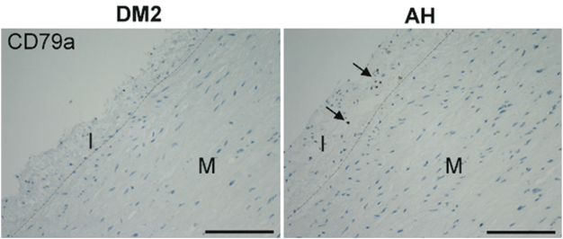
In this study, three samples of the aorta from the most expanded segment of the aneurysm were examined for MCs present in the intima, media, and adventitia. Among the patients, seven from the AH group and five from the DM group exhibited no CD117-positive cells in the intima. Within the media, 13 patients from the AH group lacked MCs, whereas only one patient from the DM group exhibited a similar absence. Furthermore, only five patients from the AH group were devoid of MCs in the adventitia, while MCs were present in all specimens from the DM group. It was observed that MCs were more abundant in the outer media and in the connective tissue adjacent to the blood vessels.
Table 2 presents the average counts of intact, degranulated, and total MCs per mm2 in the intima, media, and adventitia for both the AH and DM groups.
| AH (n ═ 42) | Intima | Media | Adventitia |
|---|---|---|---|
| Intact | 8.583/ mm2± 11.940 | 0.244/mm2 ± 0.359 | 6.497/mm2 ± 7.875 |
| Degran. | 3.015/mm2 ± 3.970 | 0.116/mm2 ± 0.196 | 2.454/mm2 ± 3.866 |
| All | 11.598/mm2 ± 15.160 | 0,359/mm2 ± 0.527 | 8.951/mm2 ± 11.539 |
| DM (n ═ 9) | Intima | Media | Adventitia |
| Intact | 0.659/mm2 ± 1.290 | 0.039/mm2 ± 0.036 | 13.638/mm2 ± 5.136 |
| Degran. | 0.520/mm2 ± 1.286 | 0.022/mm2 ± 0.033 | 5.327/mm2 ± 3.524 |
| All | 1.179/mm2 ± 2.556 | 0.061/mm2 ± 0.060 | 18.965/mm2 ± 7.688 |
MCs: Mast cells; DM: Diabetes mellitus; AH: Arterial hypertension; SD: Standard deviation; N: Number; Intact: Intact mast cells; Degran: Degranulated mast cells.
A comparison of the AH and DM groups revealed a significantly higher number of intact, degranulated, and total MCs in the intima of the AH group, alongside a significantly lower count in the adventitia (refer to Figure 1 and Table 3).
| DM (n ═ 9) | AH (n ═ 42) | |||||
|---|---|---|---|---|---|---|
| Variable | Mean rank (MCs/mm2) | Mean rank (MCs/mm2) | P | U | Z | r |
| Intima | ||||||
| Intact* | 16.56 | 28.02 | 0.032 | 104.000 | −2.140 | 0.300 |
| Degran.* | 17.67 | 27.79 | 0.050 | 114.000 | −1.932 | 0.271 |
| All* | 16.44 | 28.05 | 0.030 | 103.000 | −2.165 | 0.303 |
| Media | ||||||
| Intact | 22.83 | 26.68 | 0.471 | 160.500 | −0.720 | 0.100 |
| Degran. | 21.50 | 26.96 | 0.290 | 148.500 | −1.057 | 0.150 |
| All | 24.50 | 26.32 | 0.735 | 175.500 | −0.339 | 0.050 |
| Advnentitia | ||||||
| Intact* | 39.56 | 23.10 | 0.003 | 67.000 | −3.016 | 0.422 |
| Degran.* | 38.67 | 23.29 | 0.005 | 75.000 | −2.819 | 0.395 |
| All* | 40.00 | 23.00 | 0.002 | 63.000 | −3.115 | 0.436 |
*Significantly different, Mann–Whitney test (P< 0.05). AH: Arterial hypertension; DM: Diabetes mellitus; MCs: Mast cells.
Within both the DM and AH groups, the number of intact MCs was significantly greater than that of degranulated MCs only in the adventitia, with no significant differences observed in the intima and media (see Figure 1 and Table 4).
| Intact | Degran. | |||||
|---|---|---|---|---|---|---|
| Variable | Mean rank (MCs/mm2) | Mean rank (MCs/mm2) | P | U | Z | r |
| AH (n = 42) | ||||||
| Intima | 47.26 | 37.74 | 0.068 | 682.000 | −1.823 | 0.200 |
| Media | 45.89 | 39.11 | 0.187 | 739.500 | −1.319 | 0.144 |
| Adventitia* | 49.35 | 35.65 | 0.010 | 594.500 | −2.575 | 0.281 |
| DM (n = 9) | ||||||
| Intima | 10.11 | 8.89 | 0.580 | 35.000 | −0.553 | 0.130 |
| Media | 10.56 | 8.44 | 0.380 | 31.000 | −0.878 | 0.207 |
| Adventitia* | 13.33 | 5.67 | 0.002 | 6.000 | −3.046 | 0.718 |
*Significantly different, Mann–Whitney test (P < 0.05). AH: Arterial hypertension; DM: Diabetes mellitus; MCs: Mast cells
Assessment of vascularity in the media
The vessels were predominantly located in the outer region of the tunica media in both studied groups—AH and DM (Figure 1). No significant difference in vascularity was observed between the DM group (0.852 ± 0.747) and the AH group (1.603 ± 1.204), as determined by the Mann-Whitney test (P ═ 0.073) (Table 5).
| DM (n ═ 9) | AH (n ═ 42) | |||||
|---|---|---|---|---|---|---|
| Variable | Mean rank (MCs/mm2) | Mean rank (MCs/mm2) | P | U | Z | r |
| Vascularity in media | 18 | 27.71 | 0.073 | 117.000 | -1.791 | 0.251 |
AH: Arterial hypertension; DM: Diabetes mellitus; MCs: Mast cells.
Analysis of vascularity and MCs in media
In the DM group, a significant positive correlation was observed between vascularity and degranulated MCs (r ═ 0.815, P ═ 0.007). Conversely, in the AH group, significant positive correlations were found between vascularity and intact MCs (r ═ 0.519, P ═ 0.000), degranulated MCs (r ═ 0.531, P ═ 0.000), and the total number of MCs (r ═ 0.531, P ═ 0.000) (Figure 2).
Discussion
MCs play a crucial role in host defense against pathogens and are widely distributed throughout the loose connective tissue of the body. They are predominantly located at interfaces between the external environment and internal tissues, particularly in the gastrointestinal tract, respiratory tract, and skin, where they target and destroy antigens such as bacteria, parasites, and viruses [2, 21–23].
Progenitor cells for MCs originate from hematopoietic stem cells in the bone marrow. These cells enter the bloodstream and migrate to various tissues, where they differentiate into mature MCs under the influence of specific cytokines and stem cell factors [24]. The cytoplasm of MCs contains granules rich in pro-inflammatory mediators, including proteases (such as chymase, tryptase, and carboxypeptidase), biogenic amines (including histamine, serotonin, and dopamine), lysosomal enzymes (such as beta-hexosaminidase), glycosaminoglycans and proteoglycans (including heparin, heparan sulfate, chondroitin sulfate, and serglycin), as well as cytokines and growth factors [8, 21, 25, 26]. Upon activation, these granules are released into the environment to combat the antigen.
The most well-known mechanism of MC activation involves the binding of antigens to IgE antibodies attached to FcɛRI receptors on the cell surface [8, 25–27]. This interaction triggers the release (degranulation) of preformed granules, which is characteristic of a type I allergic reaction. In addition to IgE-mediated activation, MCs can also be activated via Fc receptors for immunoglobulin A (IgA) and immunoglobulin G (IgG), complement receptors (e.g., C3a), adenosine receptors, cytokines, chemokines, and various pathogen-associated molecular patterns (PAMPs) [2, 5, 28].
Activated MCs not only release preformed mediators but also synthesize and secrete pro-inflammatory and anti-inflammatory mediators, contingent upon the stimulus and the specific tissue context [2, 8]. In humans, seven transcriptomically distinct MC subtypes have been identified across various tissues, including the trachea, lungs, tongue, lymph nodes, small and large intestines, pancreas, skeletal muscle, skin, bladder, mammary glands, and vasculature [29, 30]. It is hypothesized that the local tissue microenvironment influences MC polarization toward either protective or pathogenic phenotypes. MCs can exert both beneficial and detrimental effects in cardiovascular diseases, such as hypertension, atherosclerosis, myocardial infarction, dilated cardiomyopathy, myocarditis, and thrombosis. One proposed beneficial role of MCs in the cardiovascular system is their promotion of angiogenesis, which is mediated by the release of VEGF-A, tryptase, and the cysteinyl leukotrienes LTC4 and LTD4 [29].
DM is a chronic systemic inflammatory disease closely linked to atherosclerosis. The inflammatory state associated with DM contributes to endothelial dysfunction, increased vascular permeability, the formation of multiple vascular thrombi, and hemodynamic disturbances [31]. MCs further exacerbate DM-related complications, including atherosclerosis [32], kidney disease [33–36], and cardiomyopathy [37, 38]. Experimental studies indicate that inhibiting MC degranulation or inducing MC deficiency in diabetic mice can prevent the progression of cardiomyopathy [39].
In damaged skin, activated MCs release a variety of mediators, including tumor necrosis factor alpha (TNFα), histamine, vascular endothelial growth factor (VEGF), interleukin 6 (IL-6) and interleukin 8 (IL-8), platelet-derived growth factor (PDGF), transforming growth factor beta (TGFβ), and nerve growth factor. These mediators play a crucial role in promoting wound healing. Activated MCs contribute to the stabilization of blood clots, enhance neoangiogenesis, fibrinogenesis, and epithelialization, increase vasodilation and endothelial permeability, facilitate the recruitment of neutrophils and monocytes to the injury site, and support the activation of keratinocytes and fibroblasts [40, 41].
Diabetic wounds are characterized by delayed closure, impaired angiogenesis, and prolonged inflammation. Experimental models demonstrate that intact skin from diabetic subjects contains a higher proportion of degranulated MCs compared to non-diabetic controls.
Moreover, while MC degranulation increases in response to injury in non-diabetic individuals, this response is absent in diabetics. Notably, pharmacological inhibition of MC degranulation in diabetic models resulted in normalized wound healing. These findings suggest that in diabetes, intact MCs support proper wound repair, whereas MCs degranulation impairs the healing process. This indicates the presence of a functionally detrimental MCs phenotype in the diabetic state [41, 42].
MCs are known to inhabit the intima of healthy aortas and early atherosclerotic lesions, such as fatty streaks; however, their prevalence significantly declines in advanced atheromas and further decreases in AAAs [9, 11, 43]. In contrast, the density of MCs in the media and adventitia increases with the progression of atherosclerosis, reaching its peak in the media and adventitia of AAAs [9, 11, 12].
Notably, a significantly higher number of degranulated MCs have been identified in the media and adventitia of AAAs compared to control and atherosclerotic aortas, where degranulated MCs are nearly absent. In the referenced study, 95% of patients had AH, while only 4% had DM [12].
In the current study, we compared the densities of intact and degranulated MCs in the intima, media, and adventitia of ascending aortic aneurysms among patients with DM and those with AH. AH is well recognized as a prevalent risk factor for aneurysm formation [14], while aneurysms are infrequent in patients with DM who do not also have AH [9, 15–18, 20].
Consequently, the DM group comprised only nine patients. In addition to the limited sample size—particularly within the DM group—our study faced several other limitations, including incomplete patient data such as glycated haemoglobin (HbA1c) levels, duration of diabetes, medication use, and smoking history, the reliance on semi-quantitative vascular scoring, and data derived from a single center.
Our findings indicated that, in comparison to the AH group, the DM group exhibited significantly fewer intact and degranulated MCs in the intima, while displaying a significantly greater number in the adventitia. Within both groups, a notably higher quantity of intact MCs was observed relative to degranulated MCs exclusively in the adventitia, with no such difference detected in the intima or media.
Drawing on data from previous studies [9, 12, 41–43], it can be inferred that the ascending aortic aneurysms in the AH group are less impacted than those in the DM group based on our findings.
It is well established that true aneurysms affect all three layers of the vessel wall, whereas false aneurysms compromise only the media and adventitia.
The thickest layer of the aorta is the tunica media, which, along with the adventitia, provides structural integrity and resistance to the vessel wall. The presence of elastic lamellae within the media allows the aorta to expand during systole as it fills with blood and to recoil during diastole, thereby stabilizing and smoothing arterial pressure in conjunction with left ventricular function. Collagen fibers in the adventitia help prevent excessive stretching of the aorta during systole [44]. The primary mechanical property of the aortic wall is its elasticity, predominantly attributed to the tunica media.
However, our previous studies revealed no significant differences in the composition of the aneurysmal wall—such as the volume density of elastic and collagen fibers, vascular smooth muscle cells, and ground substance—between the DM and AH groups [15, 16]. These findings align with our results, which indicated no statistically significant difference in aortic diameter between the AH and DM groups. It is well established that MCs density correlates with the degree of vascularity in the tunica media of the abdominal aorta under both atherosclerotic and aneurysmal conditions [2, 9, 11, 12].
In our current study, we observed a marginally greater vascularity in the tunica media of the AH group compared to the DM group. This trend of reduced vascularity in the DM group is consistent with previous research indicating that diabetes inhibits angiogenesis and impairs wound healing. In the AH group, vascularity was positively correlated with the total number of MCs, including both intact and degranulated forms. In contrast, in the DM group, a significant positive correlation was found only between vascularity and degranulated MCs.
Activated MCs secrete a diverse range of mediators, including both constitutive and inducible pro-angiogenic factors, such as VEGF [3, 45]. However, research indicates that diabetes suppresses VEGF secretion. Experimental studies reveal that a reduced number of MCs results in lower VEGF levels, leading to delayed wound healing in both diabetic and non-diabetic models. Furthermore, VEGF expression and angiogenesis were significantly diminished in diabetic animals compared to their non-diabetic counterparts during the healing process. Pharmacological inhibition of MC degranulation in diabetic models restored VEGF levels and angiogenesis to those observed in non-diabetic subjects [41, 42].
Consequently, we hypothesized a positive correlation between vascularity and intact (as opposed to degranulated) MCs in the diabetic group.
Despite the seemingly contradictory findings, we hypothesize that MCs may play a beneficial role in the wall of aortic aneurysms. MCs are present in the intima of both healthy and early atherosclerotic aortas, yet they are rare in the adventitia. As atherosclerosis progresses, the density of MCs decreases in the intima while increasing in the adventitia. In cases of advanced atherosclerosis and AAAs, MCs are scarce in the intima but abundant in the adventitia [9, 12]. Furthermore, MCs are crucial for normal wound healing in both healthy individuals and those with diabetes, as their deficiency has been linked to delayed repair [41, 42].
It is essential to recognize that MCs constitute a highly pleomorphic population of inflammatory cells whose behavior is shaped by the local tissue environment. Aneurysm formation is a chronic process, as is the progression of diabetes, and the microenvironments of the intima, media, and adventitia in ascending aortic aneurysms differ significantly from those in experimentally induced skin wounds. Therefore, further studies are warranted to clarify the role of MCs in the pathogenesis of ascending aortic aneurysms in diabetic patients.
Conclusion
Our findings show that, in diabetic patients, MCs are less abundant in the intima and more abundant in the adventitia compared to patients with AH. This altered distribution pattern suggests that diabetic patients may have a distinct pathogenesis of aneurysm progression compared to those with AH.
References
- Varricchi G, de Paulis A, Marone G, Galli SJ. Future needs in mast cell biology. Int J Mol Sci 2019;20(18):4397. https://doi.org/10.3390/ijms20184397
- Krystel-Whittemore M, Dileepan KN, Wood JG. Mast cell: a multi-functional master cell. Front Immunol 2015;6:620. https://doi.org/10.3389/fimmu.2015.00620
- Dileepan KN, Raveendran VV, Sharma R, Abraham H, Barua R, Singh V, et al. Mast cell-mediated immune regulation in health and disease. Front Med (Lausanne) 2023;10:1213320. https://doi.org/10.3389/fmed.2023.1213320
- Strauss-Albee DM, Horowitz A, Parham P, Blish CA. Coordinated regulation of NK receptor expression in the maturing human immune system. J Immunol 2014;193(10):4871–9. https://doi.org/10.4049/jimmunol.1401821
- Marshall JS. Mast-cell responses to pathogens. Nat Rev Immunol 2004;4(10):787–99. https://doi.org/10.1038/nri1460
- Metz M, Siebenhaar F, Maurer M. Mast cell functions in the innate skin immune system. Immunobiology 2008;213(3–4):251–2834. https://doi.org/10.1016/j.imbio.2007.10.017
- Varadaradjalou S, Féger F, Thieblemont N, Hamouda NB, Pleau JM, Dy M, et al. Toll-like receptor 2 (TLR2) and TLR4 differentially activate human mast cells. Eur J Immunol 2003;33(4):899–906. https://doi.org/10.1002/eji.200323830
- Vukman KV, Försönits A, Oszvald Á, Tóth EÁ, Buzás EI. Mast cell secretome: soluble and vesicular components. Semin Cell Dev Biol 2017;67:65–73. https://doi.org/10.1016/j.semcdb.2017.02.002
- Sakamoto S, Tsuruda T, Hatakeyama K, Sekita Y, Kato J, Imamura T, et al. Mast cell density and distribution in human abdominal aortic aneurysm. In: Reinhart G, editor. Etiology, pathogenesis and pathophysiology of aortic aneurysms and aneurysm rupture. Rijeka: IntechOpen; 2011. p. 55–66.
- Anvari M, Boroumand M, Mojarad E, Karimi A, Abbasi K, Shirzad M, et al. Do adventitial mast cells contribute to the pathogenesis of ascending thoracic aorta aneurysm?. Int J Surg Pathol 2012;20:474–9. https://doi.org/10.1177/1066896912440820
- Mäyränpää MI, Trosien JA, Fontaine V, Folkesson M, Kazi M, Eriksson P, et al. Mast cells associate with neovessels in the media and adventitia of abdominal aortic aneurysms. J Vasc Surg 2009;50(2):388–96. https://doi.org/10.1016/j.jvs.2009.03.055
- Tsuruda T, Kato J, Hatakeyama K, Kojima K, Yano M, Yano Y, et al. Adventitial mast cells contribute to pathogenesis in the progression of abdominal aortic aneurysm. Circ Res 2008;102(11):1368–77. https://doi.org/10.1161/CIRCRESAHA.108.173682
- Isselbacher EM. Thoracic and abdominal aortic aneurysms. Circulation 2005;111(6):816–28. https://doi.org/10.1161/01.CIR.0000154569.08857.7A
- Ganizada BH, Veltrop RJA, Akbulut AC, Koenen RR, Accord R, Lorusso R, et al. Unveiling cellular and molecular aspects of ascending thoracic aortic aneurysms and dissections. Basic Res Cardiol 2024;119(3):371–95. https://doi.org/10.1007/s00395-024-01053-1
- Milutinović A, Zivin M, Zorc-Pleskovič R. Differences between inflammatory cells infiltrated into tunica intima, media, and adventitia of ascending aortic aneurysms within diabetic and hypertensive patients. Biomol Biomed 2023;23(4):596–604. https://doi.org/10.17305/bb.2022.8565
- Milutinović A, Zorc-Pleskovič R. Inflammatory cells in the ascending aortic aneurysm in patients with type 2 diabetes versus patients with hypertension. Bosn J Basic Med Sci 2021;22:178–84. https://doi.org/10.17305/bjbms.2021.6488
- D’Cruz RT, Wee IJY, Syn NL, Choong A. The association between diabetes and thoracic aortic aneurysms. J Vasc Surg 2019;69(1):263–8.e261. https://doi.org/10.1016/j.jvs.2018.07.031
- Arun D, Munir W, Schmitt LV, Vyas R, Ravindran JI, Bashir M, et al. Exploring the correlation and protective role of diabetes mellitus in aortic aneurysm disease. Front Cardiovasc Med 2021;8:769343. https://doi.org/10.3389/fcvm.2021.769343
- Raffort J, Lareyre F, Clément M, Hassen-Khodja R, Chinetti G, Mallat Z. Monocytes and macrophages in abdominal aortic aneurysm. Nat Rev Cardiol 2017;14(8):457–71. https://doi.org/10.1038/nrcardio.2017.52
- Nordness MJ, Baxter BT, Matsumura J, Terrin M, Zhang K, Ye F, et al. The effect of diabetes on abdominal aortic aneurysm growth over 2years. J Vasc Surg 2022;75(4):1211–22.e1211. https://doi.org/10.1016/j.jvs.2021.10.019
- da Silva EZ, Jamur MC, Oliver C. Mast cell function: a new vision of an old cell. J Histochem Cytochem 2014;62(10):698–738. https://doi.org/10.1369/0022155414545334
- Galli SJ, Tsai M. Mast cells in allergy and infection: versatile effector and regulatory cells in innate and adaptive immunity. Eur J Immunol 2010;40(7):1843–51. https://doi.org/10.1002/eji.201040559
- Jamur MC, Grodzki AC, Berenstein EH, Hamawy MM, Siraganian RP, Oliver C. Identification and characterization of undifferentiated mast cells in mouse bone marrow. Blood 2005;105(11):4282–9. https://doi.org/10.1182/blood-2004-02-0756
- Collington SJ, Williams TJ, Weller CL. Mechanisms underlying the localisation of mast cells in tissues. Trends Immunol 2011;32(10):478–85. https://doi.org/10.1016/j.it.2011.08.002
- Lundequist A, Pejler G. Biological implications of preformed mast cell mediators. Cell Mol Life Sci 2011;68(6):965–75. https://doi.org/10.1007/s00018-010-0587-0
- Wernersson S, Pejler G. Mast cell secretory granules: armed for battle. Nat Rev Immunol 2014;14(7):478–94. https://doi.org/10.1038/nri3690
- Sibilano R, Frossi B, Pucillo CE. Mast cell activation: a complex interplay of positive and negative signaling pathways. Eur J Immunol 2014;44(9):2558–66. https://doi.org/10.1002/eji.201444546
- Jean SM, Christine K, Jeffrey DM. Mast cell cytokine and chemokine responses to bacterial and viral infection. Curr Pharm Des 2003;9(1):11–24. https://doi.org/10.2174/1381612033392413
- Poto R, Marone G, Galli SJ, Varricchi G. Mast cells: a novel therapeutic avenue for cardiovascular diseases?. Cardiovasc Res 2024;120(7):681–98. https://doi.org/10.1093/cvr/cvae066
- Tauber M, Basso L, Martin J, Bostan L, Pinto MM, Thierry GR, et al. Landscape of mast cell populations across organs in mice and humans. J Exp Med 2023;220(10):e20230570. https://doi.org/10.1084/jem.20230570
- Mota RI, Morgan SE, Bahnson EM. Diabetic vasculopathy: macro and microvascular injury. Curr Pathobiol Rep 2020;8(1):1–14. https://doi.org/10.1007/s40139-020-00205-x
- Spinas E, Kritas S, Saggini A, Mobili A, Caraffa A, Antinolfi P, et al. Role of mast cells in atherosclerosis: a classical inflammatory disease. Int J Immunopathol Pharmacol 2014;27(4):517–21. https://doi.org/10.1177/039463201402700407
- Hickey FB, Martin F. Role of the immune system in diabetic kidney disease. Curr Diab Rep 2018;18(4):20. https://doi.org/10.1007/s11892-018-0984-6
- Zhang J, Shi GP. Mast cells and metabolic syndrome. Biochim Biophys Acta 2012;1822(1):14–20. https://doi.org/10.1016/j.bbadis.2010.12.012
- Yin DD, Luo JH, Zhao ZY, Liao YJ, Li Y. Tranilast prevents renal interstitial fibrosis by blocking mast cell infiltration in a rat model of diabetic kidney disease. Mol Med Rep 2018;17(5):7356–64. https://doi.org/10.3892/mmr.2018.8776
- de Morais RB, do Couto Muniz VP, Nunes Costa E, Filho SRF, Nakamura Hiraki KR, Bispo-da-Silva LB, et al. Mast cell population in the development of diabetic nephropathy: effects of renin angiotensin system inhibition. Biomed Pharmacother 2018;107:1115–8. https://doi.org/10.1016/j.biopha.2018.08.066
- Patella V, Marinò I, Arbustini E, Lamparter-Schummert B, Verga L, Adt M, et al. Stem cell factor in mast cells and increased mast cell density in idiopathic and ischemic cardiomyopathy. Circulation 1998;97(10):971–8. https://doi.org/10.1161/01.CIR.97.10.971
- Huang ZG, Jin Q, Fan M, Cong XL, Han SF, Gao H, et al. Myocardial remodeling in diabetic cardiomyopathy associated with cardiac mast cell activation. PLoS One 2013;8(3):e60827. https://doi.org/10.1371/journal.pone.0060827
- He A, Fang W, Zhao K, Wang Y, Li J, Yang C, et al. Mast cell-deficiency protects mice from streptozotocin-induced diabetic cardiomyopathy. Transl Res 2019;208:1–14. https://doi.org/10.1016/j.trsl.2019.01.005
- Weller K, Foitzik K, Paus R, Syska W, Maurer M. Mast cells are required for normal healing of skin wounds in mice. FASEB J 2006;20(13):2366–8. https://doi.org/10.1096/fj.06-5837fje
- Dong J, Chen L, Zhang Y, Jayaswal N, Mezghani I, Zhang W, et al. Mast cells in diabetes and diabetic wound healing. Adv Ther 2020;37(11):4519–37. https://doi.org/10.1007/s12325-020-01499-4
- Tellechea A, Leal EC, Kafanas A, Auster ME, Kuchibhotla S, Ostrovsky Y, et al. Mast cells regulate wound healing in diabetes. Diabetes 2016;65(7):2006–19. https://doi.org/10.2337/db15-0340
- Kaartinen M, Penttilä A, Kovanen PT. Mast cells of two types differing in neutral protease composition in the human aortic intima. Demonstration of tryptase- and tryptase/chymase-containing mast cells in normal intimas, fatty streaks, and the shoulder region of atheromas. Arterioscler Thromb 1994;14(6):966–72. https://doi.org/10.1161/01.ATV.14.6.966
- Zafar MA, Peterss S, Ziganshin BA, Elefteriades JA. Chapter 3—histology of aortic disease and progression of aortic dissection from acute to chronic. In: Ţintoiu IC, Ursulescu A, Elefteriades JA, Underwood MJ, Droc I, editors. New approaches to aortic diseases from valve to abdominal bifurcation. Cambridge (MA): Academic Press; 2018. p. 41–59.
- McHale C, Mohammed Z, Gomez G. Human skin-derived mast cells spontaneously secrete several angiogenesis-related factors. Front Immunol 2019;10:1445. https://doi.org/10.3389/fimmu.2019.01445
About this article
Correspondence
*Correspondence to Aleksandra Milutinović: [email protected]
Article history
Received: 2025-06-10 · Rev-recd: 2025-07-29 · Accepted: 2025-07-03
Acknowledgements
The authors thank Ms. Petra Nussdorfer, M.Sc. from Institute of Histology and Embryology, Ms. Ajla Hajrlahović, and Ms. Miša Omerzel, both from Institute of Pathology,Faculty of Medicine, University of Ljubljana, for technical assistance. We also thank surgeon Miladin Djordjević (MC Medicor) who provided human aortic aneurysms.
Data Availability
Data supporting the findings of this study are available from the corresponding author upon reasonable request.
Conflicts of interest
Conflicts of interest: Authors declare no conflicts of interest.
Funding
Funding: This work was supported by the program grants of the Slovenian Research Agency, ARRS P3-0019, and MC Medicor d.d., No 1, Slovenia.
Copyright
© 2025 Pleskovič et al.
Downloads
References
Varricchi G, de Paulis A, Marone G, Galli SJ. Future needs in mast cell biology. Int J Mol Sci. 2019;20(18):4397. doi:10.3390/ijms20184397 DOI: https://doi.org/10.3390/ijms20184397
Krystel-Whittemore M, Dileepan KN, Wood JG. Mast cell: a multi-functional master cell. Front Immunol. 2015;6:620. doi:10.3389/fimmu.2015.00620 DOI: https://doi.org/10.3389/fimmu.2015.00620
Dileepan KN, Raveendran VV, Sharma R, Abraham H, Barua R, Singh V, et al. Mast cell-mediated immune regulation in health and disease. Front Med (Lausanne). 2023;10:1213320. doi:10.3389/fmed.2023.1213320 DOI: https://doi.org/10.3389/fmed.2023.1213320
Strauss-Albee DM, Horowitz A, Parham P, Blish CA. Coordinated regulation of NK receptor expression in the maturing human immune system. J Immunol. 2014;193(10):4871-9. doi:10.4049/jimmunol.1401821 DOI: https://doi.org/10.4049/jimmunol.1401821
Marshall JS. Mast-cell responses to pathogens. Nat Rev Immunol. 2004;4(10):787-99. doi:10.1038/nri1460 DOI: https://doi.org/10.1038/nri1460
Metz M, Siebenhaar F, Maurer M. Mast cell functions in the innate skin immune system. Immunobiology. 2008;213(3–4):251-2834. doi:10.1016/j.imbio.2007.10.017 DOI: https://doi.org/10.1016/j.imbio.2007.10.017
Varadaradjalou S, Féger F, Thieblemont N, Hamouda NB, Pleau JM, Dy M, et al. Toll-like receptor 2 (TLR2) and TLR4 differentially activate human mast cells. Eur J Immunol. 2003;33(4):899-906. doi:10.1002/eji.200323830 DOI: https://doi.org/10.1002/eji.200323830
Vukman KV, Försönits A, Oszvald Á, Tóth EÁ, Buzás EI. Mast cell secretome: soluble and vesicular components. Semin Cell Dev Biol. 2017;67:65-73. doi:10.1016/j.semcdb.2017.02.002 DOI: https://doi.org/10.1016/j.semcdb.2017.02.002
Sakamoto S, Tsuruda T, Hatakeyama K, Sekita Y, Kato J, Imamura T, et al. doi:10.5772/18561 DOI: https://doi.org/10.5772/18561
Anvari M, Boroumand M, Mojarad E, Karimi A, Abbasi K, Shirzad M, et al. Do adventitial mast cells contribute to the pathogenesis of ascending thoracic aorta aneurysm?. Int J Surg Pathol. 2012;20:474-9. doi:10.1177/1066896912440820 DOI: https://doi.org/10.1177/1066896912440820
Mäyränpää MI, Trosien JA, Fontaine V, Folkesson M, Kazi M, Eriksson P, et al. Mast cells associate with neovessels in the media and adventitia of abdominal aortic aneurysms. J Vasc Surg. 2009;50(2):388-96. doi:10.1016/j.jvs.2009.03.055 DOI: https://doi.org/10.1016/j.jvs.2009.03.055
Tsuruda T, Kato J, Hatakeyama K, Kojima K, Yano M, Yano Y, et al. Adventitial mast cells contribute to pathogenesis in the progression of abdominal aortic aneurysm. Circ Res. 2008;102(11):1368-77. doi:10.1161/CIRCRESAHA.108.173682 DOI: https://doi.org/10.1161/CIRCRESAHA.108.173682
Isselbacher EM. Thoracic and abdominal aortic aneurysms. Circulation. 2005;111(6):816-28. doi:10.1161/01.CIR.0000154569.08857.7A DOI: https://doi.org/10.1161/01.CIR.0000154569.08857.7A
Ganizada BH, Veltrop RJA, Akbulut AC, Koenen RR, Accord R, Lorusso R, et al. Unveiling cellular and molecular aspects of ascending thoracic aortic aneurysms and dissections. Basic Res Cardiol. 2024;119(3):371-95. doi:10.1007/s00395-024-01053-1 DOI: https://doi.org/10.1007/s00395-024-01053-1
Milutinović A, Zivin M, Zorc-Pleskovič R. Differences between inflammatory cells infiltrated into tunica intima, media, and adventitia of ascending aortic aneurysms within diabetic and hypertensive patients. Biomol Biomed. 2023;23(4):596-604. doi:10.17305/bb.2022.8565 DOI: https://doi.org/10.17305/bb.2022.8565
Milutinović A, Zorc-Pleskovič R. Inflammatory cells in the ascending aortic aneurysm in patients with type 2 diabetes versus patients with hypertension. Bosn J Basic Med Sci. 2021;22:178-84. doi:10.17305/bjbms.2021.6488 DOI: https://doi.org/10.17305/bjbms.2021.6488
D’Cruz RT, Wee IJY, Syn NL, Choong A. The association between diabetes and thoracic aortic aneurysms. J Vasc Surg. 2019;69(1):263-8.e261. doi:10.1016/j.jvs.2018.07.031 DOI: https://doi.org/10.1016/j.jvs.2018.07.031
Arun D, Munir W, Schmitt LV, Vyas R, Ravindran JI, Bashir M, et al. Exploring the correlation and protective role of diabetes mellitus in aortic aneurysm disease. Front Cardiovasc Med. 2021;8:769343. doi:10.3389/fcvm.2021.769343 DOI: https://doi.org/10.3389/fcvm.2021.769343
Raffort J, Lareyre F, Clément M, Hassen-Khodja R, Chinetti G, Mallat Z. Monocytes and macrophages in abdominal aortic aneurysm. Nat Rev Cardiol. 2017;14(8):457-71. doi:10.1038/nrcardio.2017.52 DOI: https://doi.org/10.1038/nrcardio.2017.52
Nordness MJ, Baxter BT, Matsumura J, Terrin M, Zhang K, Ye F, et al. The effect of diabetes on abdominal aortic aneurysm growth over 2years. J Vasc Surg. 2022;75(4):1211-22.e1211. doi:10.1016/j.jvs.2021.10.019 DOI: https://doi.org/10.1016/j.jvs.2021.10.019
da Silva EZ, Jamur MC, Oliver C. Mast cell function: a new vision of an old cell. J Histochem Cytochem. 2014;62(10):698-738. doi:10.1369/0022155414545334 DOI: https://doi.org/10.1369/0022155414545334
Galli SJ, Tsai M. Mast cells in allergy and infection: versatile effector and regulatory cells in innate and adaptive immunity. Eur J Immunol. 2010;40(7):1843-51. doi:10.1002/eji.201040559 DOI: https://doi.org/10.1002/eji.201040559
Jamur MC, Grodzki AC, Berenstein EH, Hamawy MM, Siraganian RP, Oliver C. Identification and characterization of undifferentiated mast cells in mouse bone marrow. Blood. 2005;105(11):4282-9. doi:10.1182/blood-2004-02-0756 DOI: https://doi.org/10.1182/blood-2004-02-0756
Collington SJ, Williams TJ, Weller CL. Mechanisms underlying the localisation of mast cells in tissues. Trends Immunol. 2011;32(10):478-85. doi:10.1016/j.it.2011.08.002 DOI: https://doi.org/10.1016/j.it.2011.08.002
Lundequist A, Pejler G. Biological implications of preformed mast cell mediators. Cell Mol Life Sci. 2011;68(6):965-75. doi:10.1007/s00018-010-0587-0 DOI: https://doi.org/10.1007/s00018-010-0587-0
Wernersson S, Pejler G. Mast cell secretory granules: armed for battle. Nat Rev Immunol. 2014;14(7):478-94. doi:10.1038/nri3690 DOI: https://doi.org/10.1038/nri3690
Sibilano R, Frossi B, Pucillo CE. Mast cell activation: a complex interplay of positive and negative signaling pathways. Eur J Immunol. 2014;44(9):2558-66. doi:10.1002/eji.201444546 DOI: https://doi.org/10.1002/eji.201444546
Jean SM, Christine K, Jeffrey DM. Mast cell cytokine and chemokine responses to bacterial and viral infection. Curr Pharm Des. 2003;9(1):11-24. doi:10.2174/1381612033392413 DOI: https://doi.org/10.2174/1381612033392413
Poto R, Marone G, Galli SJ, Varricchi G. Mast cells: a novel therapeutic avenue for cardiovascular diseases?. Cardiovasc Res. 2024;120(7):681-98. doi:10.1093/cvr/cvae066 DOI: https://doi.org/10.1093/cvr/cvae066
Tauber M, Basso L, Martin J, Bostan L, Pinto MM, Thierry GR, et al. Landscape of mast cell populations across organs in mice and humans. J Exp Med. 2023;220(10):e20230570. doi:10.1084/jem.20230570 DOI: https://doi.org/10.1084/jem.20230570
Mota RI, Morgan SE, Bahnson EM. Diabetic vasculopathy: macro and microvascular injury. Curr Pathobiol Rep. 2020;8(1):1-14. doi:10.1007/s40139-020-00205-x DOI: https://doi.org/10.1007/s40139-020-00205-x
Spinas E, Kritas S, Saggini A, Mobili A, Caraffa A, Antinolfi P, et al. Role of mast cells in atherosclerosis: a classical inflammatory disease. Int J Immunopathol Pharmacol. 2014;27(4):517-21. doi:10.1177/039463201402700407 DOI: https://doi.org/10.1177/039463201402700407
Hickey FB, Martin F. Role of the immune system in diabetic kidney disease. Curr Diab Rep. 2018;18(4):20. doi:10.1007/s11892-018-0984-6 DOI: https://doi.org/10.1007/s11892-018-0984-6
Zhang J, Shi GP. Mast cells and metabolic syndrome. Biochim Biophys Acta. 2012;1822(1):14-20. doi:10.1016/j.bbadis.2010.12.012 DOI: https://doi.org/10.1016/j.bbadis.2010.12.012
Yin DD, Luo JH, Zhao ZY, Liao YJ, Li Y. Tranilast prevents renal interstitial fibrosis by blocking mast cell infiltration in a rat model of diabetic kidney disease. Mol Med Rep. 2018;17(5):7356-64. doi:10.3892/mmr.2018.8776 DOI: https://doi.org/10.3892/mmr.2018.8776
de Morais RB, do Couto Muniz VP, Nunes Costa E, Filho SRF, Nakamura Hiraki KR, Bispo-da-Silva LB, et al. Mast cell population in the development of diabetic nephropathy: effects of renin angiotensin system inhibition. Biomed Pharmacother. 2018;107:1115-8. doi:10.1016/j.biopha.2018.08.066 DOI: https://doi.org/10.1016/j.biopha.2018.08.066
Patella V, Marinò I, Arbustini E, Lamparter-Schummert B, Verga L, Adt M, et al. Stem cell factor in mast cells and increased mast cell density in idiopathic and ischemic cardiomyopathy. Circulation. 1998;97(10):971-8. doi:10.1161/01.CIR.97.10.971 DOI: https://doi.org/10.1161/01.CIR.97.10.971
Huang ZG, Jin Q, Fan M, Cong XL, Han SF, Gao H, et al. Myocardial remodeling in diabetic cardiomyopathy associated with cardiac mast cell activation. PLoS One. 2013;8(3):e60827. doi:10.1371/journal.pone.0060827 DOI: https://doi.org/10.1371/journal.pone.0060827
He A, Fang W, Zhao K, Wang Y, Li J, Yang C, et al. Mast cell-deficiency protects mice from streptozotocin-induced diabetic cardiomyopathy. Transl Res. 2019;208:1-14. doi:10.1016/j.trsl.2019.01.005 DOI: https://doi.org/10.1016/j.trsl.2019.01.005
Weller K, Foitzik K, Paus R, Syska W, Maurer M. Mast cells are required for normal healing of skin wounds in mice. FASEB J. 2006;20(13):2366-8. doi:10.1096/fj.06-5837fje DOI: https://doi.org/10.1096/fj.06-5837fje
Dong J, Chen L, Zhang Y, Jayaswal N, Mezghani I, Zhang W, et al. Mast cells in diabetes and diabetic wound healing. Adv Ther. 2020;37(11):4519-37. doi:10.1007/s12325-020-01499-4 DOI: https://doi.org/10.1007/s12325-020-01499-4
Tellechea A, Leal EC, Kafanas A, Auster ME, Kuchibhotla S, Ostrovsky Y, et al. Mast cells regulate wound healing in diabetes. Diabetes. 2016;65(7):2006-19. doi:10.2337/db15-0340 DOI: https://doi.org/10.2337/db15-0340
Kaartinen M, Penttilä A, Kovanen PT. Mast cells of two types differing in neutral protease composition in the human aortic intima. Demonstration of tryptase- and tryptase/chymase-containing mast cells in normal intimas, fatty streaks, and the shoulder region of atheromas. Arterioscler Thromb. 1994;14(6):966-72. doi:10.1161/01.ATV.14.6.966 DOI: https://doi.org/10.1161/01.ATV.14.6.966
Zafar MA, Peterss S, Ziganshin BA, Elefteriades JA. doi:10.1016/B978-0-12-809979-7.00003-1 DOI: https://doi.org/10.1016/B978-0-12-809979-7.00003-1
McHale C, Mohammed Z, Gomez G. Human skin-derived mast cells spontaneously secrete several angiogenesis-related factors. Front Immunol. 2019;10:1445. doi:10.3389/fimmu.2019.01445 DOI: https://doi.org/10.3389/fimmu.2019.01445
Article files
Additional Files
Publication details
- Citation
- Vol. 25 No. 12 (2025), pp. 2827–2834
- Language
- EN
- License
- CC BY 4.0
- Section
- Research article
How to cite (Vancouver)
Pleskovič A, Zorc-Pleskovič R, Zorc M, Milutinović A. Diabetes-induced redistribution of mast cells to the adventitia in ascending aortic aneurysms. Biomol Biomed. 2025;25(12):2827–2834. https://doi.org/10.17305/bb.2025.12772