Extraovarian fibrothecomas: Two case reports and comprehensive review of ovarian sex cord-stromal fibroma-thecoma tumors
DOI:
https://doi.org/10.17305/bb.2025.12816Keywords:
Fibrothecoma, extraovarian tumors, sex cord-stromal tumors, Meigs syndrome, CA-125, differential diagnosis, ovarian neoplasmsAbstract
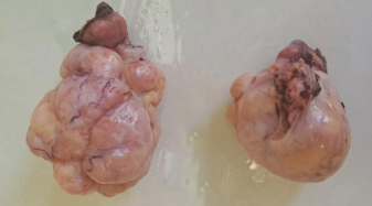
Sex cord-stromal tumors are rare ovarian neoplasms, with fibromas comprising approximately 4% and thecomas accounting for 0.5–1% of all ovarian tumors. The occurrence of these tumors outside the ovaries is exceptionally rare and diagnostically challenging, often mimicking malignancy when associated with ascites, elevated CA-125 levels, or Meigs-like syndrome. This review aims to synthesize current knowledge on the histopathological, immunohistochemical, radiological, and molecular features of ovarian fibroma-thecoma group tumors and highlight their clinical relevance. We report two postmenopausal women with large abdominal masses located extraovarian: one in the broad ligament and the other adherent to the omentum and intestines. In the first case, markedly elevated CA-125, ascites, and pleural effusion initially suggested Meigs syndrome. The second case presented with an abdominal mass and ascites. Imaging studies indicated the possibility of malignant ovarian tumors in both patients, leading to surgical excision. Histopathological examination revealed spindle-to-oval tumor cells arranged in fascicular or storiform patterns, with focal lipid-rich theca-like cells. Immunohistochemical analysis showed that the tumors were positive for vimentin, WT1, progesterone receptor (PR), and variably for estrogen receptor (ER), CD56, inhibin, and calretinin, while being negative for markers of epithelial, melanocytic, and gastrointestinal stromal tumors. A review of the literature identified only 11 well-documented cases of extraovarian fibroma-thecoma group tumors, which most commonly arise in the broad ligament or pelvic cavity. These cases are frequently associated with ascites and elevated CA-125 levels and are often misdiagnosed preoperatively as malignant disease. Our cases underscore the importance of considering extraovarian fibromas and thecomas in the differential diagnosis of pelvic and abdominal masses presenting with similar features. Accurate pathological assessment can prevent unnecessary radical surgeries and promote more favorable patient outcomes.
Citations
Downloads
Downloads
Published
Issue
Section
Categories
License
Copyright (c) 2025 Nejra Selak, Ivana Čerkez, Ermina Iljazović, Azra Sadiković, Maja Konrad Čustović , Jasminka Mustedanagić Mujanović, Edina Ahmetović Karić

This work is licensed under a Creative Commons Attribution 4.0 International License.


