Evaluation of left ventricular hypertrophy in hypertensive patients with echocardiographic myocardial videodensitometry normalized by displacement
DOI:
https://doi.org/10.17305/bjbms.2010.2674Keywords:
myocardial tissue characterization, tissue tracking, grayscale intensity, displacement, left ventricular hypertrophy, hypertensionAbstract
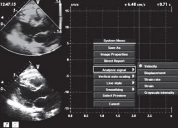
Left ventricular hypertrophy (LVH) is an important predictor of cardiovascular morbidity and mortality. To investigate the feasibility of the myocardial grayscale intensity (GI) normalized by displacement (d) to discriminate between healthy and hypertrophic myocardium in hypertensive patients, sixty hypertensive patients and sixty age and sex-matched healthy volunteers were involved in this study. The peak d and the maximal GI [GI(max)] and minimal GI [GI(min)] for the middle interventricular septal (IVS) and the middle posterior wall (PW) at the level of papillary muscle were obtained from the standard parasternal long axis views using tissue tracking (TT) and videodensitometric analysis, respectively. The GI and the cyclic variation of GI (CVGI) normalized by d were calculated. The results showed that the d both for IVS and PW the amplitude of CVGI for IVS in hypertensive patients with LVH were smaller than the ones without LVH and the normal subjects. But, the CVGI/d both for IVS and PW in hypertensive patients with LVH were all greater than the ones without LVH and the normal subjects. Moreover, the parameter, CVGI/d correlated positively with left ventricular mass index (LVMI). So, the method employed in this study, videodensitometric analysis in combination with TT allow objective and accurate determination of LVH and CVGI/d is a sensitive indicator for hypertensive patients with LVH.
Citations
Downloads

Published
Issue
Section
Categories
How to Cite
Accepted 2017-11-19
Published 2010-11-20


