

Martin-gruber Anastomosis and Transposition in Cubital Tunnel
Abstract
This describes the indivisible anastomosis of the main stem of median nerve with ulnar nerve through cubital tunnel followed by the anomaly of ulnar artery that appears as superficial ulnar artery. Both anomalies are found during the anatomical dissection of a grown-up male cadaver, on his right arm. Such case is very rare as, in the literature available to us, it has not been described.
Keywords
median nerve ulnar nerve superficial ulnar arteryFull text
Introduction
Median nerve and ulnar nerve separate from ulnar fascicle of brachial plexus and, as integral parts of the neurovascular bundle of the upper arm, go down medial bicipital sulcus so that ulnar nerve does not enter the elbow pit (cubital fossa), but rather goes through sulcus of ulnar nerve on humerus between humerale caput and ulnar caput of flexor carpi ulnaris muscle and into medial antebrachial sulcus where it joins the ulnar artery and veins bearing the same name. Dorsal branch of ulnar nerve separates in the distal part of the forearm and goes to the rear side of the hand, whereas the palmar branch follows the course of ulnar nerve and enteres palm through the Guyon tunnel. Median nerve enters the elbow pit from medial bi-cipital sulcus and then continues its cours between superficial flexor digitorum muscle and deep flexor digitorum muscle to the carpal tunnel through which it enters palm. Ulnar artery is one of the terminal branches of brachial artery from which it separates at a sharp angle and reaches the palm where it largery participates in the formation of superficial palm arch. The direction of the proximal third of ulnar artery is from the center of the elbow pit to the point on the ulnar side of the forearm, between the proximal and intermediate third of the forearm, wile the other two thirds stretch down the line thats connects medial epicondyl of humerus with pisiforme bone. In the area of the elbow, ulnar artery lies on tendon of brachial muscle and on deep flexor digitorum muscle. Median nerve and ulnar artery exit the elbow pit together, but ulnar artery changes its course towards medial and, through cubital tunnel between suprficial flexor digitorum muscle and deep flexor digitorum muscle, enters medial antebrachial sulcus. From this groove, along with ulnar nerve, it enters palm through the Guyon tunnel, and with its terminal end participates in the formation of superficial palmar arch. (1,2,3).
Anatomical dissection
This review describes the indivisible anastomosis of the main stem of median nerve with ulnar nerve as an anomaly followed by the anomaly of ulnar artery that appears as superficial ulnar artery. Both anomalies are found during the anatomical dissection of a grown-up male cadaver, on his right arm.
Results
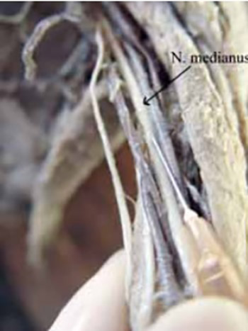
The anastomotic branch separates from median nerve and obliquely goes from proximal and lateral towards distal and medial through cubital tunel in order to join ulnar nerve. In cubital tunnel, there was no ulnar artery with its accompanying veins (Figure 1). In the area of the distal third of the medial side of the upper arm, an artery branch separated from brachial artery at a sharp angle, and as superficial ul-nar artery it went subcutaneously through the elbow pit towards the median part of the distal end of the forearm, where it joined ulnar nerve while passing through the Guyon tunnel on palma manus (Figure 2).
Discussion
Amoiridis notes the presence of Martin-Grubers anastomosis (MGA) in up to 44% of volunteers, but all the recorded anastomosis of the type median-ulnar anastomoses. Not a single anastomosis of the type ulnar-me-dian was recorded (4). Rodrigues-Niedenfiihr noted the presence of MGA on the cadaverous material in 22,9% cases. The same author systematized MGA into two groups: group I, in which anastomosis was accomplished through one anastomotic branch (89,5%); and group II, in which anastomosis was accomplished through two anastomotic branches (10,5%). He divided group I into type a (47,3%), type b (10,6%) and type c (31,6%). Within type a - the anastomotic branch for ul-nar nerve comes from the branche for superficial flexor digitorum muscle. Type b - describes the anastomoses accomplished through the branch coming from the main stem of median nerve for ulnar nerve. Within type c - the anastomotic branch comes from anterior inter-osseal nerve. In 4 cases, the anastomosis passes in front of ulnar artery; in 6 cases behind it, and in 9 ceses it is related to anterior recurrens ulnar artery (5). By EMG method, Hasegawa records the presence of MGA of 25% (6), whereas Shu notes MGA of 23,6% on the cadaverous material, out of wich the anastomosis through one branch, from the stem of median nerve towards ulnar nerve, is recorded in only 3,18% (7). In South Africa, Ta-ams described MGA on cadavers in 25% of the cases and without any statistically significant sexual differences (8), while Nakashima in 108 cadavers, find 23 extremities with MGA (9). According to the data provided by to above mentioned autors, the anastomosis described in our casuistic review is very rare (from 3,18% - 10,6% of all MGA). In addition, the case of superficial ulnar artery on the same extremity on which MGA is present, has not been described in the literature available to us.
Conclusion
The importance of the recognition of such anomalies where median nerve gives the anastomotic branch to ulnar nerve, which again suggests the type of anastomosis in which median nerve innervates the whole deep flexor digitorum muscle and flexor carpi ulnar muscle, is remarkable, especially at surgical procedures for the transposition of medial epicondyle of humerus. Taken into consideration the presence of superficial ulnar artery as well, many other surgical, therapeutic and diagnostically invasive procedures require extreme caution.
References
- Nemanić-Krmpotić J. Anatomija čovjeka JUMENA Zagreb 1982.
- Perović D. Anatomija čovjeka. 4. izdanje, Medicinska knjiga. Beo-grad-Zagreb 1964.
- Šljivić B. Sistemskai topografska anatomija-thorax i gornji exter-mitet. 3. izdanje Naučna knjiga. Beograd 1979.
- Amoiridis G, Vlachonikolis I.G. Verification of the median-to-ulnar and ulnar-to-median nerve motor fiber anastomosis in the forearm: an electrophysiological study. Clin. Neurophisiol 2003;114(1):94–98.
- Rodrgues-Niedenfuhr M, Vasquez T, Parkin I, Logan B, Sa-nudo J.R. Martin-Gruber anastomosis revisted. Clin. Anat 2002;15(2):129–134.
- Hasegawa O, Matsumoto S, Iino M, Kirigaya N, Mimura E, Wada N, Gondo G. Prevalence of Martin-Gruber anastomosis on motor nerve conduction studies. No To Shinkei 2001;53(2):161–164.
- Shu H, Chantelot C, Oberlin C, Alnot J.Y, Shao H. Anatomic study and review of the literature on the Martin-Gruber anastomosis. Morforlogie 1999;83(260):71–74.
- Taams K.O. Martin-Gruber connections in South Africa. An anatomical study. J. Hand. Surg 1997;22(3):328–330.
- Nakashima T. An anatomic study on the Martin-Gruber anastomosis. Surg. Radiol. Anat 1993;15(3):193–195.
About this article
Correspondence
*Corresponding author
Copyright
Copyright: © 2007 ABMSFBIH
Downloads
References
Nemanić-Krmpotić J. Anatomija čovjeka JUMENA Zagreb. 1982.
Perović D. Anatomija čovjeka. 4. izdanje, Medicinska knjiga. Beo-grad-Zagreb. 1964.
Šljivić B. Sistemskai topografska anatomija-thorax i gornji exter-mitet. 3. izdanje Naučna knjiga. Beograd. 1979.
Amoiridis G, Vlachonikolis I.G. Verification of the median-to-ulnar and ulnar-to-median nerve motor fiber anastomosis in the forearm: an electrophysiological study. Clin. Neurophisiol. 2003;114(1):94-98. DOI: https://doi.org/10.1016/S1388-2457(02)00328-0
Rodrgues-Niedenfuhr M, Vasquez T, Parkin I, Logan B, Sa-nudo J.R. Martin-Gruber anastomosis revisted. Clin. Anat. 2002;15(2):129-134. DOI: https://doi.org/10.1002/ca.1107
Hasegawa O, Matsumoto S, Iino M, Kirigaya N, Mimura E, Wada N, et al. Prevalence of Martin-Gruber anastomosis on motor nerve conduction studies. No To Shinkei. 2001;53(2):161-164.
Shu H, Chantelot C, Oberlin C, Alnot J.Y, Shao H. Anatomic study and review of the literature on the Martin-Gruber anastomosis. Morforlogie. 1999;83(260):71-74.
Taams K.O. Martin-Gruber connections in South Africa. An anatomical study. J. Hand. Surg. 1997;22(3):328-330. DOI: https://doi.org/10.1016/S0266-7681(97)80396-8
Nakashima T. An anatomic study on the Martin-Gruber anastomosis. Surg. Radiol. Anat. 1993;15(3):193-195. DOI: https://doi.org/10.1007/BF01627703
Article files
Additional Files
Accepted 2018-01-28
Published 2008-02-20
Publication details
- Citation
- Vol. 7 No. 1 (2007), pp. 71-73
- Language
- EN
- Section
- Case Report with review
- Categories
- Case reports
How to cite (Vancouver)
Isaković E, Delić J, Bajtarević A. Martin-gruber Anastomosis and Transposition in Cubital Tunnel. Biomol Biomed. 2008;7(1):71-73. https://doi.org/10.17305/bjbms.2007.3096