Pathohistological changes in diffuse coronary atherosclerosis and chronic infection caused by Chlamydia pneumonia.
DOI:
https://doi.org/10.17305/bjbms.2004.3455Keywords:
pathohistological changes, diffuse coronary atherosclerosis, chronic infection, Chlamydia pneumoniaAbstract
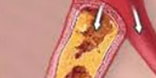
BACKGROUND AND PURPOSE: To investigate the histopathologic characteristics of atherosclerotic lessions in diffuse coronary artery disease and to evaluate the possible inflammatory role of chronic infection with Chlamydia pneumoniae (CP).
MATERIALS AND METHODS: For 10 patients (males, mean age 61 years) who were surgically treated for grave diffuse coronary artery disease, histomorphological analyses of endarterectomized segments of the coronary arteries were performed. Serological analyses for the detection of CP antibodies in peripheral blood were done, preoperatively.
RESULTS AND CONCLUSIONS: Diffuse and concentric atherosclerotic changes from VI to VIII stage according to the Stary classification were found. Immunohistochemical methods revealed infiltrates of T-lymphocytes (80% of cases), B-lymphocytes (40% of cases) and macrophages (80%). Using the nuclear marker for proliferation activity MIB-1, single MIB-1 positive cells were found in 40% of cases. Features of arteriologenesis and vasculitis of newly formed arterioles (as well as thickening of the wall of newly formed arterioles) were found in the vessel wall of 8 patients, 7 of them had chronic infection with CP (preoperative micro-immunofluorescent test results: 1:32<IgG ≥1:512 and IgA≥32), one had passed CP infection (1:32 ≤IgG<1:512, IgA negative). These features were absent in 2 patients, both recovered from CP infection and had not the chronic CP infection at the time of surgery. DNA of Chlamydia pneumoniae was detected using the polymerase chain reaction (PCR) method in the vessel wall of 3 patients who were chosen randomly for this method. This study suggests an inflammatory and proatherogenic role of CP in a high grade atherosclerotic coronary artery wall in diffuse coronary artery disease.
Citations
Downloads
Downloads
Published
Issue
Section
Categories
License
Copyright (c) 2018 Bosnian Journal of Basic Medical Sciences

This work is licensed under a Creative Commons Attribution 4.0 International License.
How to Cite
Accepted 2018-04-05
Published 2004-02-20


