Computed tomography review of the osseous structures of the orbital apex
DOI:
https://doi.org/10.17305/bjbms.2003.3529Keywords:
orbital apex, CT, optic canal, superior orbital fissure, inferior orbital fissure, pterygopalatine fossa, foramen rotundumAbstract
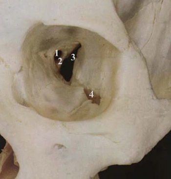
In this paper, we described osseous anatomy of the orbital apex using CT in axial and coronal projections. The main osseous landmarks facilitate the evaluation of orbital apex in radiology, especially on the axial and coronal CT scans. These landmarks include so called optic strut, small segment of the greater wing of the sphenoid bone and upper part of the pterygopalatine fossa. We also concentrate attention upon visualisation and review of the optic canal, superior and inferior orbital fissure, pterygopalatine fossa and foramen rotundum.
Citations
Downloads
Download data is not yet available.
Downloads
Published
20-08-2003
How to Cite
1.
Computed tomography review of the osseous structures of the orbital apex. Biomol Biomed [Internet]. 2003 Aug. 20 [cited 2026 Jan. 20];3(3):50-3. Available from: https://www.bjbms.org/ojs/index.php/bjbms/article/view/3529
Received 2018-04-22
Accepted 2018-04-22
Published 2003-08-20
Accepted 2018-04-22
Published 2003-08-20


