Incomplete Intestinal Metaplasia as an Indicator for Early Detection of Gastric Carcinoma in The Events of Helicobacter Pylori Positive Chronic Atrophic Gastritis
Abstract
The aim of the study was to ascertain the existence of intestinal metaplasia in gastric mucosa of patients with gastric carcinoma coupled with H. pylori positive chronic atrophic gastritis and possible connection of IM with the development of gastric carcinoma. The paper presents prospective study that included 50 patients with gastric carcinoma and 50 patients with chronic atrophic H. pylori positive gastritis. All the patients were subjected to gastroscopy as well as biopsy targeted at antrum, lesser curvature and corpus and at the area 1-2 cm removed from tumor lesion. Biopsy samples were sliced by microtome and stained. We analyzed presence, frequency and severity of inflammatory-regenerative, metaplastic and dysplastic changes in the mucosa and evaluated their prognostic value. We typed IM immunohistochemically. This study confirmed responsibility of H. pylori for inflammatory events in gastric mucosa in patients with gastriccarcinoma. According to our findings incomplete IM of types IIa and IIb as precancerous lesion is responsible for the development of gastriccarcinoma and is associated with chronic atrophic gastritis grade I and II (92% of subjects, p=0.0097, h=1, p=0.01). Thus, the finding of incomplete intestinal metaplasia may be used as an indicator for early gastric carcinoma detection. Patients with patho-histologically verified incomplete intestinal metaplasia associated with active chronic atrophic gastritis of levels I and II represent risk group for the development of gastric carcinoma of intestinal type.
Keywords
intestinal metaplasia (IM) epithelial dysplasia Helicobacter pylori (H. pylori) chronic atrophic gastritis (CAG)Full text
Introduction
H. pylori induced chronic atrophic gastritis (CAG) is a precancerous condition accompanied by gradual loss in epithelia differentiation that eventually leads towards carcinoma. Intestinal metaplasia (IM) as precancerous lesion is either partial or complete transformation of gastric glands epithelial cells into the intestinal type. It may appear either in antrum or pylorus mucosa or diffusely. Various classifications are based on morphological, histochemical, enzymehistochemical, immuno-histochemical and ultrastructural characteristics (1, 2). Complete type of mature IM is characterized by histology, mucine histochemistry, enzymes and endocrine cells identical to those found in small intestine. Patho-histological features of this type are columnar absorptive cells, multiplied goblet cells that contain predominantly acid mucines, Paneth’s non-differentiated cells and Mo-endocrine cells. Incomplete type of mature IM retains curved structure in the antrum glands. Goblet cells are less frequent and contain acid mucines while columnar cells contain traces of neutral mucines. Paneth’s and en-tero-chromophilic are either absent or extremely rare. Immature IM type is encountered in severe atrophic forms of gastritis due to rapid desquamation and regeneration as well as delayed cell differentiation. Hyperplastic glands laid with juvenile non-differentiated cells with hyperchromic nuclei and increased mitotic index develop in the process. The non-differentiated cells may differentiate into various cell types in the presence of various promoters. IM is believed to develop due to the absence of antioxidative activities in the mucosa (3, 4, 5). An issue is raised of whether Helicobacter pylori (H. pylori) presence is necessary for the development of IM. It is known that H. pylori does not dwell in the area of advanced IM as it is not found in biopsic samples collected from antrum where the atrophy is pronounced. H. pylori is found in the mucosa in B gastritis and IM. Positive correlation between H. pylori infection and IM type II is noted in patients with gastric carcinoma.
Subjects and methods
This paper presents prospective study, which includes three viewpoints at the possibility of gastric adenocar-cinoma development: clinical, pathohistological and microbiological. The study included 50 patients with gastric carcinoma that are treated at the Gastroenterohepatol-ogy Clinic. All the patients were subjected to endoscopic examination where stomach was macroscopically divided into sections: corpus, antrum, antropyloric region and pylorus. The patients with cardiac and distal esophagus carcinoma were not included in the study. Following the macroscopic verification of the tumor we performed biopsy of the tumoral formation, as well as biopsy of the mucosa in the region 1-2 cm removed from the tumor. We also collected mucosa samples from corpus, antrum and pyloric region. Biopsy mucosa samples were conserved in 10 % buffered neutral formalin, paraffin embedded and sliced with microtome into 5 μιη thick sections. The sections were stained with Heamatoxylin and Eosin (HE staining), immunohistochemically for p53 antibodies. IM typing was performed. All the patients were examined for the presence of inflammatory-regenerative changes in gastric mucosa. Based on the defined histo-logical criteria we graded inflammatory-regenerative changes as chronic superficial or chronic atrophic gastritis of the degree I, II and III. The degree of inflammation activity was determined based on leukocyte infiltration. It was defined either as active or dormant phase. When the changes were graded as inflammatory-regenerative or dysplastic the tissue was prepared for immunohis-tochemical analysis. The tissue was HE stained prior to the processing. IM was assigned to one of the three possible categories based on the detailed histochemical analysis of mucine type, PAS and HID/AB staining and morphological changes. The degree of IM dispersal was also analyzed. IM was considered focal when found in 1/3 of the biopsic sample, medium when found in 2/3 of the sample and severe when dispersed over the entire sample. The mucine type was examined within goblet and absorptive cells. Mucines may be either neutral or acid. HID/AB staining enables differentiation of acid mucines into sulfo- or sialomucin (sulfomucin – brown, sialomucin – blue). In PAS method acid mucines are stained blue while neutral mucines are stained red. We registered brush border on the surface of absorptive cells that was either well developed, underdeveloped or completely missing. Paneth’s cells were also identified and graded as numerous, reduced or missing. Villi development was also graded as: well developed, underdeveloped and undeveloped. In IM type I, mucosa architecture is mainly well preserved as well as Paneth’s cells abundance. Goblet cells mostly secrete sialomu-cines, some sulfomucines, while absorptive cells have no secretionary properties. Brush border at their surface is preserved. Mucosa surface is not villous although it may appear slightly villous in small number of cases. In IM type II mucosa architecture is significantly degraded, villi are well developed, glandular crypts are elongated and curved and coated in tall columnar cells with either underdeveloped or no brush border. Paneth’s cells are either rare or missing. Goblet cells predominantly secrete sialomucines while poorly differentiated absorptive cells secrete either sulfomucines or neutral mucines. Absorptive cells are scarce. In IM type III mucosa architecture is completely degraded while the surface is exceptionally villous. Mucosa surface glands are elongated and curved to a higher degree compared to those in the IM type II. Gland branching and distortion is evident. Goblet cells are scarce and secrete mainly acid mucines, sulfo- and sialomucines, while poorly differentiated absorptive cells contain sulfomucines. Brush border is absent from absorptive cells. Paneth’s cells are missing. The control group included 50 patients suffering from chronic H. pylori positive gastritis. We endoscopically aimed biopsy at antrum, lesser curvature and corpus mucosa. We applied the same methodology as in the cases of gastric carcinoma in conserving, embedding and staining of the samples as well as in the analysis of inflammatory-regenerative and dysplastic changes in gastric mucosa. We used immunohistochemical methods in IM typing. HUT testing was used for verification of H. pylori presence in gastric mucosa in the areas of antrum and corpus.
Results
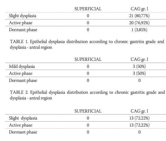
The study included 100 subjects divided into two groups. The first group consisted of 50 patients with stomach carcinoma, 28 male and 22 female. Average age of patients was 58 for males and 62 for females. Anatomically, 66 % of carcinoma formations were endo-scopically identified in the distant part of the stomach. Intestinal metaplasia was differentiated according to regions. IM in antral region was identified in 32 subjects (64%). 3,13 % of the subjects were suffering from superficial chronic gastritis in active phase. 65,63 % of the subjects had intestinal metaplasia associated with chronic atro-phic gastritis grade I. 56,25 % of those were in active phase. 32 % had intestinal metaplasia associated with chronic atrophic gastritis grade II in active phase. IM was identified in 24 patients with epithelial dysplasia. IM was associated with slight epithelial dysplasia in 18 cases and with mild epithelial dysplasia in 6 cases. Correlation is significant and high, p=0,0356, d=22. Rank correlation (Wilcox test) between the level of dysplasia and IM in antral region was significant and high (significance level p=0,05). IM in the region around carcinoma was confirmed in 30 subjects (60%). In 80 % of cases IM was associated with chronic atrophic gastritis grade I. All of those were in active phase. In 20% cases IM was associated with chronic atrophic gastritis grade II in active phase. With respect to epithelial dysplasia, it was slight in 13 IM patients and mild in 7 cases. In 10 patients epithelial dysplasia was not found. Rank correlation (Wilcox test) between IM and the degree of epithelial dysplasia was not significant (p=0,05). IM in the region of corpus was found in 7 patients (14%). In 3 patients IM was associated with superficial and in 4 patients with CAG grade I and grade II. In 14,29 % patients gastritis was in active phase. In the corpus region correlation between IM and gastritis grade was significantly high at p=0,01 level of significance. Rank correlation (Wilcox test) between IM and the degree of epithelial dysplasia was high and significant (p=0,05). Control group included 29 men and 21 women, 60 and 61 years average age respectively. In the control group 19 subjects had IM in the region of antrum (38 %). In 68,42 % subjects IM was associated with CAG grade I in active phase. In 19 subjects it was associated with epithelial dysplasia. Correlation between IM and epithelial dysplasia was significantly high at p=0,05 significance threshold. Intestinal metaplasia in the region of lesser curvature was found in 8 subjects (16%). Of those, 62 % had CAG grade I. Active phase was ascertained in 37,5 % subjects. Rank correlation was significant between the degree of epithelial dysplasia and IM presence at p=0,05 significance threshold. IM in the corpus region was found in one patient along with CAG grade I in active phase. Correlation between IM and epithelial dysplasia was not significant.
Discussion
Progression of chronic active gastritis is accompanied by changes in phenotype of gastric mucosa epithelial cells. Under the influence of the modified environment ephithelial metaplasia occurs due to gastric mucosa capacity for regenerative and metaplastic changes. H. pylori is believed to promote hyper-proliferation of epithelial cells by its presence and action upon mucosa (6, 7, 8). Prolif-erative properties of germinative cells ensure regeneration of surface layer cells in normal mucosa. These cells, referred to as base or stem cells, are located in lower segments of epithelial glands, which explains absence of mitotic activity in surface layer of mucosa (9). Known properties of these cells include auto-regulation, which accounts for adaptive reaction to tissue damage. These cells divide slowly; they are extremely sensitive to radiation and are programmed to self-destroy when their DNA pattern is damaged. They have the ability to preserve genome by selectively maintaining original, paternal DNA copy (10, 11, 12). Other cell types differentiate from those cells since they are multipotent and located in neck parts of glands in the lower crypts segments. Compensation of the lost cells in crypts requires time for regeneration. Cells migrate from the neck part upwards towards the surface. Glandular atrophy occurs in the scope of prolonged chronic atrophic gastritis as inflammation spreads and affects deeper mucosa layers. In atrophic gastritis, gastric mucosa is continuously desquamated (peeled off) and regenerated (renewed). This process is accompanied by compromised differentiation, atypical regeneration, which may lead to carcinoma. The measure of gastritis activity is presence of polymorpho-nucleous infiltrate, which correlates with H. pylori finding. It is widely believed that H. pylori is an important factor in gastritis progression. Along with the progression of atrophy, gastric mucosa suffers loss of gastric antral and corpus glands. In the process, either partial or complete transformation of gastric glands epithelial elements into intestinal type occurs. In the analysis of the mucosa we registered absorptive enterocytes that alternate with sialomucines secreting goblet cells. Such type of epithelial metaplasia is referred to as complete or intestinal metaplasia type I (13, 14). Further progression is marked with glands being coated with columnar cells, scarce presence of Paneth’s cells and sialomucines presence in goblet cells. Absorptive cells are less differentiated and secrete sulfomucine and neutral mucine. This type is denoted IM type II. In IM type III mucosa surface is extremely villous, goblet cells are scarce and secrete acid, sulfo- and sialomucines, while less differentiated absorptive cells secrete sulfomucine. Thus, changes in gastric cells phenotype accompany inflammation and atrophy of gastric mucosa. The normal gastric epithelium is replaced by cells with morphological properties resembling those of the intestinal type. IM progression generates dysplasia which may progress into gastric carcinoma (15). Gastric mucosa atrophy introduces marked increase in cell replication along with the expression of fetal antigens, which influence loss of cell differentiation. Our research targeted verification of the presence, severeness and frequency of intestinal metaplasia occurrence in various regions of gastric mucosa. We found intestinal metaplasia in antral region in 32 out of 50 patients (64%) with gastric carcinoma. In 21 subjects it was associated with chronic atrophic gastritis grade I, which was in active phase in 18 subjects. In 10 subjects we found chronic atrophic gastritis grade II in active phase. We found 30 cases of IM in the region around carcinoma (60%). 24 patients had CAG grade I while 6 had CAG grade II, all of those in active phase. In corpus region we found 7 IM (14%). In 3 cases IM was superficial, 3 had CAG grade I and one had CAG grade II. Four of those were in active phase. In the control group, for comparison, IM in antral region was found in 19 out of 50 examined patients (38%). Of those 13 (26%) had CAG grade I, 5 (10%) had CAG grade II and one had superficial gastritis. All of those were in active phase. 8 (16%) cases of IM in lesser curvature region were identified. Five of those were associated with CAG grade I, one with CAG grade II and two with superficial gastritis. We found one case of IM in corpus region associated with CAG grade I in active phase. Comparing with the group of patients with gastric carcinoma, no significant difference in distribution of intestinal metaplasia in antral region was found in our research. There is significant difference in frequency (p=0,0097, h=1 for significance threshold p=0,01). Significant difference exists in frequency of IM in corpus region. IM is more frequent in patients with gastric carcinoma. The difference is significant with p=0,00278 at significance threshold p=0,01. We used immunohis-tochemical staining to type IM. 17 (34%) subjects had complete IM type I. 20 (40%) of them had incomplete IM type IIa. Four (8%) of patients had incomplete metaplasia type IIb. According to the above, IM was found in 41 out of 50 patients with gastric carcinoma. In 27 (54%) patients it was associated with CAG grade I, of those, 48% were in active phase. In 11 (22%) patients we found CAG grade II in active phase. In 17 (34%) complete IM was focal. Incomplete IM type IIa was of focal appearance in 4 (8%) subjects. In 14% of subjects it spread over 2/3 of the mucosa sample while 18% subjects had diffuse form. Incomplete IM type IIb took up 2/3 of the sample in 2 (4%) patients. In 2 (4%) patients it was diffuse. Goblet cells more frequently secreted acid sialomucines in incomplete IM IIa and IIb. Absorptive cells secreted neutral or acid sulfomucines, with their brush border less developed and Paneth’s cells either absent or scarce. In the control group, complete IM type I was found in 13 (26%) subjects while incomplete IM type IIa was found in 7 (14%) patients. They were also associated with CAG grade I and II in active phase. IM was predominantly located in antral region – in 16 of 20 (80%) control group subjects. Four subjects had IM in corpus region. IM was focal in 4 (8%) subjects, spread over 2/3 of sample in 15 (30%) and diffuse in one subject. In 18 (36%) subjects goblet cells secreted acid sialomucine. They were found to secrete acid sulfomucine in 2 (4%) subjects. Absorptive cells secreted acid sialomucine in 2 (4%) subjects, acid sulfomucine in 5 (10%) subjects and neutral mucines in 10 (20%) subjects. Brush cover was well developed in 5 (10%), underdeveloped in 13 (26%) and absent in 2 (4%) subjects. Paneth’s cells were absent in 9 (18%) and scarce in 11 (22%) subjects. We were not able to find significant differences in Wilcox rank test between groups per regions by gastritis type (p=0,4192, h=0). There is significant difference between groups in IM occurrence in antral region with p=0,0097, h=1 at the significance threshold p=0,01. No significant difference in gastritis presence in corpus region was found. Analyzing earlier published studies we found that Reis (13) studied IM and concluded that classic sequential paths from IM I to IM III lead through IM II. In our research we found incomplete IM type IIb (type III according to certain authors) in only 4 (8%) patients with gastric carcinoma. No IM type III cases were found in the control group. Safele-Ribiero stated IM type I as the most frequent in their studies. It is suggested (16, 17) suggest that metaplastic cell lines ensue as a response of gastric mucosa to chronic damage and precede epithelial dysplasia and gastric adenocarcinoma. In our subjects, we found IM type I in 34% patients with gastric carcinoma and in 26% patients with H. pylori positive gastritis. 40% patients had incomplete IM or type II while 8% had IM type III. In the control group though, 14% subjects had incomplete IM type II. In our research IM was identified in 41 (82%) patients with gastric carcinoma. Incomplete IM type IIa was more frequent in patients with gastric carcinoma. According to the severeness of the changes incomplete IM types IIa and IIb were either diffuse or spread over 2/3 of the biopsic specimen width. In comparison with the control group no significant difference in IM frequency in antral region was found. In the region around carcinoma IM was found in 30 (60%) of the subjects and it was associated with chronic atrophic gastritis grade I and II in active phase.
Conclusion
Based on the results of our research we may conclude that IM as precancerous lesion is responsible for the development of gastric carcinoma. When associated with chronic atrophic gastritis grade I and II it can serve as an indicator of the events that lead to intestinal gastric carcinoma development. Special attention is required in patients with pathohistologically verified incomplete IM type IIa associated with slight or mild epithelial dysplasia due to its significantly frequent occurrence in our patients with gastric carcinoma.
References
- Al-Knawy B, Morad N, Jamal A, Mirdad S, Abol Fotouh M, Ahmed ME, Saydain G, Seidi O, Shatoor A. Helicobacter pylori and intestinal metaplasia with its subtypes in the gastric antrum in a Saudi population. Scand. J. Gastroenterol 1999;34(6):562–565.
- Bravo JC, Correa P. Sulphomucins favor adhesion of Helico-bacter pylori to metaplastic gastric mucosa. J. Clin. Pathol 1999;52/2:137–140.
- Correa P. Human gastric carcinogenesis: A multistep and multi-factorial Process. First American Cancer Society Award Lecture of Cancer Epidemiology and Prevention. Cancer Reasrch 1992;52:6735–6740.
- Egashira Y, Shimoda T, Ikegami M. Mucin histochemical analysis of minute gastric differentiated adenocarcinoma. Pathol. Int 1999;49(1):55–61.
- Endoh Y, Tamura G, Motayama T, Ajioka Y, Watanabe H. Well-differentiated adenocarcinoma mimicking completetype intestinal metaplasia in the stomach. Hum. Pathol 1999;30(7):826–832.
- Ernest P. Review article: The role of inflamation in the pathogenesis of gastric cancer. Aliment. Pharmacol. Ther 1999;13(Suppl 1):13–18.
- Genta MR. Helicobacter pylori as a promoter of intestinal metaplasia and gastric cancer: an alluring hypothesis in search of evidence. Eur. J. Gastroenterol. Hepatol 1995;7(Suppl.1):S25–S30.
- Gong C, Mera R, Bravo JC, Ruiz B, Diaz-Escamilla R, Fontham ET, Correa P, Hunt JD. K-RAS mutations predict progression of preneoplastic gastric lesions. Cancer Epidemiol. Biomarkers Prev 1999;8(2):167–171.
- Kim DY, Baek JY. The comparison of histologic gastritis in patients with duodenal ulcer, chronic gastritis, gastric ulcer and gastric cancer. Yonsei Med. J 1999;40(1):14–19.
- Kuipers EJ. Review article;Exploring the link between Helico-bacter pylori and gastric cancer. Aliment. Pharmacol. Ther 1999;13(Suppl 1):3–11.
- Lynch AFD, Axon TRA. Helicobacter pylori gastric cancer and gastric epithelial kinetics: Rev. Eur. J. Gastroenterol. Hepatol 1995;7(Supl.1):S17–S23.
- Nardone G, Stabiano S, Rocco A, Mezza E, D’Anniento FP, Insabato L, Coppola A, Salvatore G, Lucariello A, Figura N, De Rosa G, Budillon G. Effect of Helicobacter pylori infection and its eradication on cell proliferation, DNA status, and oncogene expression in patients with chronic gastritis. Gut 1999;44(6):789–799.
- Reis CA, David L, Correa P, Carneiro F, De Bolos C, Garcia E, Mandel U, Clausen H, Sobrinho-Simoes M. Intestinal metaplasia of human stomach displays distinct patterns of mucin (MUC1, MUC2, MUC5AC, and MUC6) expression. Cancer Res 1999;59(5):1003–1007.
- Rokkas T, Ladas S, Liatsos C, Petridou E, Papatheodoreu G, Theocharis S, Karameris A, Raptis S. Relationship of Helico-bacter pylori CagA status to gastric cell proliferation and apopto-sis. Dig. Dis. Sci 1999;44(3):487–493.
- Sasaki I, Yao T, Nawata H, Tsuneyoshi M. Minute gastric carcinoma of differentiated type with special reference to the significance of intestinal metaplasia, proliferative zone, and p53 protein during tumor development. Cancer 1999;85(8):1719–1729.
- Schmidt PH, Lee JR, Joshi V, Playford RJ, Poulsom R, Wright NA, Goldenring JR. Identification of a metaplastic cell lineage associated with human gastric adenocarcinoma. Lab. Invest 1999;79(6):639–646.
- Sethi S, Annamma MO, Preetha K. Study of gastric carcinomas with special reference to intestinal metaplasia. Indian. J. Pathol. Microbiol 1999;42(1):73–79.
About this article
Correspondence
*Corresponding author
Copyright
Copyright: © 2006 ABMSFBIH
Downloads
References
Al-Knawy B, Morad N, Jamal A, Mirdad S, Abol Fotouh M, Ahmed ME, et al. Helicobacter pylori and intestinal metaplasia with its subtypes in the gastric antrum in a Saudi population. Scand. J. Gastroenterol. 1999;34(6):562-565. DOI: https://doi.org/10.1080/003655299750026001
Bravo JC, Correa P. Sulphomucins favor adhesion of Helico-bacter pylori to metaplastic gastric mucosa. J. Clin. Pathol. 1999;52/2:137-140. DOI: https://doi.org/10.1136/jcp.52.2.137
Correa P. Human gastric carcinogenesis: A multistep and multi-factorial Process. First American Cancer Society Award Lecture of Cancer Epidemiology and Prevention. Cancer Reasrch. 1992;52:6735-6740.
Egashira Y, Shimoda T, Ikegami M. Mucin histochemical analysis of minute gastric differentiated adenocarcinoma. Pathol. Int. 1999;49(1):55-61. DOI: https://doi.org/10.1046/j.1440-1827.1999.00824.x
Endoh Y, Tamura G, Motayama T, Ajioka Y, Watanabe H. Well-differentiated adenocarcinoma mimicking completetype intestinal metaplasia in the stomach. Hum. Pathol. 1999;30(7):826-832. DOI: https://doi.org/10.1016/S0046-8177(99)90144-2
Ernest P. Review article: The role of inflamation in the pathogenesis of gastric cancer. Aliment. Pharmacol. Ther. 1999;13(Suppl 1):13-18. DOI: https://doi.org/10.1046/j.1365-2036.1999.00003.x
Genta MR. Helicobacter pylori as a promoter of intestinal metaplasia and gastric cancer: an alluring hypothesis in search of evidence. Eur. J. Gastroenterol. Hepatol. 1995;7(Suppl.1):S25-S30.
Gong C, Mera R, Bravo JC, Ruiz B, Diaz-Escamilla R, Fontham ET, et al. K-RAS mutations predict progression of preneoplastic gastric lesions. Cancer Epidemiol. Biomarkers Prev. 1999;8(2):167-171.
Kim DY, Baek JY. The comparison of histologic gastritis in patients with duodenal ulcer, chronic gastritis, gastric ulcer and gastric cancer. Yonsei Med. J. 1999;40(1):14-19. DOI: https://doi.org/10.3349/ymj.1999.40.1.14
Kuipers EJ. Review article;Exploring the link between Helico-bacter pylori and gastric cancer. Aliment. Pharmacol. Ther. 1999;13(Suppl 1):3-11. DOI: https://doi.org/10.1046/j.1365-2036.1999.00002.x
Lynch AFD, Axon TRA. Helicobacter pylori gastric cancer and gastric epithelial kinetics: Rev. Eur. J. Gastroenterol. Hepatol. 1995;7(Supl.1):S17-S23.
Nardone G, Stabiano S, Rocco A, Mezza E, D’Anniento FP, Insabato L, et al. Effect of Helicobacter pylori infection and its eradication on cell proliferation, DNA status, and oncogene expression in patients with chronic gastritis. Gut. 1999;44(6):789-799. DOI: https://doi.org/10.1136/gut.44.6.789
Reis CA, David L, Correa P, Carneiro F, De Bolos C, Garcia E, et al. Intestinal metaplasia of human stomach displays distinct patterns of mucin (MUC1, MUC2, MUC5AC, and MUC6) expression. Cancer Res. 1999;59(5):1003-1007.
Rokkas T, Ladas S, Liatsos C, Petridou E, Papatheodoreu G, Theocharis S, et al. Relationship of Helico-bacter pylori CagA status to gastric cell proliferation and apopto-sis. Dig. Dis. Sci. 1999;44(3):487-493. DOI: https://doi.org/10.1023/A:1026636803101
Sasaki I, Yao T, Nawata H, Tsuneyoshi M. Minute gastric carcinoma of differentiated type with special reference to the significance of intestinal metaplasia, proliferative zone, and p53 protein during tumor development. Cancer. 1999;85(8):1719-1729. DOI: https://doi.org/10.1002/(SICI)1097-0142(19990415)85:8<1719::AID-CNCR11>3.0.CO;2-V
Schmidt PH, Lee JR, Joshi V, Playford RJ, Poulsom R, Wright NA, et al. Identification of a metaplastic cell lineage associated with human gastric adenocarcinoma. Lab. Invest. 1999;79(6):639-646.
Sethi S, Annamma MO, Preetha K. Study of gastric carcinomas with special reference to intestinal metaplasia. Indian. J. Pathol. Microbiol. 1999;42(1):73-79.
Article files
Additional Files
Accepted 2018-02-01
Published 2006-11-20
Publication details
- Citation
- Vol. 6 No. 4 (2006), pp. 48-53
- Language
- EN
- Section
- Pathology
- Categories
- Microbiology and Microbiome, Research articles, Pathology
How to cite (Vancouver)
Vukobrat-Bijedić Z, Radović S, Husić-Selimović A, Gornjaković S. Incomplete Intestinal Metaplasia as an Indicator for Early Detection of Gastric Carcinoma in The Events of Helicobacter Pylori Positive Chronic Atrophic Gastritis. Biomol Biomed. 2006;6(4):48-53. https://doi.org/10.17305/bjbms.2006.3120