

Integrins and pulmonary fibrosis: Pathogenic roles and therapeutic opportunities
Abstract
Characterized by the formation of fibrotic scars, pulmonary fibrosis (PF) involves a complex pathogenesis, limited treatment options, and a high mortality rate. Integrins—heterodimeric transmembrane proteins composed of α and β subunits—mediate extracellular matrix remodeling and regulate the physiological functions of epithelial, mesenchymal, and immune cells through "inside-out" and "outside-in" signaling pathways. These molecules play a critical role in the initiation and progression of PF. Due to their central regulatory functions, a range of integrin-targeted therapies has been developed. However, the complex pathophysiology of PF and the structural diversity of integrins pose significant challenges to targeted treatment. In this study, we systematically delineated the signaling networks mediated by the full spectrum of integrin family members and uncovered the molecular mechanisms by which they contribute to PF through immunoregulatory pathways. We also reviewed the development of integrin-based therapies from preclinical studies to clinical trials and discussed current priorities in clinical, basic, and translational research. These insights may provide new perspectives for the diagnosis and treatment of PF.
Keywords
integrin pulmonary fibrosis PF targeted therapyFull text
Introduction
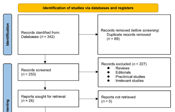
Pulmonary fibrosis (PF) represents the end stage of a diverse group of diffuse interstitial lung diseases, including nonspecific interstitial pneumonia, fibrotic hypersensitivity pneumonitis (fHP), connective tissue disease-associated interstitial lung disease (ILD), and idiopathic PF (IPF). PF is pathologically characterized by the progressive degradation of lung tissue architecture, often resulting in impaired lung function, compromised gas exchange, and, ultimately, respiratory failure and death. Studies indicate that patients with IPF have a 5-year mortality rate ranging from 30% to 50%, with a median survival of 2–3 years [1]. For patients with fHP, the reported median survival is approximately 7.1 years [2]. The interaction between cells and the extracellular matrix (ECM) plays a critical role in the initiation, progression, and prognosis of PF [3]. Integrins, which are heterodimeric transmembrane glycoproteins composed of α (120–185 kDa) and β (90–110 kDa) subunits, are central to this process. In mammals, more than 20 functionally distinct integrin molecules have been identified (Figure 1). As adhesion receptors, integrins are uniquely capable of bidirectional signaling across the plasma membrane, mediating essential interactions between cells and their surrounding microenvironment [4]. In outside-in signaling, integrins specifically bind extracellular ligands, triggering conformational changes that recruit and activate intracellular signaling molecules. This cascade regulates key cellular behaviors such as proliferation, differentiation, migration, and invasion. In inside-out signaling, intracellular activators bind to the cytoplasmic domains of integrins, causing conformational shifts that increase their affinity for extracellular ligands, promoting cell migration and altering the extracellular landscape. During the progression of PF, integrins mediate abnormal signaling among epithelial, mesenchymal, and immune cells, as well as with the ECM. This dysregulated signaling promotes the proliferation, activation, and migration of pathogenic cells, triggers the secretion of pro-fibrotic factors, and leads to excessive ECM deposition. Three primary regulatory pathways are implicated: the integrin–transforming growth factor β (TGF-β) axis, integrin-mediated mechanotransduction, and the integrin–immunity axis. For example, αv and α2β1 integrins specifically activate TGF-β signaling. Meanwhile, integrins such as αvβ3, αvβ5, α2β1, α4β1, and α5β1 facilitate epithelial–mesenchymal transition (EMT), as well as fibroblast migration, invasion, and differentiation by transmitting mechanical cues from the ECM to cells. On immune cells, integrins including α4β1, αLβ2, αMβ2, αXβ2, and αE amplify pro-inflammatory and pro-fibrotic responses via pathways such as RhoA/Rho kinase (ROCK). Given their central role in PF pathogenesis, targeting integrin-mediated signaling represents a promising therapeutic strategy to disrupt the fibrotic cascade and slow disease progression. Several integrin-targeted therapeutics are currently being evaluated in clinical trials for safety, tolerability, and efficacy. Numerous other lead compounds and candidates are undergoing preclinical development in academic and industry settings, contributing to a growing body of meaningful data. In this review, we searched PubMed using keyword combinations such as (“integrin” OR “αvβ6” OR “αvβ1,” etc.) AND (“pulmonary fibrosis” OR “IPF” OR “lung fibrosis”), focusing on publications from January 2014 to March 2025 (one month prior to submission). We included original research (both preclinical and clinical), high-quality review articles, and clinical trial registry entries (https://clinicaltrials.gov/). Our review provides a comprehensive overview of integrin-mediated pathogenesis in PF (Table 1). Specifically, we summarize integrin-related signaling across seven key cell types (Figure 2), describe the mechanisms of action of integrin inhibitors, and highlight promising candidates currently in clinical trials (Tables 2 and 3). Finally, we discuss major challenges and future directions in exploring integrin-targeted therapies for PF.
Role of integrin-dependent signaling axis in PF
Integrin/TGF-β signaling pathway in PF
The integrin αv subunit can pair with β1, β3, β5, β6, or β8 subunits to form various integrin heterodimers with distinct functions, many of which play key roles in fibrosis development. Studies have shown that αv integrins activate latent TGF-β—a classical pro-fibrotic cytokine—thereby promoting PF in multiple experimental models [5]. Galectin-3 has been found to bind both αvβ1 integrin and TGFβRII in a glycosylation-dependent manner, facilitating their spatial co-localization. This interaction enhances TGF-β1 activation and downstream signaling [6]. In lung fibroblasts, αvβ3 and αvβ5 integrins interact with periostin to activate the TGF-β/Smad3 signaling pathway, leading to upregulation of lung fibrosis-associated molecules such as SERPINE1, CTGF, IGFBP3, and IL-11 [7]. Integrin αvβ6 also engages TGF-β, contributing to both physiological and pathological responses. TGF-β1 regulates ITGB6 gene expression via the Smad pathway, which in turn influences αvβ6 integrin expression [8]. Activation of the αvβ6/TGF-β axis is implicated in TGF-α-induced pleural fibrosis and PF triggered by influenza infection [9, 10]. Additional research has shown that OGN knockdown or elevated levels of developmental endothelial locus-1 (Del-1) can inhibit integrin αv—particularly αvβ6—from binding to the latency-associated peptide (LAP), thereby suppressing TGF-β/Smad signaling [11, 12]. After lung injury, IRE1α is upregulated and modulates damage-associated transient progenitor cells (DATPs), enhancing αvβ6 expression and promoting fibrosis via TGF-β activation [13]. Thrombin has also been shown to drive fibrosis through the PAR1/αvβ6/TGF-β pathway. In vitro studies reveal that PAR1 triggers αvβ6-mediated TGF-β activation via RhoA and ROCK, identifying a potential therapeutic target in PF [14, 15]. In canine IPF, the ITGB8 gene is significantly downregulated, and TGF-β stimulation leads to decreased integrin β8 expression in MRC-5 cells [16]. These findings suggest that TGF-β may contribute to IPF progression in dogs by modulating integrin β8 expression. Researchers have also explored the heterogeneity of mesenchymal stromal cells and their roles in PF. Sciurba et al. [17] demonstrated that ITGAV promotes type 17/TGF-β-driven PF, as evidenced by reduced collagen deposition in bleomycin-induced ITGAV flox/flox (Pdgfrb-Cre+) mice. Yi et al. [18] reported decreased TGF-β1 activity, reduced p-Smad2/3 expression, and less radiographic lung fibrosis in ITGAV loxP/loxP; Pdgfrb-Cre mice following radiation exposure. Interestingly, deletion of αv integrin in α-smooth muscle actin (α-SMA)-positive cells did not protect against bleomycin-induced PF, while PDGFRβ-Cre-mediated deletion did. These findings suggest that α-SMA-positive cells are a subset of PDGFRβ-positive cells and are not the primary mediators of αv integrin–dependent TGF-β activation in PF [19].
| Integrin subunits and molecules | Models | In vivo/in vitro | Main mechanisms | References |
|---|---|---|---|---|
| αv | Cigarette smoke extract-induced and αv-silenced AECs | In vitro | Activated TGF-β1↓ | [5] |
| αv | OGN-knockdown lung fibroblasts treated with or without TGF-β | In vitro | The binding of integrin αv to LAP↓, p-Smad2↓ | [11] |
| αv | BLM-induced ITGAV flox/flox (Pdgfrb- cre+) mice | In vivo | HYP↓ | [17, 19] |
| αv | ITGAVloxP/loxP; Pdgfrb-Cre mice receiving radiation | In vivo | Activated TGF-β1↓, p-Smad2/3↓ | [18] |
| β1 | BLM-induced mice | In vivo | β1↑ | [46] |
| β1 | PQ-induced-PF rats | In vivo | β1↑ | [47] |
| β1 | Elk1-knockout mice | In vivo | β1↓, spontaneous fibrosis↑ | [48] |
| β1 | BLM-induced mice, accompanied by loss of periostin in fibrocytes; periostin -/- fibrocytes | In vivo and in vitro | β1, CTGF, collagen I↓ | [43] |
| β1 | SiO2-induced and ITGB1-knockout BEAS-2B cells | In vitro | E-cadherin↑, vimentin↓, ILK, Snail↓ | [49] |
| β1 | MWCNT-induced PF in mice; lung fibroblasts | In vivo and in vitro | TIMP1/CD63/integrin β1↑, pErk1/2↑, lung fibroblast proliferation↑ | [50] |
| β1 | PDGF-BB induced lung fibroblasts with integrin β 1 blocking antibody | In vitro | Fak activation↓, fibroblast migration on FN↓ | [20] |
| β1 | Fibroblasts in fibrotic ECM | In vitro | Integrin-β1/FAK/ERK1/2↑, fibroblasts transforming into myofibroblasts | [21] |
| β3 | ABCG1-deficient MWCNT mice | In vivo | MFG-E8, β3 in BALF cells↑ | [34] |
| β3 | LPS-induced β3-specific shRNAs in lung fibroblasts | In vitro | Activation of the PI3K-Akt-mTOR↓, LC3II/LC3I↓, p62↑ | [67] |
| β3 | Integrin β3-deficient MV mice | In vivo | PKM2, LDHA, lactate, collagen deposition↓ | [68] |
| α1 | Mice induced by adenoviral vector and BLM | In vivo | α1↑, fibrosis↑ | [51] |
| α2 | IPF patients | In vivo | α2↑ | [52] |
| α2 | DDR2-deficient mice; lung fibroblasts lacking Col1a1 | In vivo and in vitro | α2↑, fibrosis↓ | [53] |
| α2 | TC-I-15-treated lung fibroblasts | In vitro | Collagen I and α-SMA↓ | [53] |
| α2 | Floxed integrin α2 mice crossed with Col1a2-creERT mice | In vivo | Fibrosis↓ | [55] |
| α2 | Floxed integrin α2 mice crossed with SPC-rtTA/tetO-Cre mice | In vivo | Fibrosis↑ | [55] |
| α2 | MTX-A549 cells | In vitro | α2↑, mRNA of EMT-related genes↑ | [54] |
| α2, αMβ2 and αXβ2 | Lumican-induced PBMC pretreated with α2, αM, αX, and β2 inhibitors | In vitro | Lumican-induced fibrocytes differentiation↓ | [44] |
| α3 | ITGA3 missense mutation | In vivo | Fibrosis↑ | [56] |
| α4 | LPS-induced mice | In vivo | Integrin α4β1↑ | [33] |
| α4 | BLM-treated α4Y991A mice | In vivo | Rac↓, M2 macrophage markers (e.g., YM1, Fizz1, CD206) ↓ | [38] |
| α5 | Components in the supernatant secreted by IPF fibroblasts affect normal fibroblasts | In vitro | ITGA5, pIκBα↑, adhesion and migration ↑ | [57] |
| α5 | Fibroblasts were incubated with EVs | In vitro | Fibroblast invasion↑, activation of FAK and Src↑ | [22] |
| α5 | IPF-HLFs | In vitro | uPAR↑, integrin α5↑, caveolin-Fyn-Shc↑in lipid rafts, fibroblast migration↑ | [23] |
| α5 | SPCs | In vitro | Fn1, Col1a1, ITGA5↑ | [58] |
| α5 | ITGA5-silenced IPF-HLFs | In vitro | Transformation of fibroblasts to myofibroblasts, FN1, TGF-β, α-SMA and collagen I↑ | [59] |
| α6 | Human IPF lung myofibroblasts in the sclerotic matrix | In vitro | ROCK↑, c-Fos/c-Jun transcription complex↑, α6 integrin↑, collagen IV↓ | [24] |
| α6 | BLM-induced PF in mice; HLF and MLF | In vivo and in vitro | CD11c+ AREG↑, integrin α6↑, lung fibroblast motility and invasiveness↑ | [62] |
| α6 | Silica dust-induced PF mice and TGF-β1-stimulated fibroblasts | In vivo and in vitro | miR-542-5p↓, ITGA6 ↑, proliferation and migration of fibroblasts↑, lung fibrosis↑ | [63] |
| α8 | IPF patients | In vivo | CD248 low ITGA8 high fibroblast-like cells in the elastic fiber-rich connective tissue | [60] |
| α8 | ITGA5 silenced fibroblasts | In vitro | Transformation of fibroblasts to myofibroblasts, ITGA8 and ITGAV↑ | [59] |
| α10 | BLM-induced MIF knockdown in rats | In vivo | α10↓, fibrosis↓ | [64] |
| α11 | IPF patients | In vivo | α11↑ | [52] |
| α11 | IPF patients | In vivo | α11+ α-SMA myofibroblasts↑ | [65] |
| αMβ2 | Human interstitial lung lesions following neococcal pneumonia | In vivo | sITGaM and sITGb2↑ | [36] |
| αM and αX | Neutrophils under hypoxic conditions | In vitro | αM and αX↑, NETs↑ | [39] |
| αLβ2 and αMβ2 | HFD-fed mice | In vivo | ITGAM, ITGAL, ITGB2L, ITGB2, NAIP6 and NAIP5↑ | [35] |
| αvβ1 | HLFs | In vitro | Galectin-3 binds to β1 integrin, p-Smad2↑ | [6] |
| αvβ3 | BLM-induced mice | In vivo | αvβ3↑ | [66] |
| αvβ3 | Periostin or integrin silencing in lung fibroblasts | In vitro | Lung fibroblast proliferation↓ | [27] |
| αvβ3 and αvβ5 | Knockdown of integrin αv/β3/β5 in lung fibroblasts treated with TGF-β | In vitro | SERPINE1, CTGF, IGFBP3, and IL11↓ | [7] |
| αvβ3 | BLM mice lacking Thy-1 | In vivo | αvβ3 in αSMA+ myofibroblasts↑ | [28] |
| αvβ3 and αvβ5 | PDGF-BB stimulated fibroblasts | In vitro | Src activation, Src interacted with integrative αvβ3, fibroblasts migration | [31] |
| αvβ3 | Thy-1 neg fibroblasts | In vitro | RhoA activity↑, collagen matrix contraction↑ | [30] |
| αvβ3 | BLM-treated mice; CD146+ MACS-enriched primary cells seeded on fibronectin | In vivo and in vitro | Pericytes α-SMA↑, αvβ3↑ | [26] |
| β6 | TGF-β1 stimulation of iHBECs | In vitro | Time-dependent increase in ITGB6 mRNA | [8] |
| αvβ6 | TGF-α-induced PF in mice | In vivo | αvβ6↑, TGF-β ↑ | [10] |
| αvβ6 | Influenza-infected mice | In vivo | αvβ6↑, p-smad2/3↑ | [9] |
| αvβ6 | Elk1-deficient BLM-induced mice; Elk1 siRNA IHBECs | In vivo and in vitro | ITGB6↑ | [69] |
| αvβ6 | Mice treated with Del-1 following BLM induction; HSAEpC cells incubated with inactive TGF-β were treated with Del-1 | In vivo and in vitro | αvβ6↓, active TGF-β↓ | [12] |
| αvβ6 | Mice exposed to BLM and treated with the IRE1α kinase inhibitor KRA8 | In vivo | Krt7 and ITGB6 double-positive cells ↓, local TGF-β signaling and fibrosis↓ | [13] |
| αvβ6 | SFLLRN-stimulated immortomouse lung epithelial (IMLE) cells with transformed mink lung reporter (TML) cells + αvβ6 blocking antibody; SFLLRN-stimulated IMLE cells treated with the Rho kinase inhibitor Y-27632 | In vitro | PAR1, αvβ6, TGF-β↓ | [14] |
| αvβ8 | West highland white terrier with IPF; TGF-β stimulated MRC-5 cells | In vivo and in vitro | ITGB8↓ | [16] |
| αE | Mice exposed to aspergillus antigen | In vivo | CD69hiCD103lo CD4+ TRM cells: IL5 and IL13↑; CD69hiCD103hi Foxp3+ Treg cells: ITGAE, Foxp3↑ | [40] |
Abbreviations: TGF-β1: Transforming growth factor-β1; AEC: Alveolar epithelial cell; PF: Pulmonary fibrosis; MWCNT: Multiwalled carbon nanotube; TIMP1: Tissue inhibitor of metalloproteinase 1; PDGF-BB: Platelet-derived growth factor BB; FN: Fibronectin; ECM: Extracellular matrix; PI3K-Akt-mTOR: Phosphatidylinositol 3-kinase/protein kinase B/mammalian target of rapamycin; PKM2: Pyruvate kinase M2; LDHA: Lactate dehydrogenase A; IPF: Idiopathic pulmonary fibrosis; α-SMA: α-smooth muscle actin; EMT: Epithelial–mesenchymal transition; FAK: Focal adhesion kinase; SPC: Stromal progenitor cell.
Integrin-mediated mechanotransduction in PF
The mechanism of integrin-mediated mechanotransduction in PF involves multiple levels of cellular signaling regulation. In PF, various integrins, particularly those containing the αv subunit, connect to the actin cytoskeleton by recognizing the RGD sequence in the LAP. This integrin-mediated mechanical tension acts on the LAP–TGF-β complex, inducing a conformational change that leads to the rupture of the disulfide bond between LAP and TGF-β. As a result, active TGF-β is released, initiating downstream signaling pathways. When lung tissue is damaged, ECM components such as collagen and fibronectin (FN) increase, leading to heightened tissue stiffness. Integrins, functioning as transmembrane receptors, link the ECM to the intracellular cytoskeleton, enabling the sensing of mechanical signals (e.g., ECM stiffness and tensile forces) and their conversion into biochemical cues. Integrin-mediated mechanotransduction pathways—primarily involving focal adhesion kinase (FAK), Src family kinases, and Rho GTPases—promote fibroblast migration, invasion, and transformation. This process is further amplified by increased ECM deposition, creating a vicious cycle. Upon stimulation with platelet-derived growth factor BB (PDGF-BB), FAK directly binds to integrin β1, promoting fibroblast migration toward FN. The integrins α5β1 and α4β1 are the primary mediators of FAK-dependent fibroblast migration [20]. The integrin β1/FAK/ERK1/2 signaling pathway facilitates the differentiation of fibroblasts into myofibroblasts within the fibrotic ECM microenvironment [21]. Senescent fibroblasts and those derived from patients with IPF (IPF-HLFs) secrete more extracellular vesicles (EVs), which are enriched in FN. These EVs interact with α5β1 integrins on fibroblasts, activating pro-invasive signaling pathways involving FAK and Src family kinases [22]. Notably, IPF-HLFs overexpress the urokinase-type plasminogen activator receptor (uPAR), which increases their migratory capacity. Binding of uPAR to its receptor binding domain shifts integrin signaling from the lipid raft–independent FAK pathway to the lipid raft–dependent caveolin–Fyn–Shc pathway. This signaling switch represents a novel mechanism underlying the enhanced migratory ability of fibrotic fibroblasts in IPF patients [23]. As a mechanosensitive molecule, α6 integrin regulates the invasive behavior of myofibroblasts and fibroblasts in response to matrix stiffening. A sclerotic ECM activates the c-Fos/c-Jun transcriptional complex via a ROCK-dependent mechanism, leading to increased α6 integrin expression, MMP-2–mediated hydrolysis of basement membrane collagen IV, and enhanced myofibroblast invasion [24]. Increased matrix stiffness and cellular stretching also activate integrin β3, which plays a critical role in vascular remodeling, as shown by computational modeling of pulmonary artery adventitial fibroblasts (PAAFs) [25]. During fibrosis, αvβ3 integrin promotes fibroblast activation and mechanotransduction by binding to provisional matrix components such as FN [26]. Moreover, periostin–integrin αvβ3 interactions play a key role in lung fibroblast proliferation [27]. Thy-1, a cell surface glycoprotein, can physically interact with inactive αvβ3 integrins, reducing their adhesion to FN. Studies have shown that the lung environment in Thy-1–deficient mice supports persistent αvβ3 integrin activation in fibroblasts, contributing to the sustained pro-fibrotic myofibroblast phenotype in vivo [28, 29]. Under normal conditions, integrins bind high-affinity ligands and induce Rho signaling and rigidity sensing. Deletion of Thy-1 results in rapid aggregation and upregulation of c-Src signaling downstream of αvβ3 integrin, leading to stiffness-insensitive RhoA activity, fibroblast activation, and collagen matrix contraction [30]. Additionally, Src kinase primarily interacts with αvβ5 and αvβ3 integrins to regulate fibroblast migration [31].
| Name (sponsor) | Modality | Delivery route | Integrin targets | Highest human dose reported/dose | Clinical Trials gov identifiers | Safety and efficacy | Study statusa | Population or indication |
|---|---|---|---|---|---|---|---|---|
| GSK3008348 (GlaxoSmithKline ) | Small molecule | Topical inhalation | αvβ6 | 1 to 3000 ug | NCT02612051; NCT03069989; | Well tolerated | Phase I (terminated) | IPF and healthy volunteers |
| BG00011 (Biogen) | Humanized mAb | s.c. | αvβ6 | 56 mg weekly | NCT03573505 | Toxicity observed | Phase II (terminated) | IPF |
| IDL-2965 (Indalo Therapeutics) | Small molecule | Oral | pan-αv | / | NCT03949530 | / | Terminated | IPF |
| PLN-74809 (Pliant Therapeutics) | Small molecule | Oral | αvβ6, αvβ1 | 40 mg daily for 7 days [102] | NCT04396756 | Good tolerability | Phase II Recruiting | IPF |
Abbreviation: IPF: Idiopathic pulmonary fibrosis.
| Integrin inhibitor | Targeted integrins | Models | In vivo/in vitro | Main mechanisms | References |
|---|---|---|---|---|---|
| Cilengitide | Pan-αv | MRC-5 cells and mice after radiation | In vitro and in vivo | αv, active TGF-β1, Smad2/3 and p-Smad2/3↓, collagen and α-SMA protein↓ | [18] |
| Cilengitide | Pan-αv | LPS-induced lung fibroblasts | In vitro | β3↓, PI3K-Akt-mTOR pathway activation↓, autophagy inhibition↓ | [67] |
| Cilengitide | Pan-αv | MV mice | In vivo | PKM2, LDHA, lactate, collagen deposition↓ | [68] |
| CWHM12 | Pan-αv | TGF-β induced PF in PCLS | In vitro | αv↓, Aspn, Col1a1, Csrp2, Fap, Fbln2, Fbn2, Has2, Pappa and Wisp1↓ | [79] |
| CWHM12 | Pan-αv | Mechanically stretched fibrotic lung tissues | In vitro | αv↓, TGF-β1↓, p-Smad2/3↓ | [80] |
| MK-0429 | Pan-αv | BLM-induced mice | In vivo | Fibrosis↓ | [81] |
| Ab-31 | Pan-αv | IPF lung fibroblasts | In vitro | α-SMA↓ | [81] |
| C8 | αVβ1 | BLM-induced mice | In vivo | Collagen↓ | [82] |
| CP4715 | αvβ3 | Lung fibroblasts from IPF patients | in vitro | Down-regulation of the expression of proliferation- and cell cycle-related genes | [27] |
| CP4715 | αvβ3 | BLM-induced mice | In vivo | Attenuated PF and Smad3 activation | [7] |
| Cyclo(-RGDfK) | αvβ3 | Myh11 profile-positive cells cultured on fibronectin-coated Petri dishes | In vitro | Inhibition of pericyte-to-myofibroblast transformation | [26] |
| B6_BP_dslf | αvβ6 | Fibrotic lung-like organs | In vitro | α-SMA and fibronectin↓ | [84] |
| Lovastatin | αLβ2 | BLM-induced mice | In vivo | Inflammation, collagen deposition↓ | [85] |
| α-αMβ2 and CBP-α-αMβ2 | αMβ2 | BLM-induced mice; Monocyte-derived myofbroblasts | In vivo and in vitro | collagen deposition↓, de-diferentiate mouse myofbroblasts | [86] |
| BTT 3033 | α2β1 | Lumican-induced PBMC | In vitro | Lumican-induced fibrocytes differentiation↓ | [44] |
| TC-I 15 | α2β1 | TGF-β1 stimulated fibroblasts | In vitro | collagen I and α-SMA↓ | [53] |
| E7820 | α2β1 | In MTX-treated A549 cells | In vitro | α-SMA↓ | [54] |
| α-α3 and CBP-α-α3 | α3β1 | BLM-induced mice; Monocyte-derived myofibroblasts | In vivo and in vitro | Collagen deposition↓, de-differentiate mouse myofibroblasts | [86] |
| ATN-161 | α5β1 | O-PMs stimulated A549cells | In vitro | Fibronectin and vimentin↓, E-cadherin↑ | [89] |
| ATN-161 | α5β1 | PQ-induced PF in rats | In vivo | Fibrosis↓ | [21] |
| Echistatin | β1 | Silicosis rats | In vivo | Snail, AKT and β-catenin↓, EMT↓ | [90] |
| Pirfenidone | αMβ2 | Co-culture of macrophages and lung fibroblasts | In vitro | M2 polarization and its adhesion, α-SMA, active TGF-β levels↓ | [37] |
| Triptolide | β1 | BLM-induced mice | In vivo | Integrin β1-FAK activation-mediated nuclear translocation of YAP1↓ | [46] |
| IL-32 | / | MRC-5 cells treated with TGF-β | In vitro | FAK and paxillin activation↓ | [92] |
| yASCs | αv | BLM-induced PF in aged mice | In vivo | αv↓, collagen, MMP-2 activity and AKT phosphorylation, Caspase-9, TGF-β, TNF-α, VEGF-A↓, ros↓, nrf2↑ | [93] |
| PD29 | αvβ3 and αE | BLM-induced PF in rats | In vivo | αvβ3 and αE↓, TGF-β1/Smad3↓ | [94] |
Abbreviations: TGF-β1: Transforming growth factor-β; PI3K-Akt-mTOR: Phosphatidylinositol 3-kinase/protein kinase B/mammalian target of rapamycin; PKM2: Pyruvate kinase M2; LDHA: Lactate dehydrogenase A; α-SMA: α-smooth muscle actin; PF: Pulmonary fibrosis.
Integrin-mediated inflammation and fibrosis in immune cells
The inflammatory response plays a crucial role in the early stages of PF [32]. Mice that received intratracheally administered LPS exhibited increased expression of very late antigen-4 (VLA-4, α4β1) in their lungs. Moreover, VLA-4 expression was significantly correlated with several inflammatory markers, including nitric oxide synthase-2 (NOS-2), interleukin-1β (IL-1β), and tumor necrosis factor-α (TNF-α) [33]. In mice deficient in the myeloid ATP-binding cassette cholesterol transporter (ABCG1) and exposed to multiwalled carbon nanotubes (MWCNTs), increased expression of MFG-E8 and integrin β3 indicated enhanced efferocytosis. This response can intensify lung inflammation and perpetuate the production of pro-fibrotic cytokines such as TGF-β [34]. In high-fat diet (HFD)-fed mice, the expression of integrin-inflammasome pathway genes (ITGAM, ITGAL, ITGB2L, ITGB2, NAIP6, and NAIP5) was significantly upregulated in lung tissue. Conversely, deletion of p16 downregulated multiple integrin pathway genes (e.g., ITGA4, ITGA7, ITGAL, ITGAM, ITGAX, ITGB2, and ITGB2L), suggesting that p16 may influence PF progression by modulating the integrin-inflammasome signaling pathway [35]. Integrins also contribute to the abnormal secretion of pro-fibrotic mediators and promote pathological ECM deposition by regulating various signaling pathways in immune cells such as macrophages, neutrophils, and T lymphocytes. Studies have shown that long-term pulmonary complications in post-COVID-19 patients are associated with elevated concentrations of soluble integrin subunits sITGaM and sITGb2 [36]. Database analyses revealed increased expression of RHOA, ITGB2, and ITGAM in alveolar macrophages, indicating mechanical activation of these cells within the extensively remodeled fibrotic microenvironment in human IPF lungs [37]. In BLM-induced PF models, α4 integrin mutant mice displayed reduced fibrosis and impaired M2 macrophage differentiation. Integrin α4β1 appears to promote the alternative (M2) activation of macrophages, which adopt a pro-fibrotic phenotype via Rac2 activation [38]. Additionally, hypoxia was shown to increase the expression of αM and αX integrin subunits in neutrophils, stimulate neutrophil extracellular trap (NET) formation, and contribute to a pro-fibrotic environment. The dose-dependent release of NETs triggered by αMβ2-specific agonists strongly supports integrin activation as a mechanistic link between hypoxia and NET generation. The co-localization of NETs with mesenchymal remodeling regions suggests that persistent NET release may directly exacerbate fibrosis [39]. Repeated exposure to fungal antigens led researchers to identify two distinct subpopulations of CD4+ TRM cells with opposing roles in PF. CD4+ TRM cells characterized by CD69hiCD103lo promoted lung fibrosis by producing effector cytokines such as IL-4, IL-5, IL-13, and IL-17A. In contrast, CD4+ regulatory T cells (Tregs) marked by CD69hiCD103hi expressed genes like Foxp3, which suppressed the fibrotic response [40]. Fibrocytes, circulating mesenchymal precursor cells derived from hematopoietic stem cells, can be recruited to fibrotic regions to produce ECM components [41]. Periostin, a matrix protein secreted by fibrocytes, mediates cell–ECM interactions [42]. In periostin-deficient mice, β1 integrin levels in fibrocytes (CD45+Col1+) were significantly reduced, and lung fibrosis was attenuated following BLM treatment. Periostin may activate fibroblasts via β1 integrin signaling, operating in a paracrine or autocrine fashion to induce the release of CTGF and other pro-fibrotic mediators and to stimulate collagen I synthesis [43]. Additionally, Pilling et al. demonstrated that TNF-α-stimulated fibroblasts secrete lumican, which promotes fibrocyte differentiation. The integrins α2β1, αMβ2, and αXβ2 are essential for lumican-induced differentiation of fibrocytes [44].
The role of integrin molecules in PF
Integrin α1β1–α11β1
The integrin subunit β1 can form heterodimers with integrin subunits α1–α11 and αv, all of which contribute to various fibrogenic functions in the PF process [45]. Integrin β1 was found to be significantly expressed in the fibrotic foci of animal models, with upregulation observed at multiple time points (2 and 4 weeks) [46, 47]. Cairns et al. [48] reported that ELK1-deficient mice developed spontaneous lung fibrosis by one year of age, which was associated with a marked decrease in ITGB1 mRNA levels and increased collagen deposition. Li et al. [49] found that knockdown of ITGB1 in BEAS-2B cells, combined with silica exposure, reduced expression of ILK/Snail pathway proteins and enhanced EMT. In lung fibroblasts, MWCNTs induced the expression of tissue inhibitor of metalloproteinase 1 (TIMP1), which formed a complex with CD63 and integrin β1. This complex activated the ERK1/2 pathway, promoting cell proliferation and providing insight into the molecular mechanisms underlying pro-fibrotic responses [50].
Integrin α1β1
Altered expression of integrin α1, a collagen receptor, may influence cellular interactions with the ECM. Both adenoviral vectors and BLM have been shown to significantly upregulate integrin α1 mRNA expression, contributing to lung fibrosis, inflammation, and tissue damage in mice [51].
Integrin α2β1
There was a substantial two-fold increase in ITGA2 expression in IPF samples, indicating a strong correlation between integrin α2 and PF [52]. Studies have shown that mice with fibroblast-specific deletion of α2 integrins are protected against fibrosis. Further research indicates that collagen I enhances TGF-β-mediated activation of collagen synthesis (COL1A1, ACTA2) and promotes myofibroblast activation via the α2β1 integrin in fibroblasts. Interestingly, collagen I/DDR2 signaling negatively regulates α2 integrin expression, suggesting that α2 may act as a compensatory receptor to sustain fibrosis in the absence of DDR2 [53]. In alveolar epithelial cells (AECs), α2 integrin appears to have a bidirectional role in regulating PF. Treatment with methotrexate (MTX) significantly increased both mRNA and cell surface expression of ITGA2 in A549 cells, along with the expression of genes associated with EMT [54]. However, mice with AEC-specific deletion of α2 integrins showed increased lung injury and a more severe fibrotic response [55]. Mechanistic studies suggest that collagen I inhibits caspase-3/7 activation and apoptosis in AECs via the α2β1 integrin pathway. Collectively, these findings indicate that the role of α2 integrins is highly context-dependent, influenced by factors such as cell type, microenvironment, stromal composition, and disease stage. Variability in downstream signaling pathways may lead to seemingly contradictory phenotypes.
Integrin α3β1
ILNEB syndrome includes ILD, nephrotic syndrome, and epidermolysis bullosa. Two patients carrying novel ITGA3 missense mutations in exons 3 and 6 presented with severe PF and skin manifestations but did not exhibit nephrotic symptoms. Both patients survived to the ages of 9 and 13 years, respectively [56].
Integrin α5β1 and α8β1
ITGA5 is highly expressed in IPF fibroblasts and is particularly enriched in fibroblast foci. TNF-α secreted by IPF fibroblasts increases ITGA5 expression in normal fibroblasts and activates the NF-κB pathway, enhancing their adhesion and migration capacity on FN [57]. Integrin α5 expression is also dynamic during mesenchymal cell differentiation. As stromal progenitor cells (SPCs) differentiate into myofibroblasts, genes such as FN1, Col1a1, and ITGA5 are upregulated; however, the proportion of ITGA5-expressing cells decreases with cell maturation [58]. Silencing ITGA5 in IPF-derived human lung fibroblasts (IPF-HLFs) results in reduced proliferation and migration, increased cell death, and promotes fibroblast-to-myofibroblast transformation. This transition is marked by elevated levels of ITGA8, FN1, TGF-β, α-SMA, and collagen I [59]. ITGA8 is mainly localized to fibrotic regions characterized by dense collagen deposition and abundant elastic fibers [59, 60]. However, in BLM-induced PDGFRβ-Cre;ITGA8flox/flox mice, deletion of ITGA8 did not significantly worsen fibrosis, suggesting that ITGA8 expression in PDGFRβ+ cells does not play a major biological role in lung fibrosis [61].
Integrin α6β1
Amphiregulin (AREG), a ligand of the EGFR, plays a critical role in tissue repair and fibrosis. In bone marrow-derived CD11c+ cells, recombinant AREG significantly upregulated the expression of α6 integrins on lung fibroblasts and enhanced their motility and invasiveness [62]. In both silica dust-induced PFmouse models and TGF-β1-stimulated fibroblasts, the expression of miR-542-5p was downregulated, while ITGA6 mRNA levels were markedly increased. Further investigation revealed that miR-542-5p directly targets the 3′-UTR of ITGA6, thereby inhibiting its expression and suppressing fibroblast proliferation and migration [63].
Integrin α10β1
Integrin α10 mRNA levels were significantly elevated in rats with BLM-induced PF. Inhibition of macrophage migration inhibitory factor (MIF) reduced the expression of ITGA10, suggesting that ITGA10 may serve as a potential therapeutic target for PF [64].
Integrin α11β1
The expression level of ITGA11 was significantly upregulated in samples from patients with IPF [52]. Further studies demonstrated that ITGA11 expression was also significantly elevated and co-localized with α-SMA–positive myofibroblasts in lung tissues from IPF patients [65].
Integrin αvβ3 and αvβ6
Integrin αvβ3
The β3 subunit can bind to either the αv or αIIb subunit to form the αvβ3 and αIIbβ3 integrins. Integrin αvβ3 is significantly elevated in various subtypes of human ILD and in BLM-induced PF in mice [66]. In vitro experiments have shown that LPS inhibits autophagy in lung fibroblasts and promotes fibrosis by reducing the binding of Thy-1 to integrin β3 and activating the phosphatidylinositol 3-kinase/protein kinase B/mammalian target of rapamycin (PI3K-Akt-mTOR) pathway [67]. Downregulation of integrin β3 attenuates mechanical ventilation (MV)-induced aerobic glycolysis and subsequent PF, accompanied by decreased expression of pyruvate kinase M2 (PKM2) and lactate dehydrogenase A (LDHA) in lung tissue, as well as reduced lactate levels in BALF [68]. Interestingly, the binding of αvβ3 integrin to FN induces pericyte differentiation into a myofibroblast phenotype in BLM-treated mice [26].
Integrin αvβ6
αvβ6 is predominantly expressed in epithelial cells. Dysregulation of Elk1 has been observed in the epithelium of patients with IPF. In vitro experiments have demonstrated that Elk1 protein binding to the ITGB6 promoter inhibits transcription of the ITGB6 gene, resulting in reduced expression of integrin αvβ6 [69].
Integrin-based therapies
Therapeutic agents targeting integrins have been actively developed due to their diversity and potential for targeted treatment. To date, seven integrin-targeting drugs have been successfully marketed [70]. Although none are currently approved specifically for PF, the promising therapeutic outcomes observed in both clinical and preclinical studies support the potential of integrin-based therapies for PF. A range of integrin-targeted therapeuticsincluding small molecules, antibodies, synthetic mimetic peptides, and natural compounds—have been investigated and developed (Figure 3).
Clinical studies of targeted integrin therapy
GSK3008348 is the first inhaled αvβ6 integrin inhibitor developed for the treatment of IPF. In human lung epithelial cells, GSK3008348 induces rapid internalization and lysosomal degradation of the αvβ6 integrin. It binds to αvβ6 with high affinity and effectively reduces downstream pro-fibrotic TGFβ signaling to normal levels in human IPF lungs, demonstrating good tolerability. Positive effects and safety were observed in a Phase Ib clinical study. However, GlaxoSmithKline discontinued development of GSK3008348 in 2018 following the Phase II clinical trial (NCT03069989), which failed to meet its efficacy endpoints. The decision likely also reflected a broader assessment of the drug’s commercial viability [71]. BG0001, an αvβ6-targeting monoclonal antibody, demonstrated antifibrotic activity in murine models. However, in a Phase IIb clinical study, the least squares mean change in forced vital capacity (FVC) was −0.097 L in the BG0001 group vs −0.056 L in the placebo group (P ═ 0.268). Additionally, a greater proportion of patients in the BG0001 group exhibited PF exacerbation on chest CT (44.4% vs 18.2%), along with more serious adverse events [72]. These effects may stem from the antibody’s prolonged half-life and high target affinity, potentially leading to excessive suppression of TGFβ1 and triggering acute exacerbations. Development of BG0001 was discontinued in early 2019 due to these safety concerns [73]. Development of IDL-2965 (NCT03949530), a pan-αv integrin inhibitor, was also halted in 2021 due to operational challenges and concerning non-clinical data [74, 75]. Bexotegrast (PLN-74809) is an oral small-molecule inhibitor targeting both αvβ6 and αvβ1 integrins. In studies using precision-cut lung slices from IPF patients, PLN-74809 significantly reduced type I collagen gene expression and TGF-β signaling [76]. In 2022, Pliant Therapeutics, Inc. reported encouraging results from the INTEGRIS-IPF Phase IIa trial, showing a modest reduction in FVC decline compared to placebo. Biomarker analysis (ITGB6 and PRO-C3) indicated a dose-dependent response. No drug-related serious adverse events or deaths were observed. PLN-74809 is currently considered the most promising αv integrin-targeting candidate in clinical development for IPF.
Preclinical studies on integrin-based therapy
Targeted integrin therapy
pan-αv
Cilengitide is a potent integrin antagonist. Its IC50 values against the integrins αvβ3, αvβ5, and α5β1 are 0.61, 8.4, and 14.9 nM, respectively [77]. In vivo, cilengitide inhibited radiation-induced excess collagen and α-SMA production, and in vitro, it reduced the adhesion of lung fibroblasts to FN and hyaluronan by inhibiting the integrin αv/TGF-β1/Smad2/3 signaling pathway [18]. Additionally, cilengitide suppressed autophagy and inhibited LPS-induced activation of the PI3K-Akt-mTOR pathway in lung fibroblasts [67]. In MV mice, cilengitide reduced levels of PKM2, LDHA, and lactate, along with collagen deposition [68]. CWHM-12 is another potent inhibitor of αv integrins, specifically targeting αvβ8, αvβ3, αvβ6, and αvβ1, with IC50 values of 0.2, 0.8, 1.5, and 1.8 nM, respectively [78]. In a rat PCLS fibrosis model, CWHM-12 significantly suppressed the expression of fibrotic genes and the secretion of proteins such as Col1a1 and Wisp1 [79]. It also attenuated mechanical stretch-induced activation of TGF-β1 and phosphorylation of Smad2/3 in fibrotic lung strips over a range of inhibitory concentrations [80]. Zhang et al. identified integrin antibodies MK-0429 and Ab-31, which exhibit cross-reactivity in both human and mouse models and demonstrate significant anti-fibrotic effects. In mice, MK-0429 significantly inhibited the progression of BLM-induced PF. In lung fibroblasts derived from patients with IPF, Ab-31 markedly reduced α-SMA expression. MK-0429 strongly inhibited human integrins αvβ1, αvβ3, αvβ5, αvβ6, αvβ8, and α5β1, with IC50 values of 0.46, 0.15, 9.9, 3.8, 58.3, and 17.3 nM, respectively. Ab-31 also significantly inhibited mouse integrin-mediated cell adhesion for αvβ1, αvβ3, and αvβ5, with IC50 values of 1.5, 1.0, and 5.6 nM, respectively [81].
αvβ1, αvβ3 and αvβ6
Reed et al. [82] developed a potent and highly specific small-molecule inhibitor of αvβ1, known as C8, which significantly reduced collagen deposition in mice with BLM-induced PF. CP4715, a strong inhibitor of integrin αvβ3, effectively suppressed the proliferation of IPF fibroblasts [27]. Additionally, CP4715 reduced Smad3 activation and attenuated fibrosis in BLM-induced mouse models [7]. Cyclo(-RGDfK), a potent and selective αvβ3 integrin inhibitor with an IC50 of 2.25 nM [83], blocked the transition of pericytes to myofibroblasts and reduced α-SMA expression in FN-coated Myh11 lineage-positive cells [26]. Roy et al. [84] designed B6_BP_dslf to selectively inhibit αvβ6-mediated TGF-β activation, achieving IC50 values of 1.84 nM for αvβ6 and 32.8 nM for TGF-β. In a human lung-like organ model, B6_BP_dslf significantly reduced pro-fibrotic markers, including α-SMA and FN, demonstrating its anti-fibrotic potential.
αLβ2 and αMβ2
Leukocyte functional antigen-1 (LFA-1), an integrin expressed on the surface of leukocytes, interacts with hyaluronic acid receptors such as CD44 and TLR2, playing a crucial role in the regulation of inflammatory cytokine expression. Lovastatin uniquely binds to LFA-1 and significantly inhibits the expression of MIP-1α and other inflammatory cytokines induced by LMW HA in mouse alveolar macrophages. It also reduces fibrosis in a BLM-induced lung injury model [85]. Moreover, blocking antibodies targeting integrin αM (α-αM) and αMβ2 (α-αMβ2) promoted the dedifferentiation of monocytes into myofibroblasts and significantly reduced the secretion of profibrotic factors by these cells. When decorin’s collagen-binding peptide (CBP) was conjugated to these antibodies—creating CBP-α-αM and CBP-α-αMβ2—their accumulation in fibrotic lungs increased, thereby enhancing their antifibrotic efficacy and inhibiting the progression of lung fibrosis [86].
α2β1, α3β1, α5β1 and α6β1
BTT3033, an integrin α2 inhibitor, significantly inhibited lumican-induced fibrocyte differentiation [44]. Under specific experimental conditions, TC-I 15 inhibited α2β1 integrin with an IC50 of 0.4 µM for GLOGEN peptides and 26.8 µM for GFOGER peptides [87]. Treatment with TC-I15 also reduced the expression of collagen I and α-SMA in fibroblasts [53]. Similarly, the ITGA2 inhibitor E7820 inhibited MTX-induced EMT-associated phenotypic changes, including alterations in cellular morphology and decreased α-SMA expression [54]. Integrin α3 plays a critical role in the differentiation of monocytes into myofibroblasts. Like αMβ2, integrin α3 was shown to reverse myofibroblast differentiation and reduce the secretion of profibrotic factors [86]. The integrin α5β1 antagonist ATN-161—a short, five-amino-acid peptide derived from the synergistic region of FN—has a reported IC50 of 4.2 µM [88]. Administration of ATN-161 reduced the expression of FN and ETS-1, thereby inhibiting EMT progression in A549 cells treated with organic solvent–soluble PMs (OPMs) [89]. Moreover, ATN-161 attenuated paraquat (PQ)-induced PF and improved survival in rats [21]. Finally, echistatin, an integrin β1 inhibitor, was found to block EMT and lung fibrosis by interfering with various stages of the integrin β1/ILK/PI3K signaling pathway [90].
Non-specific therapy based on integrins
In macrophage–lung fibroblast co-culture microtissues, inhibition of ROCK2 and integrin αMβ2 by PFD prevented macrophage mechanical activation and reduced their ability to adhere, align, and spread. This, in turn, affected M2 polarization and pro-fibrotic activity [37]. Triptolide, a diterpenoid compound derived from the traditional Chinese medicine Tripterygium wilfordii Hook.f., was shown to inhibit integrin β1 expression, prevent FAK phosphorylation, block YAP1 translocation from the cytoplasm to the nucleus, and attenuate fibrosis progression [46]. Artemisinins, known for their potential clinical use in modulating immune responses and reducing inflammation, inhibited nuclear factor-κB (NF-κB) activity by targeting receptor-coupled signaling pathways—including β3 integrin—and downregulated numerous NF-κB-regulated genes such as cytokines, chemokines, and immune receptors [91]. IL-32γ reduced fibrosis marker expression by inhibiting the integrin–FAK–paxillin signaling axis [92]. In aged mice treated with young donor adipose-derived mesenchymal stem cells (yASCs), αv-integrin mRNA expression in the lungs was significantly reduced, leading to decreased lung fibrosis [93]. PD29, a 29-amino acid peptide with high biological activity and low toxicity, may prevent the onset and progression of PF through antiangiogenic effects, inhibition of matrix metalloproteinase activity, and suppression of integrin signaling. Specifically, PD29 significantly reduced expression of integrins αvβ3 and αE, thereby inhibiting PF development in rats with BLM-induced fibrosis [94].
Discussion
PF is a multifactorial, end-stage lung disease associated with ILD, characterized by pathological features such as fibroblast proliferation and ECM deposition. These changes lead to the destruction of lung architecture and substantial loss of function, posing a serious global health concern. In investigating the underlying mechanisms, we found that integrin alterations in PF are influenced by temporal dynamics, spatial context (cellular microenvironment), and cell type, resulting in distinct functional outcomes. Notably, α4β1 integrins exhibit time-dependent dual roles: they promote inflammation in the early stages of PF and fibrosis in later stages. During fibroblast-to-myofibroblast transition, we observed a functional switch between integrin α5β1 and α8β1, highlighting spatial specificity in PF progression. Expression of α2β1 integrins in PF is modulated by ECM ligand type (laminin vs collagen I) and by cell identity (epithelial vs. fibroblast). The spatiotemporal behavior of α2β1 may reflect preferential regulation of distinct downstream signaling pathways in different model systems. Additionally, compensatory and alternative signaling within the integrin family contributes to the complexity of PF. Both αv and α8 integrins are upregulated in PDGFRβ-positive cells. However, knockdown of αv significantly inhibited PF progression, while α8 knockdown had a milder effect, suggesting that ITGAV plays a more prominent pro-fibrotic role than ITGA8 in these cells. Notably, deletion of αv integrins using α-SMA-Cre did not confer protection against BLM-induced PF, whereas deletion via PDGFRβ-Cre did. This indicates that targeting αv integrins in PDGFRβ+ precursor cells, rather than α-SMA+ terminal cells, may hold greater therapeutic promise. The contribution of immune cells to PF via integrin-mediated pathways remains incompletely understood. Future research should employ co-culture systems to investigate interactions between immune cells and both lung epithelial and fibroblast lineage cells. The repeated failure of integrin inhibitors to translate from preclinical models to clinical efficacy in IPF underscores systemic challenges. We propose that overcoming these hurdles will require advances in target validation, biomarker development, and optimized drug delivery strategies. Currently, the BLM-induced model commonly used in preclinical studies has notable limitations. This model exhibits a self-limiting fibrotic response following acute injury, which fundamentally differs from the chronic, progressive nature of human IPF. Future optimization of this model could include simulating the pathomechanical microenvironment through dynamic matrix stiffness modulation systems, using aged mice or genetically modified models that better reflect human disease progression, and constructing lung-like organoids derived from IPF patients to evaluate the efficacy of integrin inhibitors. Furthermore, existing mechanistic studies are largely confined to phenotypic correlation analyses between integrins and fibrotic progression. However, the validation of causal relationships using gene-editing technologies remains insufficient. While targeting specific integrins such as αvβ6 has shown antifibrotic potential in preclinical models, the effectiveness of single-target inhibition may be undermined by compensatory signaling pathways within the complex pathological microenvironment of IPF. Therefore, the development of more conditional knockout models is essential to elucidate the causal roles of integrins in PF. A combination approach, using integrin inhibitors alongside inhibitors of key downstream signaling molecules, may offer a synergistic strategy to suppress fibrosis more effectively. Clinical trial designs also present challenges. They currently lack a biomarker-based stratification system for integrin expression and fail to implement precise intervention strategies that account for spatiotemporal heterogeneity and intercellular variability. This shortcoming may dilute efficacy signals across heterogeneous patient populations. Elevated αvβ6 integrin levels could serve as a stratification criterion in clinical trials, improving their sensitivity to therapeutic effects [95]. Comparing integrin levels in blood or bronchoalveolar lavage fluid across different ILD types may help identify patients most likely to benefit from integrin-targeted therapies. However, practical challenges in integrating biomarkers into clinical decision-making remain. Standardized workflows for sample collection, isolation, processing, and follow-up testing must be established. Moreover, potential sources of bias in data analysis need to be addressed. The integration of spatial transcriptomics with single-cell sequencing may help identify cellular subtypes with high integrin expression and clarify their functional roles in distinct temporal and spatial contexts, paving the way for novel PF treatments. Future research should also consider targeting integrins such as α4β1, α5β1, α8β1, and α2β2, building on insights from current mechanistic studies. Finally, integrin inhibitors face the challenge of off-target effects. Drugs such as vedolizumab, eptifibatide, and tirofiban have been approved for Crohn’s disease and acute coronary syndromes [70], but their intravenous administration is associated with systemic toxicities, including viral infections [96], hepatotoxicity [97], and increased bleeding risk [98]. In the context of PF, inhalation formulations offer localized delivery to the lungs, improving target specificity and reducing systemic exposure and toxicity. However, inhaler development is technically demanding, requiring precise control of particle size and stability for effective lung deposition. Patient misuse and lower dosing accuracy compared to systemic delivery also present obstacles. Emerging drug delivery systems offer promising solutions due to their enhanced targeting accuracy, improved drug specificity and bioavailability, extended duration of action, and reduced side effects [99]. For instance, a study synthesized and tested three new covalent conjugates combining the small cyclic peptide c(AmpLRGDL), which targets αvβ6 integrins, with the tyrosine kinase inhibitor nintedanib. These conjugates exhibited selective uptake by cells overexpressing αvβ6 and enhanced the antifibrotic effects of nintedanib [100]. Similarly, in liver fibrosis models, bionanoparticles cloaked with hepatic stellate cell (HSC) membranes (HSC-PLGA-BAY) significantly improved delivery of the antifibrotic agent BAY 11-7082 to activated HSCs. This targeting was mediated by homologous adhesion molecules such as integrin αvβ3 and N-cadherin on the cell surface, offering a new direction for integrin-targeted therapy [101].
Conclusion
By deepening our understanding of integrin specificity across various cell types—their interactions with TGF-β and other signaling pathways, as well as their engagement with the extracellular environment—we aim to translate these insights into innovative therapeutic strategies. Effective integrin-based therapies for PF will require a combination of targeted inhibition, precise patient stratification, and advanced delivery technologies, underscoring the need for balanced, combinatorial approaches that prioritize safety. Future progress will likely depend on detailed analyses of spatiotemporal dynamics within the fibrotic microenvironment and strong interdisciplinary collaboration.
References
- Raghu G, Collard HR, Egan JJ, Martinez FJ, Behr J, Brown KK, et al. An official ATS/ERS/JRS/ALAT statement: idiopathic pulmonary fibrosis: evidence-based guidelines for diagnosis and management. Am J Respir Crit Care Med 2011;183:788–824. https://doi.org/10.1164/rccm.2009-040GL
- Vourlekis JS, Schwarz MI, Cherniack RM, Curran-Everett D, Cool CD, Tuder RM, et al. The effect of pulmonary fibrosis on survival in patients with hypersensitivity pneumonitis. Am J Med 2004;116:662–8. https://doi.org/10.1016/j.amjmed.2003.12.030
- Razzaque MS, Taguchi T. Pulmonary fibrosis: cellular and molecular events. Pathol Int 2003;53:133–45. https://doi.org/10.1046/j.1440-1827.2003.01446.x
- Slack RJ, Macdonald SJF, Roper JA, Jenkins RG, Hatley RJD. Emerging therapeutic opportunities for integrin inhibitors. Nat Rev Drug Discov 2022;21:60–78. https://doi.org/10.1038/s41573-021-00284-4
- Checa M, Hagood JS, Velazquez-Cruz R, Ruiz V, García-De-Alba C, Rangel-Escareño C, et al. Cigarette smoke enhances the expression of profibrotic molecules in alveolar epithelial cells. PLoS One 2016;11:e0150383. https://doi.org/10.1371/journal.pone.0150383
- Calver JF, Parmar NR, Harris G, Lithgo RM, Stylianou P, Zetterberg FR, et al. Defining the mechanism of galectin-3–mediated TGF-β1 activation and its role in lung fibrosis. J Biol Chem 2024;300:107300. https://doi.org/10.1016/j.jbc.2024.107300
- Nanri Y, Nunomura S, Terasaki Y, Yoshihara T, Hirano Y, Yokosaki Y, et al. Cross-talk between transforming growth factor-b and periostin can be targeted for pulmonary fibrosis. Am J Respir Cell Mol Biol 2020;62:204–16. https://doi.org/10.1165/rcmb.2019-0245OC
- Tatler AL, Goodwin AT, Gbolahan O, Saini G, Porte J, John AE, et al. Amplification of TGFβ induced ITGB6 gene transcription may promote pulmonary fibrosis. PLoS One 2016;11:1–19. https://doi.org/10.1371/journal.pone.0158047
- Jolly L, Stavrou A, Vanderstoken G, Meliopoulos VA, Habgood A, Tatler AL, et al. Influenza promotes collagen deposition via αvβ6 integrin-mediated transforming growth factor β activation. J Biol Chem 2014;289:35246–63. https://doi.org/10.1074/jbc.M114.582262
- Madala SK, Korfhagen TR, Schmidt S, Davidson C, Edukulla R, Ikegami M, et al. Inhibition of the αvβ6 integrin leads to limited alteration of TGF-α-induced pulmonary fibrosis. Am J Physiol Lung Cell Mol Physiol 2014;306:726–35. https://doi.org/10.1152/ajplung.00357.2013
- Huang S, Lin Y, Deng Q, Zhang Y, Peng S, Qiu Y, et al. Suppression of OGN in lung myofibroblasts attenuates pulmonary fibrosis by inhibiting integrin αv-mediated TGF-β/Smad pathway activation. Matrix Biol 2024;132:87–97. https://doi.org/10.1016/j.matbio.2024.07.001
- Kim DY, Lee SH, Fu Y, Jing F, Kim WY, Hong SB, et al. Del-1, an endogenous inhibitor of TGF-β activation, attenuates fibrosis. Front Immunol 2020;11:1–16. https://doi.org/10.3389/fimmu.2020.00068
- Auyeung VC, Downey MS, Thamsen M, Wenger TA, Backes BJ, Sheppard D, et al. IRE1a drives lung epithelial progenitor dysfunction to establish a niche for pulmonary fibrosis. Am J Physiol Lung Cell Mol Physiol 2022;322:L564–80. https://doi.org/10.1152/ajplung.00408.2021
- Jenkins RG, Su X, Su G, Scotton CJ, Camerer E, Laurent GJ, et al. Ligation of protease-activated receptor 1 enhances alpha(v)beta6 integrin-dependent TGF-beta activation and promotes acute lung injury. J Clin Invest 2006;116:1606–14. https://doi.org/10.1172/JCI27183
- Shea BS, Probst CK, Brazee PL, Rotile NJ, Blasi F, Weinreb PH, et al. Uncoupling of the profibrotic and hemostatic effects of thrombin in lung fibrosis. JCI Insight 2017;2:1–15. https://doi.org/10.1172/jci.insight.86608
- Krafft E, Lybaert P, Roels E, Laurila HP, Rajamäki MM, Farnir F, et al. Transforming growth factor beta 1 activation, storage, and signaling pathways in idiopathic pulmonary fibrosis in dogs. J Vet Intern Med 2014;28:1666–75. https://doi.org/10.1111/jvim.12432
- Sciurba JC, Gieseck RL, Jiwrajka N, White SD, Karmele EP, Redes J, et al. Fibroblast-specific integrin-alpha V differentially regulates type 17 and type 2 driven inflammation and fibrosis. J Pathol 2019;248:16–29. https://doi.org/10.1002/path.5215
- Yi M, Yuan Y, Ma L, Li L, Qin W, Wu B, et al. Inhibition of TGFβ1 activation prevents radiation-induced lung fibrosis. Clin Transl Med 2024;14:e1546. https://doi.org/10.1002/ctm2.1546
- Sun KH, Chang Y, Reed NI, Sheppard D. α-Smooth muscle actin is an inconsistent marker of fibroblasts responsible for force-dependent TGFβ activation or collagen production across multiple models of organ fibrosis. Am J Physiol Lung Cell Mol Physiol 2016;310:L824–36. https://doi.org/10.1152/ajplung.00350.2015
- Zhao XK, Cheng Y, Liang Cheng M, Yu L, Mu M, Li H, et al. Focal adhesion kinase regulates fibroblast migration via integrin beta-1 and plays a central role in fibrosis. Sci Rep 2016;6:1–12. https://doi.org/10.1038/srep19276
- Wang Z, Yang J, Tu M, Zhang R, Ma Y, Jin H, et al. Integrin-β(1) aggravates paraquat-induced pulmonary fibrosis by activation of FAK/ ERK1/2 pathway depending on fibrotic ECM. Int Immunopharmacol 2024;141:112947. https://doi.org/10.1016/j.intimp.2024.112947
- Chanda D, Otoupalova E, Hough KP, Locy ML, Bernard K, Deshane JS, et al. Fibronectin on the surface of extracellular vesicles mediates fibroblast invasion. Am J Respir Cell Mol Biol 2019;60:279–88. https://doi.org/10.1165/rcmb.2018-0062OC
- Grove LM, Southern BD, Jin TH, White KE, Paruchuri S, Harel E, et al. Urokinase-type plasminogen activator receptor (uPAR) ligation induces a raft-localized integrin signaling switch that mediates the hypermotile phenotype of fibrotic fibroblasts. J Biol Chem 2014;289:12791–804. https://doi.org/10.1074/jbc.M113.498576
- Chen H, Qu J, Huang X, Kurundkar A, Zhu L, Yang N, et al. Mechanosensing by the α 6-integrin confers an invasive fibroblast phenotype and mediates lung fibrosis. Nat Commun 2016;7:1–12. https://doi.org/10.1038/ncomms12564
- Wang A, Cao S, Stowe JC, Valdez-jasso D. Substrate stiffness and stretch regulate profibrotic mechanosignaling in pulmonary arterial adventitial fibroblasts. Cells 2021;10:1000. https://doi.org/10.3390/cells10051000
- Hannan RT, Miller AE, Hung RC, Sano C, Peirce SM, Barker TH. Extracellular matrix remodeling associated with bleomycin-induced lung injury supports pericyte-to-myofibroblast transition. Matrix Biol Plus 2021;10:100056. https://doi.org/10.1016/j.mbplus.2020.100056
- Yoshihara T, Nanri Y, Nunomura S, Yamaguchi Y, Feghali-Bostwick C, Ajito K, et al. Periostin plays a critical role in the cell cycle in lung fibroblasts. Respir Res 2020;21:1–12. https://doi.org/10.1186/s12931-020-1299-0
- Tan C, Jiang M, Wong SS, Espinoza CR, Kim C, Li X, et al. Soluble Thy-1 reverses lung fibrosis via its integrin-binding motif. JCI Insight 2019;4:1–13. https://doi.org/10.1172/jci.insight.131152
- Fiore VF, Wong SS, Tran C, Tan C, Xu W, Sulchek T, et al. αvβ3 Integrin drives fibroblast contraction and strain stiffening of soft provisional matrix during progressive fibrosis. JCI Insight 2018;3:1–15. https://doi.org/10.1172/jci.insight.97597
- Fiore VF, Strane PW, Bryksin AV, White ES, Hagood JS, Barker TH. Conformational coupling of integrin and Thy-1 regulates Fyn priming and fibroblast mechanotransduction. J Cell Biol 2015;211:173–90. https://doi.org/10.1083/jcb.201505007
- Lu YY, Zhao XK, Yu L, Qi F, Zhai B, Gao CQ, et al. Interaction of Src and alpha-V integrin regulates fibroblast migration and modulates lung fibrosis in a preclinical model of lung fibrosis. Sci Rep 2017;7:1–11. https://doi.org/10.1038/srep46357
- Wilson MS, Wynn TA. Pulmonary fibrosis: pathogenesis, etiology and regulation. Mucosal Immunol 2009;2:103–21. https://doi.org/10.1038/mi.2008.85
- Haddad J, Latoche JD, Nigam S, Bellavia MC, Day KE, Zhu Q, et al. Molecular imaging of very late antigen-4 in acute lung injury. J Nucl Med 2021;62:280–6. https://doi.org/10.2967/jnumed.120.242347
- Soliman E, Bhalla S, Elhassanny AEM, Malur A, Ogburn D, Leffler N, et al. Myeloid ABCG1 deficiency enhances apoptosis and initiates efferocytosis in bronchoalveolar lavage cells of murine multi-walled carbon nanotube-induced granuloma model. Int J Mol Sci 2022;23:1–18. https://doi.org/10.3390/ijms23010047
- Gu X, Meng H, Peng C, Lin S, Li B, Zhao L, et al. Inflammasome activation and metabolic remodelling in p16-positive aging cells aggravates high-fat diet-induced lung fibrosis by inhibiting NEDD4L-mediated K48-polyubiquitin-dependent degradation of SGK1. Clin Transl Med 2023;13:e1308. https://doi.org/10.1002/ctm2.1308
- Siekacz K, Kumor-Kisielewska A, Miłkowska-Dymanowska J, Pietrusińska M, Bartczak K, Majewski S, et al. Soluble ITGaM and ITGb2 integrin subunits are involved in long-term pulmonary complications after COVID-19 infection. J Clin Med 2023;12:342. https://doi.org/10.3390/jcm12010342
- Xu Y, Ying L, Lang JK, Hinz B, Zhao R. Modeling mechanical activation of macrophages during pulmonary fibrogenesis for targeted anti-fibrosis therapy. Sci Adv. 2024;10:eadj9559. https://doi.org/10.1126/sciadv.adj9559
- Joshi S, Singh AR, Wong SS, Zulcic M, Jiang M, Pardo A, et al. Rac2 is required for alternative macrophage activation and bleomycin induced pulmonary fibrosis; a macrophage autonomous phenotype. PLoS One 2017;12:1–18. https://doi.org/10.1371/journal.pone.0182851
- Khawaja AA, Chong DLW, Sahota J, Mikolasch TA, Pericleous C, Ripoll VM, et al. Identification of a novel HIF-1α-αMβ2 integrin-NET axis in fibrotic interstitial lung disease. Front Immunol 2020;11:1–17. https://doi.org/10.3389/fimmu.2020.02190
- Ichikawa T, Hirahara K, Kokubo K, Kiuchi M, Aoki A, Morimoto Y, et al. CD103hi Treg cells constrain lung fibrosis induced by CD103lo tissue-resident pathogenic CD4 T cells. Nat Immunol 2019;20:1469–80. https://doi.org/10.1038/s41590-019-0494-y
- Rieder F, Fiocchi C. Intestinal fibrosis in inflammatory bowel disease: progress in basic and clinical science. Curr Opin Gastroenterol 2008;24:462–8. https://doi.org/10.1097/MOG.0b013e3282ff8b36
- Naik PK, Bozyk PD, Bentley JK, Popova AP, Birch CM, Wilke CA, et al. Periostin promotes fibrosis and predicts progression in patients with idiopathic pulmonary fibrosis. Am J Physiol Lung Cell Mol Physiol 2012;303:L1046–56. https://doi.org/10.1152/ajplung.00139.2012
- Ashley SL, Wilke CA, Kim KK, Moore BB. Periostin regulates fibrocyte function to promote myofibroblast differentiation and lung fibrosis. Mucosal Immunol 2017;10:341–51. https://doi.org/10.1038/mi.2016.61
- Pilling D, Vakil V, Cox N, Gomer RH, Herzog E. TNF-α-stimulated fibroblasts secrete lumican to promote fibrocyte differentiation. Proc Natl Acad Sci U S A 2015;112:11929–34. https://doi.org/10.1073/pnas.1507387112
- Brakebusch C, Fässler R. Beta 1 integrin function in vivo: adhesion, migration and more. Cancer Metastasis Rev 2005;24:403–11. https://doi.org/10.1007/s10555-005-5132-5
- Lin W, Song Y, Li T, Yan J, Zhang R, Han L, et al. Triptolide attenuates pulmonary fibrosis by inhibiting fibrotic extracellular matrix remodeling mediated by MMPs/LOX/integrin. Biomed Pharmacother 2023;166:115394. https://doi.org/10.1016/j.biopha.2023.115394
- Wan XL, Zhou ZL, Wang P, Zhou XM, Xie MY, Mei J, et al. Small molecule proteomics quantifies differences between normal and fibrotic pulmonary extracellular matrices. Chin Med J (Engl) 2020;133:1192–202. https://doi.org/10.1097/CM9.0000000000000754
- Cairns JT, Habgood A, Edwards-Pritchard RC, Joseph C, John AE, Wilkinson C, et al. Loss of ELK1 has differential effects on age-dependent organ fibrosis. Int J Biochem Cell Biol 2020;120:1–18. https://doi.org/10.1016/j.biocel.2019.105668
- Li H, Xu S, Li X, Wang P, Hu M, Li N, et al. Inhibition of the ITGB1 gene attenuates crystalline silica-induced pulmonary fibrosis via epithelial-mesenchymal transformation. Brazilian J Med Biol Res 2024;57:e13486. https://doi.org/10.1590/1414-431X2024e13486
- Dong J, Ma Q. TIMP1 promotes multi-walled carbon nanotube-induced lung fibrosis by stimulating fibroblast activation and proliferation. Nanotoxicology 2017;11:41–51. https://doi.org/10.1080/17435390.2016.1262919
- Zhou Q, Chen T, Bozkanat M, Ibe JCF, Christman JW, Raj JU, et al. Intratracheal instillation of high dose adenoviral vectors is sufficient to induce lung injury and fibrosis in mice. PLoS One 2014;9:e116142. https://doi.org/10.1371/journal.pone.0116142
- Bian H, Nie X, Bu X, Tian F, Yao L, Chen J, et al. The pronounced high expression of discoidin domain receptor 2 in human interstitial lung diseases. ERJ Open Res 2018;4:136–2016. https://doi.org/10.1183/23120541.00138-2016
- Jia S, Agarwal M, Yang J, Horowitz JC, White ES, Kim KK. Discoidin domain receptor 2 signaling regulates fibroblast apoptosis through PDK1/akt. Am J Respir Cell Mol Biol 2018;59:295–305. https://doi.org/10.1165/rcmb.2017-0419OC
- Kawami M, Ojima T, Yumoto R, Takano M. Role of integrin α2 in methotrexate-induced epithelial-mesenchymal transition in alveolar epithelial A549 cells. Toxicol Res 2022;38:449–58. https://doi.org/10.1007/s43188-022-00127-3
- Agarwal M, Goheen M, Jia S, Ling S, White ES, Kim KK. Type I collagen signaling regulates opposing fibrotic pathways through a2b1 integrin. Am J Respir Cell Mol Biol 2020;63:613–22. https://doi.org/10.1165/RCMB.2020-0150OC
- Colombo EA, Spaccini L, Volpi L, Negri G, Cittaro D, Lazarevic D, et al. Viable phenotype of ILNEB syndrome without nephrotic impairment in siblings heterozygous for unreported integrin alpha3 mutations. Orphanet J Rare Dis 2016;11:1–9. https://doi.org/10.1186/s13023-016-0514-z
- Epstein Shochet G, Brook E, Israeli-Shani L, Edelstein E, Shitrit D. Fibroblast paracrine TNF-α signaling elevates integrin A5 expression in idiopathic pulmonary fibrosis (IPF). Respir Res 2017;18:1–12. https://doi.org/10.1186/s12931-017-0606-x
- Sen N, Weingarten M, Peter Y. Very late antigen-5 facilitates stromal progenitor cell differentiation into myofibroblast. Stem Cells Transl Med 2014;3:1342–53. https://doi.org/10.5966/sctm.2014-0014
- Shochet GE, Brook E, Bardenstein-Wald B, Grobe H, Edelstein E, Israeli-Shani L, et al. Integrin alpha-5 silencing leads to myofibroblastic differentiation in IPF-derived human lung fibroblasts. Ther Adv Chronic Dis 2020;11:2040622320936023. https://doi.org/10.1177/2040622320936023
- Matsushima S, Aoshima Y, Akamatsu T, Enomoto Y, Meguro S, Kosugi I, et al. CD248 and integrin alpha-8 are candidate markers for differentiating lung fibroblast subtypes. BMC Pulm Med 2020;9:1–15. https://doi.org/10.1186/s12890-020-1054-9
- Hung CF, Wilson CL, Chow Y, Schnapp LM. Role of integrin alpha8 in murine model of lung fibrosis. PLoS One 2018;13:1–13. https://doi.org/10.1371/journal.pone.0197937
- Ding L, Liu T, Wu Z, Hu B, Nakashima T, Ullenbruch M, et al. Bone marrow CD11c+ cell-derived amphiregulin promotes pulmonary fibrosis. J Immunol 2016;197:303–12. https://doi.org/10.4049/jimmunol.1502479
- Yuan J, Li P, Pan H, Li Y, Xu Q, Xu T, et al. miR-542-5p attenuates fibroblast activation by targeting integrin α6 in silica-induced pulmonary fibrosis. Int J Mol Sci 2018;19:1–17. https://doi.org/10.3390/ijms19123717
- Luo Y, Yi H, Huang X, Lin G, Kuang Y, Guo Y, et al. Inhibition of macrophage migration inhibitory factor (MIF) as a therapeutic target in bleomycin-induced pulmonary fibrosis rats. Am J Physiol Lung Cell Mol Physiol 2021;321:L6–16. https://doi.org/10.1152/AJPLUNG.00288.2020
- Bansal R, Nakagawa S, Yazdani S, van Baarlen J, Venkatesh A, Koh AP, et al. Integrin alpha 11 in the regulation of the myofibroblast phenotype: implications for fibrotic diseases. Exp Mol Med 2017;49:e396. https://doi.org/10.1038/emm.2017.213
- Schniering J, Benešová M, Brunner M, Haller S, Cohrs S, Frauenfelder T, et al. Visualisation of interstitial lung disease by molecular imaging of integrin αvβ3 and somatostatin receptor 2. Ann Rheum Dis 2019;78:218–27. https://doi.org/10.1136/annrheumdis-2018-214322
- Wan H, Xie T, Xu Q, Hu X, Xing S, Yang H, et al. Thy-1 depletion and integrin β3 upregulation-mediated PI3K-Akt-mTOR pathway activation inhibits lung fibroblast autophagy in lipopolysaccharide-induced pulmonary fibrosis. Lab Investig 2019;99:1636–49. https://doi.org/10.1038/s41374-019-0281-2
- Mei S, Xu Q, Hu Y, Tang R, Feng J, Zhou Y, et al. Integrin β3-PKM2 pathway-mediated aerobic glycolysis contributes to mechanical ventilation-induced pulmonary fibrosis. Theranostics 2022;12:6057–68. https://doi.org/10.7150/thno.72328
- Tatler AL, Habgood A, Porte J, John AE, Stavrou A, Hodge E, et al. Reduced Ets domain-containing protein Elk1 promotes pulmonary fibrosis via increased integrin αvβ6 expression. J Biol Chem 2016;291:9540–53. https://doi.org/10.1074/jbc.M115.692368
- Pang X, He X, Qiu Z, Zhang H, Xie R, Liu Z, et al. Targeting integrin pathways: mechanisms and advances in therapy. Signal Transduct Target Ther 2023;8:1. https://doi.org/10.1038/s41392-022-01259-6
- John AE, Graves RH, Pun KT, Vitulli G, Forty EJ, Mercer PF, et al. Translational pharmacology of an inhaled small molecule αvβ6 integrin inhibitor for idiopathic pulmonary fibrosis. Nat Commun 2020;11:1–14. https://doi.org/10.1038/s41467-020-18397-6
- Raghu G, Mouded M, Chambers DC, Martinez FJ, Richeldi L, Lancaster LH, et al. A phase IIb randomized clinical study of an anti-α(v)α(6) monoclonal antibody in idiopathic pulmonary fibrosis. Am J Respir Crit Care Med 2022;206:1128–39. https://doi.org/10.1164/rccm.202112-2824OC
- Idrus A Al.. Biogen axes phase 2 study of lung-scarring med due to. “safety concerns.” Fierce Biotech. [Internet]. 2019.
- Wang F, Zhu M, Luo F. INTEGRIS-IPF: a new hope for tomorrow. Am J Respir Crit Care Med 2024;10:374–5. https://doi.org/10.1164/rccm.202407-1295ED
- Kossen K, Schaefer C, Lim S, Michener M, Ruminiski P, Griggs D, et al. IDL-2965: a selective, highly-potent, oral integrin antagonist for IPF. Eur Respir J 2019;54:PA5374. https://doi.org/10.1183/13993003.congress-2019.PA5374
- Decaris ML, Schaub JR, Chen C, Cha J, Lee GG, Rexhepaj M, et al. Dual inhibition of α(v)β(6) and α(v)β(1) reduces fibrogenesis in lung tissue explants from patients with IPF. Respir Res 2021;22:265. https://doi.org/10.1186/s12931-021-01863-0
- Kapp TG, Rechenmacher F, Neubauer S, Maltsev OV, Cavalcanti-Adam EA, Zarka R, et al. A comprehensive evaluation of the activity and selectivity profile of ligands for RGD-binding integrins. Sci Rep 2017;7:1–13. https://doi.org/10.1038/srep39805
- Henderson N, Arnold T, Katamura Y, Giacomini M, Rodriguez J, McCarty J, et al. OC-002 selective alpha V integrin deletion identifies a core, targetable molecular pathway that regulates fibrosis across solid organs. Gut 2013;62:A1.2–A1. https://doi.org/10.1136/gutjnl-2013-304907.002
- Huang X, Li L, Ammar R, Zhang Y, Wang Y, Ravi K, et al. Molecular characterization of a precision-cut rat lung slice model for the evaluation of antifibrotic drugs. Am J Physiol Lung Cell Mol Physiol 2019;316:L348–57. https://doi.org/10.1152/ajplung.00339.2018
- Froese AR, Shimbori C, Bellaye P, Kolb M. Stretch induced activation of TGF-β1 in pulmonary fibrosis. Am J Respir Crit Care Med. 2016:1–41. https://doi.org/10.1164/rccm.201508-1638OC
- Zhang J, Wang T, Saigal A, Johnson J, Morrisson J, Tabrizifard S, et al. Discovery of a new class of integrin antibodies for fibrosis. Sci Rep 2021;11:1–15. https://doi.org/10.1038/s41598-021-81253-0
- Reed NI, Tang YZ, McIntosh J, Wu Y, Molnar KS, Civitavecchia A, et al. Exploring N-Arylsulfonyl-L-proline scaffold as a platform for potent and selective αvβ1 integrin inhibitors. ACS Med Chem Lett 2016;7:902–7. https://doi.org/10.1021/acsmedchemlett.6b00196
- Notni J, Kapp TG, Kessler H. Benefits of NOPO as chelator in gallium-68 peptides, exempli fi ed by preclinical characterization of 68 Ga-NOPO-c(RGDfK). Mol Pharm 2014;11(5):1687–95. https://doi.org/10.1021/mp5000746
- Roy A, Shi L, Chang A, Dong X, Fernandez A, Kraft JC, et al. De novo design of highly selective miniprotein inhibitors of integrins αvβ6 and αvβ8. Nat Commun 2023;14:5660. https://doi.org/10.1038/s41467-023-41272-z
- Hamblin MJ, Eberlein M, Black K, Hallowell R, Collins S, Chan-Li Y, et al. Lovastatin inhibits low molecular weight hyaluronan induced chemokine expression via LFA-1 and decreases bleomycin-induced pulmonary fibrosis. Int J Biomed Sci 2014;10:146–57. https://doi.org/10.59566/ijbs.2014.10146
- White MJV, Ozkan M, Gomez-Medellin JE, Raczy MM, Koss KM, Solanki A, et al. Blocking antibodies against integrin-α3, -αM, and -αMβ2 de-differentiate myofibroblasts, and improve lung fibrosis and kidney fibrosis. Sci Rep 2024;14:1–19. https://doi.org/10.1038/s41598-024-70737-4
- Hunter EJ, Hamaia SW, Gullberg D, Malcor JD, Farndale RW. Selectivity of the collagen-binding integrin inhibitors, TC-I-15 and obtustatin. Toxicol Appl Pharmacol 2021;428:115669. https://doi.org/10.1016/j.taap.2021.115669
- Stoeltzing O, Liu W, Reinmuth N, Fan F, Parry GC, Parikh AA, et al. Inhibition of integrin alpha5beta1 function with a small peptide (ATN-161) plus continuous 5-FU infusion reduces colorectal liver metastases and improves survival in mice. Int J cancer 2003;104:496–503. https://doi.org/10.1002/ijc.10958
- Chen YC, Chuang TY, Liu CW, Liu CW, Lee TL, Lai TC, et al. Particulate matters increase epithelial-mesenchymal transition and lung fibrosis through the ETS-1/NF-κB-dependent pathway in lung epithelial cells. Part Fibre Toxicol 2020;17:1–14. https://doi.org/10.1186/s12989-020-00373-z
- Li H, Wang P, Hu M, Xu S, Li X, Xu D, et al. Echistatin/BYL-719 impedes epithelial-mesenchymal transition in pulmonary fibrosis induced by silica through modulation of the Integrin β1/ILK/PI3K signaling pathway. Int Immunopharmacol 2024;136:112368. https://doi.org/10.1016/j.intimp.2024.112368
- Efferth T, Oesch F. The immunosuppressive activity of artemisinin-type drugs towards inflammatory and autoimmune diseases. Med Res Rev 2021;41:3023–61. https://doi.org/10.1002/med.21842
- Hong GH, Park SY, Kwon HS, Bang BR, Lee J, Kim SY, et al. IL-32γ attenuates airway fibrosis by modulating the integrin-FAK signaling pathway in fibroblasts. Respir Res 2018;19:188. https://doi.org/10.1186/s12931-018-0863-3
- Tashiro J, Elliot SJ, Gerth DJ, Xia X, Pereira-Simon S, Choi R, et al. Therapeutic benefits of young, but not old, adipose-derived mesenchymal stem cells in a chronic mouse model of bleomycin-induced pulmonary fibrosis. Transl Res 2015;166:554–67. https://doi.org/10.1016/j.trsl.2015.09.004
- Sun Q, Hu J, Yu P, Zhu Z, Yu R, Ge C, et al. Peptide PD29 treats bleomycin-induced pulmonary fibrosis by inhibiting the TGF-β/smad signaling pathway. Exp Lung Res 2019;45:123–34. https://doi.org/10.1080/01902148.2019.1614696
- Saini G, Porte J, Weinreb PH, Violette SM, Wallace WA, McKeever TM, et al. vβ6 integrin may be a potential prognostic biomarker in interstitial lung disease. Eur Respir J 2015;46:486–94. https://doi.org/10.1183/09031936.00210414
- Gregory M, Weaver KN, Hoversten P, Hicks SB, Patel D, Ciorba MA, et al. Efficacy of vedolizumab for refractory pouchitis of the ileo-anal pouch: results from a multicenter US cohort. Inflamm Bowel Dis 2019;25:1569–76. https://doi.org/10.1093/ibd/izz030
- Stine JG, Wang J, Behm BW. Chronic cholestatic liver injury attributable to vedolizumab. J Clin Transl Hepatol 2016;4:277–80. https://doi.org/10.14218/JCTH.2016.00018
- Dannenberg L, Wolff G, Naguib D, Pöhl M, Zako S, Helten C, et al. Safety and efficacy of Tirofiban in STEMI-patients. Int J Cardiol 2019;274:35–9. https://doi.org/10.1016/j.ijcard.2018.09.052
- Lan J. Overview of application of nanomaterials in medical domain. Contrast Media Mol Imag 2022;2022:3507383. https://doi.org/10.1155/2022/3507383
- Bugatti K, Andreucci E, Monaco N, Battistini L, Peppicelli S, Ruzzolini J, et al. Nintedanib-containing dual conjugates targeting αVβ6 integrin and tyrosine kinase receptors as potential antifibrotic agents. ACS Omega 2022;7:17658–69. https://doi.org/10.1021/acsomega.2c00535
- Cheng Z, Li F, Qie Y, Sun J, Wang Y, Zhao Y, et al. Hepatic stellate cell membrane-camouflaged nanoparticles for targeted delivery of an antifibrotic agent to hepatic stellate cells with enhanced antifibrosis efficacy. Nano Lett 2024;24:15827–36. https://doi.org/10.1021/acs.nanolett.4c04820
- Turner S, Lepist EI, Rock F, Decaris M, Schaub J, Chen C, et al. Identification and PK/PD assessment of an oral, selective, a v b 6 / a v b 1 dual antagonist for the treatment of idiopathic pulmonary fibrosis. Am J Respir Crit Care Med 2019;199:A5236. https://doi.org/10.1164/ajrccm-conference.2019.199.1/_meetingabstracts.a5236
About this article
Correspondence
*Correspondence to Wei Zhang: [email protected]
Article history
Received: 2025-04-17 · Rev-recd: 2025-06-19 · Accepted: 2025-05-29
Acknowledgements
Figure 2 was partly created using figures provided by Servier Medical Art (https://smart.servier.com/), licensed under a Creative Commons Attribution 4.0 Unported License.
Conflicts of interest
Conflicts of interest: Authors declare no conflicts of interest.
Funding
Funding: This study was supported by The Natural Science Foundation of Shandong Province (ZR2021LZY031), and National Natural Science Foundation of China Youth Fund (53202345).
Copyright
© 2025 Bi et al.
Downloads
References
Raghu G, Collard HR, Egan JJ, Martinez FJ, Behr J, Brown KK, et al. An official ATS/ERS/JRS/ALAT statement: idiopathic pulmonary fibrosis: evidence-based guidelines for diagnosis and management. Am J Respir Crit Care Med. 2011;183:788-824. doi:10.1164/rccm.2009-040GL DOI: https://doi.org/10.1164/rccm.2009-040GL
Vourlekis JS, Schwarz MI, Cherniack RM, Curran-Everett D, Cool CD, Tuder RM, et al. The effect of pulmonary fibrosis on survival in patients with hypersensitivity pneumonitis. Am J Med. 2004;116:662-8. doi:10.1016/j.amjmed.2003.12.030 DOI: https://doi.org/10.1016/j.amjmed.2003.12.030
Razzaque MS, Taguchi T. Pulmonary fibrosis: cellular and molecular events. Pathol Int. 2003;53:133-45. doi:10.1046/j.1440-1827.2003.01446.x DOI: https://doi.org/10.1046/j.1440-1827.2003.01446.x
Slack RJ, Macdonald SJF, Roper JA, Jenkins RG, Hatley RJD. Emerging therapeutic opportunities for integrin inhibitors. Nat Rev Drug Discov. 2022;21:60-78. doi:10.1038/s41573-021-00284-4 DOI: https://doi.org/10.1038/s41573-021-00284-4
Checa M, Hagood JS, Velazquez-Cruz R, Ruiz V, García-De-Alba C, Rangel-Escareño C, et al. Cigarette smoke enhances the expression of profibrotic molecules in alveolar epithelial cells. PLoS One. 2016;11:e0150383. doi:10.1371/journal.pone.0150383 DOI: https://doi.org/10.1371/journal.pone.0150383
Calver JF, Parmar NR, Harris G, Lithgo RM, Stylianou P, Zetterberg FR, et al. Defining the mechanism of galectin-3–mediated TGF-β1 activation and its role in lung fibrosis. J Biol Chem. 2024;300:107300. doi:10.1016/j.jbc.2024.107300 DOI: https://doi.org/10.1016/j.jbc.2024.107300
Nanri Y, Nunomura S, Terasaki Y, Yoshihara T, Hirano Y, Yokosaki Y, et al. Cross-talk between transforming growth factor-b and periostin can be targeted for pulmonary fibrosis. Am J Respir Cell Mol Biol. 2020;62:204-16. doi:10.1165/rcmb.2019-0245OC DOI: https://doi.org/10.1165/rcmb.2019-0245OC
Tatler AL, Goodwin AT, Gbolahan O, Saini G, Porte J, John AE, et al. Amplification of TGFβ induced ITGB6 gene transcription may promote pulmonary fibrosis. PLoS One. 2016;11:1-19. doi:10.1371/journal.pone.0158047 DOI: https://doi.org/10.1371/journal.pone.0158047
Jolly L, Stavrou A, Vanderstoken G, Meliopoulos VA, Habgood A, Tatler AL, et al. Influenza promotes collagen deposition via αvβ6 integrin-mediated transforming growth factor β activation. J Biol Chem. 2014;289:35246-63. doi:10.1074/jbc.M114.582262 DOI: https://doi.org/10.1074/jbc.M114.582262
Madala SK, Korfhagen TR, Schmidt S, Davidson C, Edukulla R, Ikegami M, et al. Inhibition of the αvβ6 integrin leads to limited alteration of TGF-α-induced pulmonary fibrosis. Am J Physiol Lung Cell Mol Physiol. 2014;306:726-35. doi:10.1152/ajplung.00357.2013 DOI: https://doi.org/10.1152/ajplung.00357.2013
Huang S, Lin Y, Deng Q, Zhang Y, Peng S, Qiu Y, et al. Suppression of OGN in lung myofibroblasts attenuates pulmonary fibrosis by inhibiting integrin αv-mediated TGF-β/Smad pathway activation. Matrix Biol. 2024;132:87-97. doi:10.1016/j.matbio.2024.07.001 DOI: https://doi.org/10.1016/j.matbio.2024.07.001
Kim DY, Lee SH, Fu Y, Jing F, Kim WY, Hong SB, et al. Del-1, an endogenous inhibitor of TGF-β activation, attenuates fibrosis. Front Immunol. 2020;11:1-16. doi:10.3389/fimmu.2020.00068 DOI: https://doi.org/10.3389/fimmu.2020.00068
Auyeung VC, Downey MS, Thamsen M, Wenger TA, Backes BJ, Sheppard D, et al. IRE1a drives lung epithelial progenitor dysfunction to establish a niche for pulmonary fibrosis. Am J Physiol Lung Cell Mol Physiol. 2022;322:L564-80. doi:10.1152/ajplung.00408.2021 DOI: https://doi.org/10.1152/ajplung.00408.2021
Jenkins RG, Su X, Su G, Scotton CJ, Camerer E, Laurent GJ, et al. Ligation of protease-activated receptor 1 enhances alpha(v)beta6 integrin-dependent TGF-beta activation and promotes acute lung injury. J Clin Invest. 2006;116:1606-14. doi:10.1172/JCI27183 DOI: https://doi.org/10.1172/JCI27183
Shea BS, Probst CK, Brazee PL, Rotile NJ, Blasi F, Weinreb PH, et al. Uncoupling of the profibrotic and hemostatic effects of thrombin in lung fibrosis. JCI Insight. 2017;2:1-15. doi:10.1172/jci.insight.86608 DOI: https://doi.org/10.1172/jci.insight.86608
Krafft E, Lybaert P, Roels E, Laurila HP, Rajamäki MM, Farnir F, et al. Transforming growth factor beta 1 activation, storage, and signaling pathways in idiopathic pulmonary fibrosis in dogs. J Vet Intern Med. 2014;28:1666-75. doi:10.1111/jvim.12432 DOI: https://doi.org/10.1111/jvim.12432
Sciurba JC, Gieseck RL, Jiwrajka N, White SD, Karmele EP, Redes J, et al. Fibroblast-specific integrin-alpha V differentially regulates type 17 and type 2 driven inflammation and fibrosis. J Pathol. 2019;248:16-29. doi:10.1002/path.5215 DOI: https://doi.org/10.1002/path.5215
Yi M, Yuan Y, Ma L, Li L, Qin W, Wu B, et al. Inhibition of TGFβ1 activation prevents radiation-induced lung fibrosis. Clin Transl Med. 2024;14:e1546. doi:10.1002/ctm2.1546 DOI: https://doi.org/10.1002/ctm2.1546
Sun KH, Chang Y, Reed NI, Sheppard D. α-Smooth muscle actin is an inconsistent marker of fibroblasts responsible for force-dependent TGFβ activation or collagen production across multiple models of organ fibrosis. Am J Physiol Lung Cell Mol Physiol. 2016;310:L824-36. doi:10.1152/ajplung.00350.2015 DOI: https://doi.org/10.1152/ajplung.00350.2015
Zhao XK, Cheng Y, Liang Cheng M, Yu L, Mu M, Li H, et al. Focal adhesion kinase regulates fibroblast migration via integrin beta-1 and plays a central role in fibrosis. Sci Rep. 2016;6:1-12. doi:10.1038/srep19276 DOI: https://doi.org/10.1038/srep19276
Wang Z, Yang J, Tu M, Zhang R, Ma Y, Jin H, et al. Integrin-β(1) aggravates paraquat-induced pulmonary fibrosis by activation of FAK/ ERK1/2 pathway depending on fibrotic ECM. Int Immunopharmacol. 2024;141:112947. doi:10.1016/j.intimp.2024.112947 DOI: https://doi.org/10.1016/j.intimp.2024.112947
Chanda D, Otoupalova E, Hough KP, Locy ML, Bernard K, Deshane JS, et al. Fibronectin on the surface of extracellular vesicles mediates fibroblast invasion. Am J Respir Cell Mol Biol. 2019;60:279-88. doi:10.1165/rcmb.2018-0062OC DOI: https://doi.org/10.1165/rcmb.2018-0062OC
Grove LM, Southern BD, Jin TH, White KE, Paruchuri S, Harel E, et al. Urokinase-type plasminogen activator receptor (uPAR) ligation induces a raft-localized integrin signaling switch that mediates the hypermotile phenotype of fibrotic fibroblasts. J Biol Chem. 2014;289:12791-804. doi:10.1074/jbc.M113.498576 DOI: https://doi.org/10.1074/jbc.M113.498576
Chen H, Qu J, Huang X, Kurundkar A, Zhu L, Yang N, et al. Mechanosensing by the α 6-integrin confers an invasive fibroblast phenotype and mediates lung fibrosis. Nat Commun. 2016;7:1-12. doi:10.1038/ncomms12564 DOI: https://doi.org/10.1038/ncomms12564
Wang A, Cao S, Stowe JC, Valdez-jasso D. Substrate stiffness and stretch regulate profibrotic mechanosignaling in pulmonary arterial adventitial fibroblasts. Cells. 2021;10:1000. doi:10.3390/cells10051000 DOI: https://doi.org/10.3390/cells10051000
Hannan RT, Miller AE, Hung RC, Sano C, Peirce SM, Barker TH. Extracellular matrix remodeling associated with bleomycin-induced lung injury supports pericyte-to-myofibroblast transition. Matrix Biol Plus. 2021;10:100056. doi:10.1016/j.mbplus.2020.100056 DOI: https://doi.org/10.1016/j.mbplus.2020.100056
Yoshihara T, Nanri Y, Nunomura S, Yamaguchi Y, Feghali-Bostwick C, Ajito K, et al. Periostin plays a critical role in the cell cycle in lung fibroblasts. Respir Res. 2020;21:1-12. doi:10.1186/s12931-020-1299-0 DOI: https://doi.org/10.1186/s12931-020-1299-0
Tan C, Jiang M, Wong SS, Espinoza CR, Kim C, Li X, et al. Soluble Thy-1 reverses lung fibrosis via its integrin-binding motif. JCI Insight. 2019;4:1-13. doi:10.1172/jci.insight.131152 DOI: https://doi.org/10.1172/jci.insight.131152
Fiore VF, Wong SS, Tran C, Tan C, Xu W, Sulchek T, et al. αvβ3 Integrin drives fibroblast contraction and strain stiffening of soft provisional matrix during progressive fibrosis. JCI Insight. 2018;3:1-15. doi:10.1172/jci.insight.97597 DOI: https://doi.org/10.1172/jci.insight.97597
Fiore VF, Strane PW, Bryksin AV, White ES, Hagood JS, Barker TH. Conformational coupling of integrin and Thy-1 regulates Fyn priming and fibroblast mechanotransduction. J Cell Biol. 2015;211:173-90. doi:10.1083/jcb.201505007 DOI: https://doi.org/10.1083/jcb.201505007
Lu YY, Zhao XK, Yu L, Qi F, Zhai B, Gao CQ, et al. Interaction of Src and alpha-V integrin regulates fibroblast migration and modulates lung fibrosis in a preclinical model of lung fibrosis. Sci Rep. 2017;7:1-11. doi:10.1038/srep46357 DOI: https://doi.org/10.1038/srep46357
Wilson MS, Wynn TA. Pulmonary fibrosis: pathogenesis, etiology and regulation. Mucosal Immunol. 2009;2:103-21. doi:10.1038/mi.2008.85 DOI: https://doi.org/10.1038/mi.2008.85
Haddad J, Latoche JD, Nigam S, Bellavia MC, Day KE, Zhu Q, et al. Molecular imaging of very late antigen-4 in acute lung injury. J Nucl Med. 2021;62:280-6. doi:10.2967/jnumed.120.242347 DOI: https://doi.org/10.2967/jnumed.120.242347
Soliman E, Bhalla S, Elhassanny AEM, Malur A, Ogburn D, Leffler N, et al. Myeloid ABCG1 deficiency enhances apoptosis and initiates efferocytosis in bronchoalveolar lavage cells of murine multi-walled carbon nanotube-induced granuloma model. Int J Mol Sci. 2022;23:1-18. doi:10.3390/ijms23010047 DOI: https://doi.org/10.3390/ijms23010047
Gu X, Meng H, Peng C, Lin S, Li B, Zhao L, et al. Inflammasome activation and metabolic remodelling in p16-positive aging cells aggravates high-fat diet-induced lung fibrosis by inhibiting NEDD4L-mediated K48-polyubiquitin-dependent degradation of SGK1. Clin Transl Med. 2023;13:e1308. doi:10.1002/ctm2.1308 DOI: https://doi.org/10.1002/ctm2.1308
Siekacz K, Kumor-Kisielewska A, Miłkowska-Dymanowska J, Pietrusińska M, Bartczak K, Majewski S, et al. Soluble ITGaM and ITGb2 integrin subunits are involved in long-term pulmonary complications after COVID-19 infection. J Clin Med. 2023;12:342. doi:10.3390/jcm12010342 DOI: https://doi.org/10.3390/jcm12010342
Xu Y, Ying L, Lang JK, Hinz B, Zhao R. Modeling mechanical activation of macrophages during pulmonary fibrogenesis for targeted anti-fibrosis therapy. Sci Adv. 2024;10:eadj9559. doi:10.1126/sciadv.adj9559 DOI: https://doi.org/10.1126/sciadv.adj9559
Joshi S, Singh AR, Wong SS, Zulcic M, Jiang M, Pardo A, et al. Rac2 is required for alternative macrophage activation and bleomycin induced pulmonary fibrosis; a macrophage autonomous phenotype. PLoS One. 2017;12:1-18. doi:10.1371/journal.pone.0182851 DOI: https://doi.org/10.1371/journal.pone.0182851
Khawaja AA, Chong DLW, Sahota J, Mikolasch TA, Pericleous C, Ripoll VM, et al. Identification of a novel HIF-1α-αMβ2 integrin-NET axis in fibrotic interstitial lung disease. Front Immunol. 2020;11:1-17. doi:10.3389/fimmu.2020.02190 DOI: https://doi.org/10.3389/fimmu.2020.02190
Ichikawa T, Hirahara K, Kokubo K, Kiuchi M, Aoki A, Morimoto Y, et al. CD103hi Treg cells constrain lung fibrosis induced by CD103lo tissue-resident pathogenic CD4 T cells. Nat Immunol. 2019;20:1469-80. doi:10.1038/s41590-019-0494-y DOI: https://doi.org/10.1038/s41590-019-0494-y
Rieder F, Fiocchi C. Intestinal fibrosis in inflammatory bowel disease: progress in basic and clinical science. Curr Opin Gastroenterol. 2008;24:462-8. doi:10.1097/MOG.0b013e3282ff8b36 DOI: https://doi.org/10.1097/MOG.0b013e3282ff8b36
Naik PK, Bozyk PD, Bentley JK, Popova AP, Birch CM, Wilke CA, et al. Periostin promotes fibrosis and predicts progression in patients with idiopathic pulmonary fibrosis. Am J Physiol Lung Cell Mol Physiol. 2012;303:L1046-56. doi:10.1152/ajplung.00139.2012 DOI: https://doi.org/10.1152/ajplung.00139.2012
Ashley SL, Wilke CA, Kim KK, Moore BB. Periostin regulates fibrocyte function to promote myofibroblast differentiation and lung fibrosis. Mucosal Immunol. 2017;10:341-51. doi:10.1038/mi.2016.61 DOI: https://doi.org/10.1038/mi.2016.61
Pilling D, Vakil V, Cox N, Gomer RH, Herzog E. TNF-α-stimulated fibroblasts secrete lumican to promote fibrocyte differentiation. Proc Natl Acad Sci U S A. 2015;112:11929-34. doi:10.1073/pnas.1507387112 DOI: https://doi.org/10.1073/pnas.1507387112
Brakebusch C, Fässler R. Beta 1 integrin function in vivo: adhesion, migration and more. Cancer Metastasis Rev. 2005;24:403-11. doi:10.1007/s10555-005-5132-5 DOI: https://doi.org/10.1007/s10555-005-5132-5
Lin W, Song Y, Li T, Yan J, Zhang R, Han L, et al. Triptolide attenuates pulmonary fibrosis by inhibiting fibrotic extracellular matrix remodeling mediated by MMPs/LOX/integrin. Biomed Pharmacother. 2023;166:115394. doi:10.1016/j.biopha.2023.115394 DOI: https://doi.org/10.1016/j.biopha.2023.115394
Wan XL, Zhou ZL, Wang P, Zhou XM, Xie MY, Mei J, et al. Small molecule proteomics quantifies differences between normal and fibrotic pulmonary extracellular matrices. Chin Med J (Engl). 2020;133:1192-202. doi:10.1097/CM9.0000000000000754 DOI: https://doi.org/10.1097/CM9.0000000000000754
Cairns JT, Habgood A, Edwards-Pritchard RC, Joseph C, John AE, Wilkinson C, et al. Loss of ELK1 has differential effects on age-dependent organ fibrosis. Int J Biochem Cell Biol. 2020;120:1-18. doi:10.1016/j.biocel.2019.105668 DOI: https://doi.org/10.1016/j.biocel.2019.105668
Li H, Xu S, Li X, Wang P, Hu M, Li N, et al. Inhibition of the ITGB1 gene attenuates crystalline silica-induced pulmonary fibrosis via epithelial-mesenchymal transformation. Brazilian J Med Biol Res. 2024;57:e13486. doi:10.1590/1414-431X2024e13486
Dong J, Ma Q. TIMP1 promotes multi-walled carbon nanotube-induced lung fibrosis by stimulating fibroblast activation and proliferation. Nanotoxicology. 2017;11:41-51. doi:10.1080/17435390.2016.1262919 DOI: https://doi.org/10.1080/17435390.2016.1262919
Zhou Q, Chen T, Bozkanat M, Ibe JCF, Christman JW, Raj JU, et al. Intratracheal instillation of high dose adenoviral vectors is sufficient to induce lung injury and fibrosis in mice. PLoS One. 2014;9:e116142. doi:10.1371/journal.pone.0116142 DOI: https://doi.org/10.1371/journal.pone.0116142
Bian H, Nie X, Bu X, Tian F, Yao L, Chen J, et al. The pronounced high expression of discoidin domain receptor 2 in human interstitial lung diseases. ERJ Open Res. 2018;4:136-2016. doi:10.1183/23120541.00138-2016 DOI: https://doi.org/10.1183/23120541.00138-2016
Jia S, Agarwal M, Yang J, Horowitz JC, White ES, Kim KK. Discoidin domain receptor 2 signaling regulates fibroblast apoptosis through PDK1/akt. Am J Respir Cell Mol Biol. 2018;59:295-305. doi:10.1165/rcmb.2017-0419OC DOI: https://doi.org/10.1165/rcmb.2017-0419OC
Kawami M, Ojima T, Yumoto R, Takano M. Role of integrin α2 in methotrexate-induced epithelial-mesenchymal transition in alveolar epithelial A549 cells. Toxicol Res. 2022;38:449-58. doi:10.1007/s43188-022-00127-3 DOI: https://doi.org/10.1007/s43188-022-00127-3
Agarwal M, Goheen M, Jia S, Ling S, White ES, Kim KK. Type I collagen signaling regulates opposing fibrotic pathways through a2b1 integrin. Am J Respir Cell Mol Biol. 2020;63:613-22. doi:10.1165/RCMB.2020-0150OC DOI: https://doi.org/10.1165/rcmb.2020-0150OC
Colombo EA, Spaccini L, Volpi L, Negri G, Cittaro D, Lazarevic D, et al. Viable phenotype of ILNEB syndrome without nephrotic impairment in siblings heterozygous for unreported integrin alpha3 mutations. Orphanet J Rare Dis. 2016;11:1-9. doi:10.1186/s13023-016-0514-z DOI: https://doi.org/10.1186/s13023-016-0514-z
Epstein Shochet G, Brook E, Israeli-Shani L, Edelstein E, Shitrit D. Fibroblast paracrine TNF-α signaling elevates integrin A5 expression in idiopathic pulmonary fibrosis (IPF). Respir Res. 2017;18:1-12. doi:10.1186/s12931-017-0606-x DOI: https://doi.org/10.1186/s12931-017-0606-x
Sen N, Weingarten M, Peter Y. Very late antigen-5 facilitates stromal progenitor cell differentiation into myofibroblast. Stem Cells Transl Med. 2014;3:1342-53. doi:10.5966/sctm.2014-0014 DOI: https://doi.org/10.5966/sctm.2014-0014
Shochet GE, Brook E, Bardenstein-Wald B, Grobe H, Edelstein E, Israeli-Shani L, et al. Integrin alpha-5 silencing leads to myofibroblastic differentiation in IPF-derived human lung fibroblasts. Ther Adv Chronic Dis. 2020;11:2040622320936023. doi:10.1177/2040622320936023 DOI: https://doi.org/10.1177/2040622320936023
Matsushima S, Aoshima Y, Akamatsu T, Enomoto Y, Meguro S, Kosugi I, et al. CD248 and integrin alpha-8 are candidate markers for differentiating lung fibroblast subtypes. BMC Pulm Med. 2020;9:1-15. doi:10.1186/s12890-020-1054-9 DOI: https://doi.org/10.1186/s12890-020-1054-9
Hung CF, Wilson CL, Chow Y, Schnapp LM. Role of integrin alpha8 in murine model of lung fibrosis. PLoS One. 2018;13:1-13. doi:10.1371/journal.pone.0197937 DOI: https://doi.org/10.1371/journal.pone.0197937
Ding L, Liu T, Wu Z, Hu B, Nakashima T, Ullenbruch M, et al. Bone marrow CD11c+ cell-derived amphiregulin promotes pulmonary fibrosis. J Immunol. 2016;197:303-12. doi:10.4049/jimmunol.1502479 DOI: https://doi.org/10.4049/jimmunol.1502479
Yuan J, Li P, Pan H, Li Y, Xu Q, Xu T, et al. miR-542-5p attenuates fibroblast activation by targeting integrin α6 in silica-induced pulmonary fibrosis. Int J Mol Sci. 2018;19:1-17. doi:10.3390/ijms19123717 DOI: https://doi.org/10.3390/ijms19123717
Luo Y, Yi H, Huang X, Lin G, Kuang Y, Guo Y, et al. Inhibition of macrophage migration inhibitory factor (MIF) as a therapeutic target in bleomycin-induced pulmonary fibrosis rats. Am J Physiol Lung Cell Mol Physiol. 2021;321:L6-16. doi:10.1152/AJPLUNG.00288.2020 DOI: https://doi.org/10.1152/ajplung.00288.2020
Bansal R, Nakagawa S, Yazdani S, van Baarlen J, Venkatesh A, Koh AP, et al. Integrin alpha 11 in the regulation of the myofibroblast phenotype: implications for fibrotic diseases. Exp Mol Med. 2017;49:e396. doi:10.1038/emm.2017.213 DOI: https://doi.org/10.1038/emm.2017.213
Schniering J, Benešová M, Brunner M, Haller S, Cohrs S, Frauenfelder T, et al. Visualisation of interstitial lung disease by molecular imaging of integrin αvβ3 and somatostatin receptor 2. Ann Rheum Dis. 2019;78:218-27. doi:10.1136/annrheumdis-2018-214322 DOI: https://doi.org/10.1136/annrheumdis-2018-214322
Wan H, Xie T, Xu Q, Hu X, Xing S, Yang H, et al. Thy-1 depletion and integrin β3 upregulation-mediated PI3K-Akt-mTOR pathway activation inhibits lung fibroblast autophagy in lipopolysaccharide-induced pulmonary fibrosis. Lab Investig. 2019;99:1636-49. doi:10.1038/s41374-019-0281-2 DOI: https://doi.org/10.1038/s41374-019-0281-2
Mei S, Xu Q, Hu Y, Tang R, Feng J, Zhou Y, et al. Integrin β3-PKM2 pathway-mediated aerobic glycolysis contributes to mechanical ventilation-induced pulmonary fibrosis. Theranostics. 2022;12:6057-68. doi:10.7150/thno.72328 DOI: https://doi.org/10.7150/thno.72328
Tatler AL, Habgood A, Porte J, John AE, Stavrou A, Hodge E, et al. Reduced Ets domain-containing protein Elk1 promotes pulmonary fibrosis via increased integrin αvβ6 expression. J Biol Chem. 2016;291:9540-53. doi:10.1074/jbc.M115.692368 DOI: https://doi.org/10.1074/jbc.M115.692368
Pang X, He X, Qiu Z, Zhang H, Xie R, Liu Z, et al. Targeting integrin pathways: mechanisms and advances in therapy. Signal Transduct Target Ther. 2023;8:1. doi:10.1038/s41392-022-01259-6 DOI: https://doi.org/10.1038/s41392-022-01259-6
John AE, Graves RH, Pun KT, Vitulli G, Forty EJ, Mercer PF, et al. Translational pharmacology of an inhaled small molecule αvβ6 integrin inhibitor for idiopathic pulmonary fibrosis. Nat Commun. 2020;11:1-14. doi:10.1038/s41467-020-18397-6 DOI: https://doi.org/10.1038/s41467-020-18397-6
Raghu G, Mouded M, Chambers DC, Martinez FJ, Richeldi L, Lancaster LH, et al. A phase IIb randomized clinical study of an anti-α(v)α(6) monoclonal antibody in idiopathic pulmonary fibrosis. Am J Respir Crit Care Med. 2022;206:1128-39. doi:10.1164/rccm.202112-2824OC DOI: https://doi.org/10.1164/rccm.202112-2824OC
Idrus A Al.
Wang F, Zhu M, Luo F. INTEGRIS-IPF: a new hope for tomorrow. Am J Respir Crit Care Med. 2024;10:374-5. doi:10.1164/rccm.202407-1295ED DOI: https://doi.org/10.1164/rccm.202407-1295ED
Kossen K, Schaefer C, Lim S, Michener M, Ruminiski P, Griggs D, et al. IDL-2965: a selective, highly-potent, oral integrin antagonist for IPF. Eur Respir J. 2019;54:PA5374. doi:10.1183/13993003.congress-2019.PA5374 DOI: https://doi.org/10.1183/13993003.congress-2019.PA5374
Decaris ML, Schaub JR, Chen C, Cha J, Lee GG, Rexhepaj M, et al. Dual inhibition of α(v)β(6) and α(v)β(1) reduces fibrogenesis in lung tissue explants from patients with IPF. Respir Res. 2021;22:265. doi:10.1186/s12931-021-01863-0 DOI: https://doi.org/10.1186/s12931-021-01863-0
Kapp TG, Rechenmacher F, Neubauer S, Maltsev OV, Cavalcanti-Adam EA, Zarka R, et al. A comprehensive evaluation of the activity and selectivity profile of ligands for RGD-binding integrins. Sci Rep. 2017;7:1-13. doi:10.1038/srep39805 DOI: https://doi.org/10.1038/srep39805
Henderson N, Arnold T, Katamura Y, Giacomini M, Rodriguez J, McCarty J, et al. OC-002 selective alpha V integrin deletion identifies a core, targetable molecular pathway that regulates fibrosis across solid organs. Gut. 2013;62:A1.2-A1. doi:10.1136/gutjnl-2013-304907.002 DOI: https://doi.org/10.1136/gutjnl-2013-304907.002
Huang X, Li L, Ammar R, Zhang Y, Wang Y, Ravi K, et al. Molecular characterization of a precision-cut rat lung slice model for the evaluation of antifibrotic drugs. Am J Physiol Lung Cell Mol Physiol. 2019;316:L348-57. doi:10.1152/ajplung.00339.2018 DOI: https://doi.org/10.1152/ajplung.00339.2018
Froese AR, Shimbori C, Bellaye P, Kolb M. Stretch induced activation of TGF-β1 in pulmonary fibrosis. Am J Respir Crit Care Med. 2016:1-41. doi:10.1164/rccm.201508-1638OC DOI: https://doi.org/10.1164/rccm.201508-1638OC
Zhang J, Wang T, Saigal A, Johnson J, Morrisson J, Tabrizifard S, et al. Discovery of a new class of integrin antibodies for fibrosis. Sci Rep. 2021;11:1-15. doi:10.1038/s41598-021-81253-0 DOI: https://doi.org/10.1038/s41598-021-81253-0
Reed NI, Tang YZ, McIntosh J, Wu Y, Molnar KS, Civitavecchia A, et al. Exploring N-Arylsulfonyl-L-proline scaffold as a platform for potent and selective αvβ1 integrin inhibitors. ACS Med Chem Lett. 2016;7:902-7. doi:10.1021/acsmedchemlett.6b00196 DOI: https://doi.org/10.1021/acsmedchemlett.6b00196
Notni J, Kapp TG, Kessler H. Benefits of NOPO as chelator in gallium-68 peptides, exempli fi ed by preclinical characterization of 68 Ga-NOPO-c(RGDfK). Mol Pharm. 2014;11(5):1687-95. doi:10.1021/mp5000746 DOI: https://doi.org/10.1021/mp5000746
Roy A, Shi L, Chang A, Dong X, Fernandez A, Kraft JC, et al. De novo design of highly selective miniprotein inhibitors of integrins αvβ6 and αvβ8. Nat Commun. 2023;14:5660. doi:10.1038/s41467-023-41272-z DOI: https://doi.org/10.1038/s41467-023-41272-z
Hamblin MJ, Eberlein M, Black K, Hallowell R, Collins S, Chan-Li Y, et al. Lovastatin inhibits low molecular weight hyaluronan induced chemokine expression via LFA-1 and decreases bleomycin-induced pulmonary fibrosis. Int J Biomed Sci. 2014;10:146-57. doi:10.59566/ijbs.2014.10146 DOI: https://doi.org/10.59566/IJBS.2014.10146
White MJV, Ozkan M, Gomez-Medellin JE, Raczy MM, Koss KM, Solanki A, et al. Blocking antibodies against integrin-α3, -αM, and -αMβ2 de-differentiate myofibroblasts, and improve lung fibrosis and kidney fibrosis. Sci Rep. 2024;14:1-19. doi:10.1038/s41598-024-70737-4 DOI: https://doi.org/10.1038/s41598-024-70737-4
Hunter EJ, Hamaia SW, Gullberg D, Malcor JD, Farndale RW. Selectivity of the collagen-binding integrin inhibitors, TC-I-15 and obtustatin. Toxicol Appl Pharmacol. 2021;428:115669. doi:10.1016/j.taap.2021.115669 DOI: https://doi.org/10.1016/j.taap.2021.115669
Stoeltzing O, Liu W, Reinmuth N, Fan F, Parry GC, Parikh AA, et al. Inhibition of integrin alpha5beta1 function with a small peptide (ATN-161) plus continuous 5-FU infusion reduces colorectal liver metastases and improves survival in mice. Int J cancer. 2003;104:496-503. doi:10.1002/ijc.10958 DOI: https://doi.org/10.1002/ijc.10958
Chen YC, Chuang TY, Liu CW, Liu CW, Lee TL, Lai TC, et al. Particulate matters increase epithelial-mesenchymal transition and lung fibrosis through the ETS-1/NF-κB-dependent pathway in lung epithelial cells. Part Fibre Toxicol. 2020;17:1-14. doi:10.1186/s12989-020-00373-z DOI: https://doi.org/10.1186/s12989-020-00373-z
Li H, Wang P, Hu M, Xu S, Li X, Xu D, et al. Echistatin/BYL-719 impedes epithelial-mesenchymal transition in pulmonary fibrosis induced by silica through modulation of the Integrin β1/ILK/PI3K signaling pathway. Int Immunopharmacol. 2024;136:112368. doi:10.1016/j.intimp.2024.112368 DOI: https://doi.org/10.1016/j.intimp.2024.112368
Efferth T, Oesch F. The immunosuppressive activity of artemisinin-type drugs towards inflammatory and autoimmune diseases. Med Res Rev. 2021;41:3023-61. doi:10.1002/med.21842 DOI: https://doi.org/10.1002/med.21842
Hong GH, Park SY, Kwon HS, Bang BR, Lee J, Kim SY, et al. IL-32γ attenuates airway fibrosis by modulating the integrin-FAK signaling pathway in fibroblasts. Respir Res. 2018;19:188. doi:10.1186/s12931-018-0863-3 DOI: https://doi.org/10.1186/s12931-018-0863-3
Tashiro J, Elliot SJ, Gerth DJ, Xia X, Pereira-Simon S, Choi R, et al. Therapeutic benefits of young, but not old, adipose-derived mesenchymal stem cells in a chronic mouse model of bleomycin-induced pulmonary fibrosis. Transl Res. 2015;166:554-67. doi:10.1016/j.trsl.2015.09.004 DOI: https://doi.org/10.1016/j.trsl.2015.09.004
Sun Q, Hu J, Yu P, Zhu Z, Yu R, Ge C, et al. Peptide PD29 treats bleomycin-induced pulmonary fibrosis by inhibiting the TGF-β/smad signaling pathway. Exp Lung Res. 2019;45:123-34. doi:10.1080/01902148.2019.1614696 DOI: https://doi.org/10.1080/01902148.2019.1614696
Saini G, Porte J, Weinreb PH, Violette SM, Wallace WA, McKeever TM, et al. vβ6 integrin may be a potential prognostic biomarker in interstitial lung disease. Eur Respir J. 2015;46:486-94. doi:10.1183/09031936.00210414 DOI: https://doi.org/10.1183/09031936.00210414
Gregory M, Weaver KN, Hoversten P, Hicks SB, Patel D, Ciorba MA, et al. Efficacy of vedolizumab for refractory pouchitis of the ileo-anal pouch: results from a multicenter US cohort. Inflamm Bowel Dis. 2019;25:1569-76. doi:10.1093/ibd/izz030 DOI: https://doi.org/10.1093/ibd/izz030
Stine JG, Wang J, Behm BW. Chronic cholestatic liver injury attributable to vedolizumab. J Clin Transl Hepatol. 2016;4:277-80. doi:10.14218/JCTH.2016.00018 DOI: https://doi.org/10.14218/JCTH.2016.00018
Dannenberg L, Wolff G, Naguib D, Pöhl M, Zako S, Helten C, et al. Safety and efficacy of Tirofiban in STEMI-patients. Int J Cardiol. 2019;274:35-9. doi:10.1016/j.ijcard.2018.09.052 DOI: https://doi.org/10.1016/j.ijcard.2018.09.052
Lan J. Overview of application of nanomaterials in medical domain. Contrast Media Mol Imag. 2022;2022:3507383. doi:10.1155/2022/3507383 DOI: https://doi.org/10.1155/2022/3507383
Bugatti K, Andreucci E, Monaco N, Battistini L, Peppicelli S, Ruzzolini J, et al. Nintedanib-containing dual conjugates targeting αVβ6 integrin and tyrosine kinase receptors as potential antifibrotic agents. ACS Omega. 2022;7:17658-69. doi:10.1021/acsomega.2c00535 DOI: https://doi.org/10.1021/acsomega.2c00535
Cheng Z, Li F, Qie Y, Sun J, Wang Y, Zhao Y, et al. Hepatic stellate cell membrane-camouflaged nanoparticles for targeted delivery of an antifibrotic agent to hepatic stellate cells with enhanced antifibrosis efficacy. Nano Lett. 2024;24:15827-36. doi:10.1021/acs.nanolett.4c04820 DOI: https://doi.org/10.1021/acs.nanolett.4c04820
Turner S, Lepist EI, Rock F, Decaris M, Schaub J, Chen C, et al. Identification and PK/PD assessment of an oral, selective, a v b 6 / a v b 1 dual antagonist for the treatment of idiopathic pulmonary fibrosis. Am J Respir Crit Care Med. 2019;199:A5236. doi:10.1164/ajrccm-conference.2019.199.1/_meetingabstracts.a5236 DOI: https://doi.org/10.1164/ajrccm-conference.2019.199.1_MeetingAbstracts.A5236
Article files
Additional Files
Publication details
- Citation
- Vol. 26 No. 2 (2026), pp. 200–214
- Language
- EN
- License
- CC BY 4.0
- Section
- Review
How to cite (Vancouver)
Bi Z, Zang G, Wang X, Tian L, Zhang W. Integrins and pulmonary fibrosis: Pathogenic roles and therapeutic opportunities. Biomol Biomed. 2025;26(2):200–214. https://doi.org/10.17305/bb.2025.12545